INTRODUCTION
Cell micropatterning to place the living cells on the defin-ed area of the substrate have attractdefin-ed a signigicant attention over the past decade since it is important for a wide range of applications such as biosensors, microarrays, tissue engi-neering and cellular studies (Park and Shuler 2003; Liu and Chen 2005; Karp et al. 2006; Nakanishi et al. 2008). A vari-ety of micropatterning techniques including photolithogra-phy, soft lithographotolithogra-phy, electron beam lithographotolithogra-phy, ion implan-tation, photochemical techniques, plasma treatment, and laser lithography have been developed to produce well-defin-ed cell patterns (Nishizawa et al. 2002; Yamato et al. 2003; Falconnet et al. 2006). Among them, ion implantation is an interesting technique for biological applications because it is a controllable, reproducible, and eco-friendly as well as the
dimensions of the material are not affected due to a low processing temperature (Jagielski et al. 2006; Park et al. 2006; Kim et al. 2008).
Poly(4-hydroxystyrene) (PHS) with proper protecting groups has been used widely as a matrix resin for deep UV lithography. Ion implantation leads to the formation of reac-tive intermediates in polymeric materials such as free radi-cals, ions and excited states. These intermediates can induce several chemical reactions that result in degradation and/or crosslinking. Both crosslinking and degradation occur during irradiation, but one or the other of these effects may be pre-dominant in some polymeric materials. Generally, polysty-rene-derivatives are crosslinked by irradiation without using any crosslinkers (Yan and Harnish 2003; Bhuvana and Kul-karni 2008). Thus, polystyrene-derivatives can be a suitable candidate to produce negative-type patterns by ion implanta-tion which is applicable to control cellular behavior.
In this study, we report on a simple method for patterning PHS on a wafer by using ion beam contact lithography and
Journal of Radiation Industry 3 (2) : 67~70 (2009)
─ ─ 67 ──
Micropatterning of Poly(4-hydroxystyrene) by Ion Beam
Contact Lithography for the Control of Cell Adhesion
In-Tae Hwang, Byoung-Min Lee1, Chan-Hee Jung, Jae-Hak Choi*, Young-Chang Nho and Sung-Kwon Hong1
Advanced Radiation Technology Institute, Korea Atomic Energy Research Institute 1Department of Polymer Science and Engineering, Chungnam National University
Abstract-- In this study, we report on a simple method of micropatterning of cells by using ion beam contact lithography. Thin poly(4-hydroxystyrene) (PHS) films spin-coated on a silicon wafer were irradiated through a pattern mask in a contact mode with proton ions and then developed to generate the patterns of the PHS. Well-defined 50μμm line (pitch 150 μμm) patterns were obtained without using any additives. The remaining thickness after development was increased with an increasing fluence up to 3××1014ions cm--2after which it leveled off. The in-vitro cell culture test revealed that the cells were preferentially adhered to and proliferated only on the space regions between the PHS line patterns. Inhibition of cell adhesion on the PHS patterns could be due to antifouling property of the irradiated PHS.
Key words : Poly(4-hydroxystyrene), Ion beam contact lithography, Pattern, Cell adhesion
* Corresponding authors: Jae-Hak Choi, Tel. +82-63-570-3062, Fax. +82-63-570-3090, E-mail. [email protected]
a cell adhesion on a PHS-patterned wafer. Thin PHS films spin-coated on a wafer irradiated through a pattern mask in a contact mode with H++
ions under various conditions. The formed patterns of the PHS were investigated by a scanning electron microscope and a surface profiler. The cellular behavior on the PHS-patterned surfaces was investigated by in-vitro cell culture test.
MATERIALS AND METHODS
1. Materials and measurementsPHS (Mn: 10,000, MWD: 1.12) was donated from Dong-woo FineChem Company. The film thickness was measured with an AS-500 surface profiler (KLA Tencor, USA). Ion beam irradiation was carried out on a 300-keV ion implanter. The pressure in the implanter’s target chamber was 10-5~ 10-6Torr, and the ion beam current density was about 1.0 μA cm-2. The penetration depth of the ions in polymeric materials as a function of their acceleration energy was cal-culated by the TRIM98 simulation program. The PHS pat-terns were investigated by using a JSM-6390 scanning elec-tron microscope (Jeol, Japan).
2. Micropatterning of PHS
The resist solutions were prepared by dissolving 1.0 g of PHS in 9.0 g of cyclohexanone. The solutions were filtered through a 0.2μm Teflon filter. The resist thin films with around 300 nm thickness were prepared by spin-coating the prepared solutions on a well-cleaned silicon wafer at 2,500 ~3,000 rpm and baking on a hot plate at 100�C for 3 min. The prepared thin films were irradiated through a pattern mask having 50μm spaces (pitch 150 μm) in a contact mode with 200-keV proton ions at fluences ranging from 1×10¹⁴ to 1×10¹⁶ions cm-2. The films were then post-baked on a hot plate at 100�C for 3 min. The irradiated PHS films were developed in a conventional 2.38 wt% tetramethylammoni-um hydroxide (TMAH) aqueous solution for 90 s.
3. Cell culture
HaCaT cells (human keratinocyte) were grown on culture dishes in Dulbecco’s modified eagle medium (DMEM, Gib-co) containing 10% fetal bovine serum (FBS) and 1% peni-cillin-streptomycin. Prior to cell culture, the PHS-patterned
wafers were sterilized with 70% EtOH. The pre-confluent cells were detached by 0.25% trypsin-EDTA and pipetted several times to disperse into a single cell. The single cell suspension with a density of 1×10⁵cells ml-1 was plated over the PHS-patterned wafers and incubated at 37�C and 5% CO2for 3 days. After washing the PBS buffer, cells were fixed with 2.5% paraformaldehyde for 15 min and stained with 2μg ml-1DAPI nuclear staining solution for 10 min. Cell micropatterns were observed with a fluorescence micro-scope.
RESULTS AND DISCUSSION
The Monte Carlo TRIM (transport of ions in matters) pro-gram has been generally used to estimate the energy deposi-tion profiles, linear energy transfer, and ion penetradeposi-tions in
In-Tae Hwang, Byoung-Min Lee, Chan-Hee Jung, Jae-Hak Choi, Young-Chang Nho and Sung-Kwon Hong 68
Fig. 1. Penetration depth profiles of 200-keV H++ions in the PHS
obtained by TRIM98 simulation program.
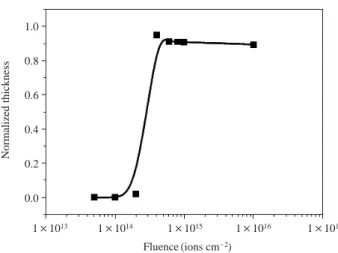
Fig. 2. Contrast curves of the PHS as a function of the fluence. 7×10¹² 6×10¹² 5×10¹² 4×10¹² 3×10¹² 2×10¹² 1×10¹² 0 1×10¹⁴ions cm-2 1×10¹⁵ions cm-2 1×10¹⁶ions cm-2 1500 2000 2500 3000
Depth from the surface (nm)
Concentration (atoms cm -2) 1×10¹³ 1×10¹⁴ 1×10¹⁵ 1×10¹⁶ 1×10¹⁷ Fluence (ions cm-2) 1.0 0.8 0.6 0.4 0.2 0.0 Normalized thickness
polymeric materials. The penetration depth of 200-keV H++
ions in the PHS was simulated by means of the TRIM98 pro-gram and the results are shown in Fig. 1. As shown in Fig. 1, the penetration depth of the 200-keV H++
ion into the PHS was around 2μm. Therefore, the 200-keV H++
ions can pen-etrate the PHS with a 300 nm thickness which can result in a homogeneous chemical reaction by ion irradiation.
To estimate the performance of the PHS as a negative type resist for ion beam lithography, the PHS was characterized by its contrast curve. Fig. 2 shows the contrast curves of the PHS. The remaining thickness of the PHS patterns after the development was increased with an increasing fluence up to 4×10¹⁴ions cm-2after which it leveled off. The measured
contrast and sensitivity of the PHS were 3.32 and 4×10¹⁴
ions cm-2, respectively.
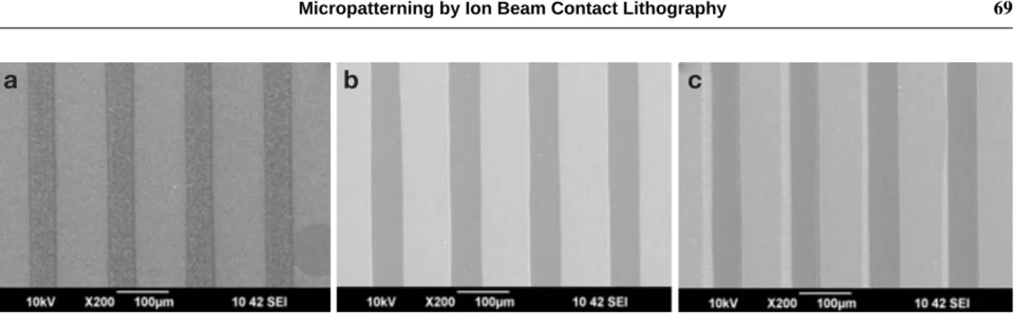
Fig. 2 shows the SEM images of the negative type patterns on a wafer prepared by ion irradiation at various fluences.
The surface of the patterns obtained at a fluence of 1×10¹⁴
ions cm-2was rough and had a lager number of pores (Fig.
3(a)). This implies that the fluence was not enough to cross-link the PHS, and thus uncrosscross-linked PHS in the irradiated regions was removed during the development process. How-ever, the patterns prepared at a fluence of 1×10¹⁵ions cm-2
were well-defined as shown in Fig. 3(b). For the patterns at a fluence of 1×10¹⁶ions cm-2, the surface of the patterns
was very smooth but the patterns were not well resolved (Fig. 3(c)). Therefore, the fluence of 1×10¹⁵ions cm-2was
the optimum point to resolve the negative patterns of the PHS within our experimental range.
Cell culture on the PHS-patterned wafers was carried out to investigate the cell adhesion and the results are presented in Fig. 4. Cultured cells were stained with DAPI nuclear staining solution and observed by a fluorescence microscope. On the control wafer, HaCaT human keratinocytes were ad-hered to all over the surface as shown in Fig. 4(a). However, as shown in Fig. 4(b), (c) and (d), the cells exhibited the pre-ferential adhesion only to the wafer surfaces, not to the pat-terned PHS surfaces. This result demonstrated that the PHS-patterned surfaces inhibited cell adhesion and proliferation regardless of the given fluence. Inhibition of cell adhesion on the irradiated PHS surface can be due to antifouling pro-perty of the irradiated PHS (Ertel et al. 1991; Adden et al. 2007; Choi et al. 2008).
CONCLUSION
In this study, PHS as one of the possible candidates for an ion beam contact lithography was introduced in order to con-trol cellular behavior. The PHS showed a reasonable sensi-tivity (4×10¹⁴ions cm-2) and contrast (3.32). Well-defined
Micropatterning by Ion Beam Contact Lithography 69
Fig. 3. SEM images of the PHS patterns obtained at fluences of 1×10¹⁴ (a), 1×10¹⁵(b), and 1×10¹⁶(c) ions cm-2.
Fig. 4. Fluorescence microscope images of the cell patterns on the
control (a) and PHS-patterned wafers at fluences of 1×10¹⁴
(b), 1×10¹⁵ (c), and 1×10¹⁶(d) ions cm-2.
a
b
c
a
b
50μm (pitch 150 μm) negative type patterns of the PHS were obtained successfully. The results of the cell culture test revealed that the cells were preferentially adhered to and proliferated only on the space regions between the PHS line patterns. Therefore, the irradiated PHS can be used as a antifouling material to control cellular behavior.
ACKNOWLEDGMENT
This research was supported by the Nuclear R & D pro-gram through the Korea Science and Engineering Founda-tion funded by the Ministry of EducaFounda-tion, Science and Tech-nology, Korea.
REFERENCES
Adden N, Hoffmann A, Gross G, Windhagen H, Thorey F and Menzel H. 2007. Screening of photochemically grafted polymer films for compatibility with osteogenic precursor cells. J. Biomater. Sci. Polymer Edn. 18:303-316.
Bhuvana T and Kulkarni GU. 2008. Polystyrene as a zwitter resist in electron beam lithography based electroless pattern-ing of gold. Bull. Mater. Sci. 31:201-206.
Choi YH, Kim JC, Ahn JK, Ko SY, Kim DH and Lee TY. 2008. Anti-biofouling behavior of natural unsaturated hyd-rocarbon phenols impregnated in PDMS matrix. J. Ind. Eng. Chem. 14:292-296.
Ertel SI, Chilkoti A, Horbett TA and Ratner BD. 1991. Endothe-lial cell growth on oxygen-containing films deposited by radio-frequency plasmas: the role of surface carbonyl groups. J. Biomater. Sci. Polymer Edn. 3:163-183.
Falconnet D, Csucs G, Grandin HM and Textor M. 2006. Sur-face engineering approaches to micropattern surSur-faces for
cell-based assays. Biomaterials 27:3044-3063.
Jagielski J, Piatkowska A, Aubert P, Thome L, Turos A and Kader AA. 2006. Ion implantation for surface modification of biomaterials. Surf. Coat. Technol. 200:6355-6361. Karp JM, Yeo Y, Geng W, Cannizarro C, Yan K, Kohane DS,
Vunjak-Novakovic G, Langer RS and Radisic M. 2006. A photolithographic method to creat cellular micropatterns. Biomaterials 27:4755-4764.
Kim DK, Jung CH, Kwon HJ, Choi JH, Nho YC, Suh DH, Ga-nesan R and Kim JB. 2009. Patterned grafting of acrylic acid onto polymer substrates. Polym. Adv. Technol. 20: 173-177.
Liu WF and Chen SC. 2005. Engineering biomaterials to cont-rol cell function. Mater. Today 8:28-35.
Nakanishi J, Takarada T, Yamaguchi K and Maeda M. 2008. Recent advances in cell micropatterning techniques for bioanalytical and biomedical sciences. Anal. Sci. 24:67-72. Nishizawa M, Takoh K and Matsue T. 2002. Micropatterning of HeLa cells on glass substrates and evaluation of respira-tory activity using microelectrodes. Langmuir 18:3645-3649.
Park JW, Sohn CW and Choi BH. 2006. Some characteristics of materials surface-modified by ions beam bombardment. Curr. Appl. Phys. 6:188-193.
Park TH and Shuler ML. 2003. Integrating of cell culture and microfabrication technology. Biotechnol. Prog. 19:243-253. Yamato M, Konno C, Koike S, Isoi Y, Shimizu T, Kikuchi A, Makino K and Okano T. 2003. Nanofabrication for micro-patterned cell arrays by combining electron beam-irradiated polymer grafting and localized laser ablation. J. Biomed. Mater. Res. 65A:1065-1071.
Yan M and Harnish B. 2003. A simple method for the attach-ment of polymer films on solid substrates. Adv. Mater. 15: 244-248.
Manuscript Received: May 25, 2009 Revision Accepted: June 29, 2009
In-Tae Hwang, Byoung-Min Lee, Chan-Hee Jung, Jae-Hak Choi, Young-Chang Nho and Sung-Kwon Hong