Research Article
Cyclooxygenase-2 Expression Is a Predictive Marker for Late
Recurrence in Colorectal Cancer
Sung Hoo Kim
,
1Byung Kyu Ahn,
1Seung Sam Paik
,
2and Kang Hong Lee
1 1Department of Surgery, Hanyang University College of Medicine, Seoul, Republic of Korea2Department of Pathology, Hanyang University College of Medicine, Seoul, Republic of Korea
Correspondence should be addressed to Kang Hong Lee; [email protected]
Received 27 December 2017; Revised 26 May 2018; Accepted 6 June 2018; Published 24 June 2018 Academic Editor: Gianluca Pellino
Copyright © 2018 Sung Hoo Kim et al. This is an open access article distributed under the Creative Commons Attribution License, which permits unrestricted use, distribution, and reproduction in any medium, provided the original work is properly cited. Introduction. Cyclooxygenase-2 (COX-2) expression is elevated in colorectal cancer (CRC). However, data about the relation between COX-2 expression and the impact on the biologic behavior of recurrent disease are inconclusive as yet. The aim of this study is to investigate the relationship between the status of COX-2 expression in the primary CRC and the characteristics of recurrence after curative resection of stage I to III CRC. Materials and Methods. Ninety-eight patients with recurrence in 376 CRC patients, who underwent curative surgery between January 1991 and August 2001, were retrospectively assessed. Immunohistochemical staining, performed for the presence of COX-2 on tissue microarrays, was analyzed. Results. Forty-six patients showed elevated COX-2 expression, and 52 patients did not. The mean time to recurrence was significantly longer in the positive group than in the negative group (34.1 months± 30.0 versus 21.9 months ± 17.4; P = 0 019). Positive COX-2 expression was correlated with late recurrence (>3 years after surgery) [43.5% versus 13.5%; P = 0 001]. In multivariate analysis, COX-2 expression was an independent factor associated with late recurrence (OR 4.656; 95% CI, 1.696 to 12.779;P = 0 003). Recurrence pattern and postrecurrence survival were not different between the two groups. Conclusions. Elevated COX-2 expression in itself is not a prognostic factor, but COX-2 expression in tumor tissue may be an independent predictive marker of late recurrence for patients with stage I to III CRC.
1. Introduction
The mainstay of colorectal cancer (CRC) treatment is curative resection, and tumor recurrence is a major concern after surgery. There have been several attempts to identify molecular markers that can predict recurrence and survival rates, but still, none is approved for clinical application.
Cyclooxygenase-2 (COX-2) is a rate-limiting enzyme involved in the conversion of arachidonic acid to prostaglan-dins and thromboxanes. These products play crucial roles in cell proliferation, immune response, angiogenesis, and inflammatory reaction, which may involve tumor develop-ment and progression [1, 2]. Previous studies have reported that COX-2 overexpression is detected in colorectal, gastric, breast, pulmonary, esophageal, and pancreatic cancer [3–7]. Increased COX-2 gene expression has been reported in human colorectal adenocarcinoma and in
carcinogen-induced rat colonic tumors [8–12]. However, the molecular mechanisms by which COX-2 contributes to CRC progres-sion and metastasis remain unclear. In addition, it remains controversial whether COX-2 expression is a prognostic factor for the survival of CRC patients or not [13–15].
The aim of this study is to investigate the relationship between the status of COX-2 expression and the characteris-tics of recurrent disease in stage I–III CRC patients after curative resection.
2. Methods
2.1. Selection of Patients. We retrospectively reviewed, between January 1991 and August 2001, 492 patients with the diagnosis of CRC and treated in one tertiary care center. 44 patients were excluded by the inclusion criteria. The inclu-sion criteria were stage I–III patients with curative resection Volume 2018, Article ID 7968149, 8 pages
(R0). Patients with (a) distant metastasis (n = 29), (b)
incom-plete resection (n = 2), and (c) metachronous cancers (n = 3) were excluded. The patients who expired due to other causes (n = 10) were also excluded. As a result, formalin-fixed paraffin-embedded samples from 376 patients were available. Patients with lymph node-positive disease received 5-FU-based adjuvant chemotherapy, and none of the patients received preoperative chemotherapy or radiotherapy. The median follow-up period was 56 months (range, 3 to 192 months). Of the 376 patients, COX-2 expression was elevated in 211 patients (56.0%, 211 patients), and the overall recur-rence rate was 26.0% (98/376 patients).
2.2. Tissue Microarrays (TMAs). After the histological examination of H&E-stained samples by an experienced pathologist, parts containing a high proportion of tumor cells were assembled. TMAs were constructed with a tissue arrayer (AccuMac Arrayer, ISU ABXIS Co. Ltd., Seoul, Korea). The assembled TMAs were held in an X-Y position guide with 1 mm increments between individual samples and a 3 mm punch-depth stop device. Briefly, this instrument was utilized to make holes in a recipient block with defined array cores, and a solid stylet, which fitted the needle closely, was used to transfer the tissue cores into the recip-ient block. Due to the limited size of representative areas of the tumors, triplicate 1 mm diameter tissue cores were made from each donor block.
2.3. Immunohistochemical Staining. We obtained multiple 4μm cut sections using a Leica microtome in immunohisto-chemical staining. The obtained sections were shifted to adhesive-coated slides. Dewaxing was performed with the TMA slides by heating at 55°C for 30 min and by three washes, of 5 min each, with xylene. Rehydration was done with the tissues by 5 min washes in 100%, 90%, and 70% eth-anol and phosphate-buffered saline (PBS). Antigen was retrieved by heating the samples for 4 min 20 s in a micro-wave at full power in 250 ml 10 mM sodium citrate (pH 6.0). Endogenous peroxidase activity was blocked with 0.3% hydrogen peroxidase for 20 min. The sections were incubated with primary goat polyclonal COX-2 anti-body (N-20; Santa Cruz Biotechnology, Santa Cruz, CA, USA) diluted 1 : 100 in goat serum at room temperature for 1 h. After three washes of 2 min each with PBS, the sections were incubated with biotinylated goat secondary anti-body for 30 min (DAKO, Carpinteria, CA, USA). After three further washes with PBS, horseradish peroxidase streptavidin (DAKO) was added to the section for 30 min, followed by another three washes. The samples were developed for 1 min with 3,3′-diaminobenzidine substrate (Vector Labora-tories, Burlington, Ontario, Canada) and counterstained with Mayer’s hematoxylin. They were dehydrated according to standard procedures and closed with coverslips.
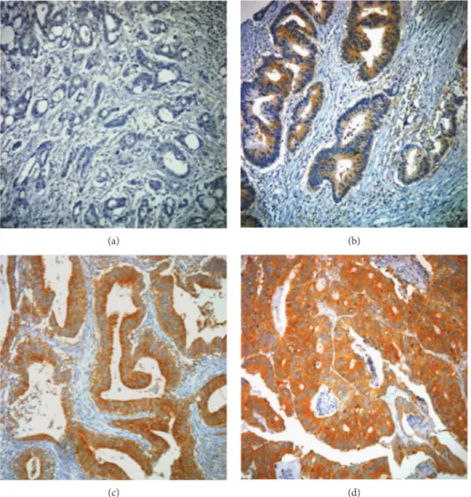
2.4. Interpretation of COX-2 Expression. COX-2 expression was interpreted independently by two experienced pathol-ogists (SS Paik and SH Jang) on the basis of staining intensity and extent. Three punches per case were evalu-ated and considered a whole. Staining intensity was scored
as 0 to 3 (0 = negative; 1 = weak; 2 = moderate; and 3 = strong) (Figure 1). Staining extent was scored as 0 to 4 based on the percentage of positive-stained cells (0 = 0%; 1 = 1–25%; 2 = 26–50%; 3 = 51–75%; and 4 = 76– 100%). The final staining score was determined with a sum of the intensity and extent score. We divided all cases into four expression groups based on their sum of scores (0 = negative; 1–3 = low; 4-5 = moderate; and 6-7 = high). If the sum of scores was ≥4, we classified the cases as ele-vated COX-2 expression (positive). If the sum of scores was ≤3, we classified the cases as COX-2 negative. When there was a disagreement between the two pathologists, reinvestigation of the slide was performed with a multi-headed microscope and the final agreement was achieved. 2.5. Statistical Analysis. Statistical analysis was conducted with SPSS ver. 19.0 (SPSS Inc., Chicago, IL, USA). The chi-square test and Student’s t-test were used to examine the association between COX-2 expression and clinical and path-ological features including age, gender, tumor location, tumor size, gross type, cell type, differentiation, lymphatic invasion, vascular invasion, T category, N category, AJCC stage, and time to recurrence. Survival curves were con-structed using the Kaplan-Meier method, and survival differ-ences were analyzed by the log-rank test.P values < 0.05 were considered statistically significant.
3. Results
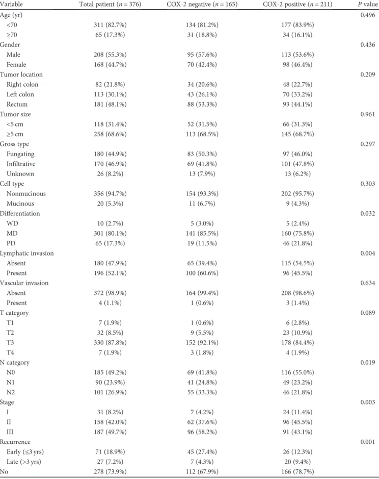
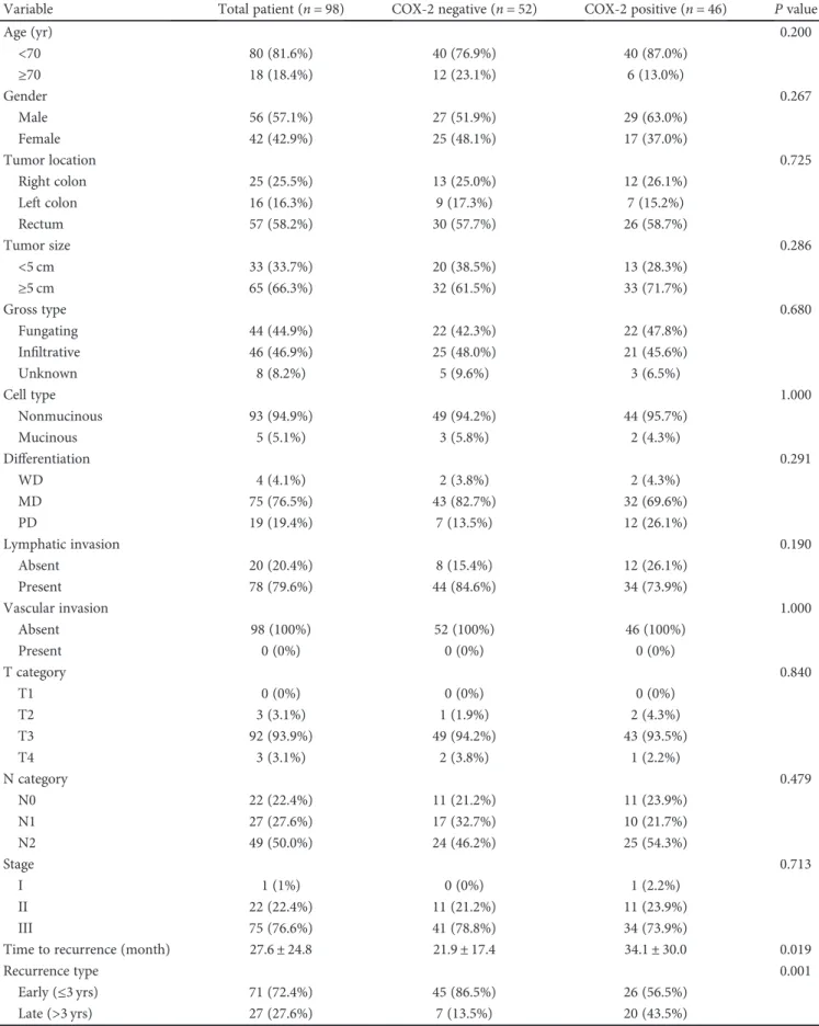
3.1. COX-2 Expression Is Related with Time to Recurrence. The clinical and pathological characteristics of the 376 patients are described in Table 1 according to the COX-2 expression status. In univariate analysis, the COX-2-positive group was different from the COX-2-negative group in terms of differentiation, lymphatic invasion, N category, AJCC stage, and recurrence rate. The clinical and pathologic findings of the 98 patients with tumor recurrence are included in Table 2. There were no statistically significant differences in terms of age, gender, tumor location, tumor size, gross type, cell type, differentiation, lymphatic invasion, vascular invasion, T category, N category, or AJCC stage between the two groups except for the time to recurrence. This was significantly longer in the COX-2-positive group than in the COX-2-negative group (34.1 months± 30.0 versus 21.9 months± 17.4, P = 0 019).
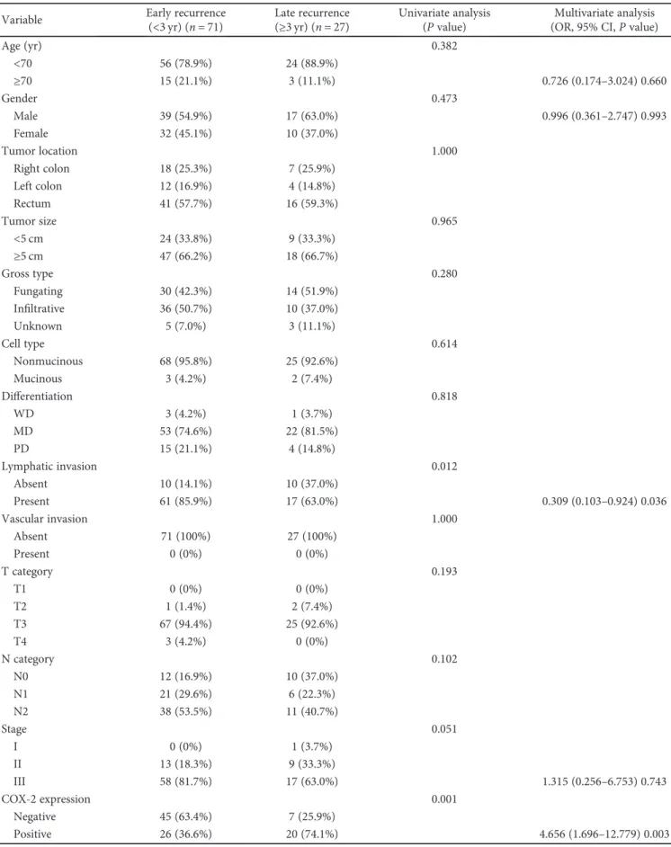
71 out of 98 patients with recurrence experienced early recurrence (72.4%), and 27 patients had late recurrence (27.6%). In univariate analysis, lymphatic invasion and positive COX-2 expression were significantly different between the two groups (P = 0 012 and P = 0 001, resp.) (Table 3). Positive lymphatic invasion was significantly correlated with early recurrence (P = 0 012), while positive COX-2 expression was significantly related with late recur-rence (P = 0 001). Multivariate analysis revealed that lym-phatic invasion was an independent factor for the early recurrence (OR 0.309; 95% CI, 0.103 to 0.924; P = 0 036), and positive COX-2 expression was an independent factor for the late recurrence (OR 4.656; 95% CI, 1.696 to 12.779; P = 0 003) (Table 3).
3.2. Recurrence Patterns and Postrecurrence Survival according to the COX-2 Expression Status. Thirty-two (8.5%) of the 376 patients experienced local recurrence, and 66 (17.5%) had distant metastasis. The most common site of distant metastasis was the liver (n = 25, 6.6%) followed by the lung (n = 19, 5.1%). The patterns of recurrence in the positive and the negative COX-2 expression groups were not different (Table 4). In the 98 patients with recurrence, there was no relation on postrecurrence survival according to the COX-2 expression status (Figure 2).
4. Discussion
Our results suggest that COX-2 expression in CRC is associ-ated with late recurrence (>3 years after surgery) during the postsurgery follow-up period, which may not mean that COX-2 expression prevents early recurrence. In this study, lymphatic invasion was a significant factor for the early recurrence but COX-2 expression was a significant factor for the late recurrence. We assumed that positive COX-2 expression do not prevent early recurrence but induce late recurrence. Maybe there are other mechanisms which
contribute to recurrence in the positive COX-2 expression group different from the lymphatic invasion group. A num-ber of mechanisms may be involved in the process of late recurrence. COX-2 overexpression increases the migration and proliferation of intestinal epithelial cell and inhibits pro-grammed cell death, so prolonging the survival of abnormal cells [16]. Interestingly, another study found that COX-2 overexpression was correlated with elevated intracellular tel-omerase and reduced apoptosis [17]. In nonsmall cell lung cancer, COX-2 overexpression has been shown to stabilize survivin, an inhibitor of apoptosis [18]. In breast cancer, the presence of cytoplasmic survivin positively correlates with COX-2 expression [19]. COX-2 and survivin are overex-pressed and positively correlated in endometrial adenocarci-noma [20]. Although our data do not have a bearing on molecular mechanisms, we suspect that the relation between COX-2 overexpression and late recurrence may be due to a decreased rate of apoptosis of surviving tumor cells. We sus-pect that surviving tumor cells in the stromal compartment may grow and migrate to other organs, and time may be required for surviving tumor cells to acquire resistance to adjuvant treatment.
(a) (b)
(c) (d)
Figure 1: The microphotographs of COX-2 immunostaining by intensity in colorectal cancer: (a) negative, (b) weak, (c) moderate, and (d) strong.
Table 1: Clinical and pathological characteristics according to COX-2 expression in 376 patients with R0 resection.
Variable Total patient (n = 376) COX-2 negative (n = 165) COX-2 positive (n = 211) P value
Age (yr) 0.496 <70 311 (82.7%) 134 (81.2%) 177 (83.9%) ≥70 65 (17.3%) 31 (18.8%) 34 (16.1%) Gender 0.436 Male 208 (55.3%) 95 (57.6%) 113 (53.6%) Female 168 (44.7%) 70 (42.4%) 98 (46.4%) Tumor location 0.209 Right colon 82 (21.8%) 34 (20.6%) 48 (22.7%) Left colon 113 (30.1%) 43 (26.1%) 70 (33.2%) Rectum 181 (48.1%) 88 (53.3%) 93 (44.1%) Tumor size 0.961 <5 cm 118 (31.4%) 52 (31.5%) 66 (31.3%) ≥5 cm 258 (68.6%) 113 (68.5%) 145 (68.7%) Gross type 0.297 Fungating 180 (44.9%) 83 (50.3%) 97 (46.0%) Infiltrative 170 (46.9%) 69 (41.8%) 101 (47.8%) Unknown 26 (8.2%) 13 (7.9%) 13 (6.2%) Cell type 0.303 Nonmucinous 356 (94.7%) 154 (93.3%) 202 (95.7%) Mucinous 20 (5.3%) 11 (6.7%) 9 (4.3%) Differentiation 0.032 WD 10 (2.7%) 5 (3.0%) 5 (2.4%) MD 301 (80.1%) 141 (85.5%) 160 (75.8%) PD 65 (17.3%) 19 (11.5%) 46 (21.8%) Lymphatic invasion 0.004 Absent 180 (47.9%) 65 (39.4%) 115 (54.5%) Present 196 (52.1%) 100 (60.6%) 96 (45.5%) Vascular invasion 0.634 Absent 372 (98.9%) 164 (99.4%) 208 (98.6%) Present 4 (1.1%) 1 (0.6%) 3 (1.4%) T category 0.089 T1 7 (1.9%) 1 (0.6%) 6 (2.8%) T2 32 (8.5%) 9 (5.5%) 23 (10.9%) T3 330 (87.8%) 152 (92.1%) 178 (84.4%) T4 7 (1.9%) 3 (1.8%) 4 (1.9%) N category 0.019 N0 185 (49.2%) 69 (41.8%) 116 (55.0%) N1 90 (23.9%) 41 (24.8%) 49 (23.2%) N2 101 (26.9%) 55 (33.3%) 46 (21.8%) Stage 0.003 I 31 (8.2%) 7 (4.2%) 24 (11.4%) II 158 (42.0%) 62 (37.6%) 96 (45.5%) III 187 (49.7%) 96 (58.2%) 91 (43.1%) Recurrence 0.001 Early (≤3 yrs) 71 (18.9%) 45 (27.4%) 26 (12.3%) Late (>3 yrs) 27 (7.2%) 7 (4.3%) 20 (9.4%) No 278 (73.9%) 112 (67.9%) 166 (78.7%)
Table 2: Clinical and pathological characteristics according to COX-2 expression in patients with recurrence.
Variable Total patient (n = 98) COX-2 negative (n = 52) COX-2 positive (n = 46) P value
Age (yr) 0.200 <70 80 (81.6%) 40 (76.9%) 40 (87.0%) ≥70 18 (18.4%) 12 (23.1%) 6 (13.0%) Gender 0.267 Male 56 (57.1%) 27 (51.9%) 29 (63.0%) Female 42 (42.9%) 25 (48.1%) 17 (37.0%) Tumor location 0.725 Right colon 25 (25.5%) 13 (25.0%) 12 (26.1%) Left colon 16 (16.3%) 9 (17.3%) 7 (15.2%) Rectum 57 (58.2%) 30 (57.7%) 26 (58.7%) Tumor size 0.286 <5 cm 33 (33.7%) 20 (38.5%) 13 (28.3%) ≥5 cm 65 (66.3%) 32 (61.5%) 33 (71.7%) Gross type 0.680 Fungating 44 (44.9%) 22 (42.3%) 22 (47.8%) Infiltrative 46 (46.9%) 25 (48.0%) 21 (45.6%) Unknown 8 (8.2%) 5 (9.6%) 3 (6.5%) Cell type 1.000 Nonmucinous 93 (94.9%) 49 (94.2%) 44 (95.7%) Mucinous 5 (5.1%) 3 (5.8%) 2 (4.3%) Differentiation 0.291 WD 4 (4.1%) 2 (3.8%) 2 (4.3%) MD 75 (76.5%) 43 (82.7%) 32 (69.6%) PD 19 (19.4%) 7 (13.5%) 12 (26.1%) Lymphatic invasion 0.190 Absent 20 (20.4%) 8 (15.4%) 12 (26.1%) Present 78 (79.6%) 44 (84.6%) 34 (73.9%) Vascular invasion 1.000 Absent 98 (100%) 52 (100%) 46 (100%) Present 0 (0%) 0 (0%) 0 (0%) T category 0.840 T1 0 (0%) 0 (0%) 0 (0%) T2 3 (3.1%) 1 (1.9%) 2 (4.3%) T3 92 (93.9%) 49 (94.2%) 43 (93.5%) T4 3 (3.1%) 2 (3.8%) 1 (2.2%) N category 0.479 N0 22 (22.4%) 11 (21.2%) 11 (23.9%) N1 27 (27.6%) 17 (32.7%) 10 (21.7%) N2 49 (50.0%) 24 (46.2%) 25 (54.3%) Stage 0.713 I 1 (1%) 0 (0%) 1 (2.2%) II 22 (22.4%) 11 (21.2%) 11 (23.9%) III 75 (76.6%) 41 (78.8%) 34 (73.9%)
Time to recurrence (month) 27.6± 24.8 21.9± 17.4 34.1± 30.0 0.019
Recurrence type 0.001
Early (≤3 yrs) 71 (72.4%) 45 (86.5%) 26 (56.5%)
Table 3: Univariate and multivariate analysis of independent risk factors associated with late recurrence. Variable Early recurrence(<3 yr) (n = 71) Late recurrence(≥3 yr) (n = 27) Univariate analysis
(P value)
Multivariate analysis (OR, 95% CI,P value)
Age (yr) 0.382 <70 56 (78.9%) 24 (88.9%) ≥70 15 (21.1%) 3 (11.1%) 0.726 (0.174–3.024) 0.660 Gender 0.473 Male 39 (54.9%) 17 (63.0%) 0.996 (0.361–2.747) 0.993 Female 32 (45.1%) 10 (37.0%) Tumor location 1.000 Right colon 18 (25.3%) 7 (25.9%) Left colon 12 (16.9%) 4 (14.8%) Rectum 41 (57.7%) 16 (59.3%) Tumor size 0.965 <5 cm 24 (33.8%) 9 (33.3%) ≥5 cm 47 (66.2%) 18 (66.7%) Gross type 0.280 Fungating 30 (42.3%) 14 (51.9%) Infiltrative 36 (50.7%) 10 (37.0%) Unknown 5 (7.0%) 3 (11.1%) Cell type 0.614 Nonmucinous 68 (95.8%) 25 (92.6%) Mucinous 3 (4.2%) 2 (7.4%) Differentiation 0.818 WD 3 (4.2%) 1 (3.7%) MD 53 (74.6%) 22 (81.5%) PD 15 (21.1%) 4 (14.8%) Lymphatic invasion 0.012 Absent 10 (14.1%) 10 (37.0%) Present 61 (85.9%) 17 (63.0%) 0.309 (0.103–0.924) 0.036 Vascular invasion 1.000 Absent 71 (100%) 27 (100%) Present 0 (0%) 0 (0%) T category 0.193 T1 0 (0%) 0 (0%) T2 1 (1.4%) 2 (7.4%) T3 67 (94.4%) 25 (92.6%) T4 3 (4.2%) 0 (0%) N category 0.102 N0 12 (16.9%) 10 (37.0%) N1 21 (29.6%) 6 (22.3%) N2 38 (53.5%) 11 (40.7%) Stage 0.051 I 0 (0%) 1 (3.7%) II 13 (18.3%) 9 (33.3%) III 58 (81.7%) 17 (63.0%) 1.315 (0.256–6.753) 0.743 COX-2 expression 0.001 Negative 45 (63.4%) 7 (25.9%) Positive 26 (36.6%) 20 (74.1%) 4.656 (1.696–12.779) 0.003
Some studies have suggested that elevated COX-2 expression of CRC patients is related with reduced survival [15, 21]. However, others found that the elevated expres-sion of COX-2 protein had no significant impact on disease-specific survival and overall survival in CRC patients [22, 23]. We observed no significant postrecur-rence survival diffepostrecur-rence according to COX-2 expression status. Thus, COX-2 expression is not likely a prognostic factor for postrecurrence in CRC.
After curative surgery, CRC patients with positive COX-2 expression have an increased probability of late tumor recur-rence based on the result of this study. Therefore, the positive COX-2 patients should be considered candidates for more frequent testing after 3 years of follow-up and extend follow-up period longer than 5 years after surgery. In proto-cols for postsurgery surveillance, there is a tendency for the frequency of follow-up and testing to be reduced after 3 years. We suggest that since COX-2 expression may be a
marker for late recurrence, the frequency of follow-up and testing should not be reduced after 3 years. Furthermore, sus-pending follow-up after 5 years from the initial operation may be inappropriate especially in COX-2-positive patients. A further prospective randomized study is required to iden-tify optimal surveillance methods and follow-up intervals.
As smoking habit and body mass index may modify the risk of CRC in COX-2 genotype, this bias could affect our conclusions regarding the predictive marker [24]. A further well-designed, large, sample-sized study is mandatory.
A limitation of this study was its retrospective design, which is subject to selection bias. Also, the cases were all from a single institution. As no molecular biological study was per-formed, it was not clear how COX-2 expression contributed to late recurrence. But it is meaningful that the study revealed a novel finding about the relationship between elevated COX-2 expression and late recurrence of CRC: we were able to demonstrate the possibility of COX-2 expression as a bio-logic marker predicting late recurrence in CRC patients.
5. Conclusions
Elevated COX-2 expression in itself is not a prognostic factor, but COX-2 expression in tumor tissue may be an indepen-dent predictive marker of late recurrence for patients with stage I to III CRC. A further well-designed study is required to demonstrate the regulatory mechanism of COX-2 expres-sion on CRC recurrence.
Conflicts of Interest
The authors declare that they have no conflicts of interest.
Authors
’ Contributions
Sung Hoo Kim contributed to the collection of data, analysis, interpretation, and writing the article. Byung Kyu Ahn con-tributed to the critical revision of the article. Seung Sam Paik contributed to the critical revision of the article. Kang Hong Lee contributed to the conceiving of the design and approved thefinal version of the manuscript.
References
[1] J. R. Vane, Y. S. Bakhle, and R. M. Botting,“Cyclooxygenases 1 and 2,” Annual Review of Pharmacology and Toxicology, vol. 38, no. 1, pp. 97–120, 1998.
[2] K. Subbaramaiah and A. J. Dannenberg,“Cyclooxygenase-2: a molecular target for cancer prevention and treatment,” Trends in Pharmacological Sciences, vol. 24, no. 2, pp. 96–102, 2003. [3] A. Ristimäki, N. Honkanen, H. Jänkälä, P. Sipponen, and
M. Härkönen,“Expression of cyclooxygenase-2 in human gas-tric carcinoma,” Cancer Research, vol. 57, no. 7, pp. 1276– 1280, 1997.
[4] T. Hida, Y. Yatabe, H. Achiwa et al.,“Increased expression of cyclooxygenase 2 occurs frequently in human lung cancers, specifically in adenocarcinomas,” Cancer Research, vol. 58, no. 17, pp. 3761–3764, 1998.
[5] D. Hwang, J. Byrne, D. Scollard, and E. Levine,“Expression of cyclooxygenase-1 and cyclooxygenase-2 in human breast Table 4: Recurrence pattern according to the status of COX-2
expression in the 98 patients with recurrence. COX-2 negative (n = 52) COX-2 positive (n = 46) P value 0.256 Local recurrence 14 (26.9%) 18 (39.1%) Liver 13 (25.0%) 12 (26.1%) Lung 9 (17.3%) 10 (21.7%) Peritoneal seeding 5 (9.6%) 3 (6.5%) Others (brain, bone, skin) 11 (21.2%) 3 (6.5%) COX-2 negative COX-2 positive 1.0 0.8 0.6 0.4 0.2 0.0 .0 50.0 100.0 150.0 200.0 Follow-up period (month)
O ve rall s u rv iv al ra te
Figure 2: Postrecurrence survival was not significantly different according to the COX-2 status of the 98 patients with recurrence
cancer,” Journal of the National Cancer Institute, vol. 90, no. 6, pp. 455–460, 1998.
[6] K. C. Zimmermann, M. Sarbia, A. A. Weber, F. Borchard, H. E. Gabbert, and K. Schrör, “Cyclooxygenase-2 expression in human esophageal carcinoma,” Cancer Research, vol. 59, no. 1, pp. 198–204, 1999.
[7] O. N. Tucker, A. J. Dannenberg, E. K. Yang et al., “Cyclooxy-genase-2 expression is up-regulated in human pancreatic cancer,” Cancer Research, vol. 59, no. 5, pp. 987–990, 1999. [8] J. Dimberg, A. Samuelsson, A. Hugander, and P. Soderkvist,
“Differential expression of cyclooxygenase 2 in human colo-rectal cancer,” Gut, vol. 45, no. 5, pp. 730–732, 1999. [9] C. E. Eberhart, R. J. Coffey, A. Radhika, F. M. Giardiello,
S. Ferrenbach, and R. N. Dubois,“Up-regulation of cyclooxy-genase 2 gene expression in human colorectal adenomas and adenocarcinomas,” Gastroenterology, vol. 107, no. 4, pp. 1183–1188, 1994.
[10] H. Sano, Y. Kawahito, R. L. Wilder et al., “Expression of cyclooxygenase-1 and -2 in human colorectal cancer,” Cancer Research, vol. 55, no. 17, pp. 3785–3789, 1995.
[11] R. N. DuBois, A. Radhika, B. S. Reddy, and A. J. Entingh, “Increased cyclooxygenase-2 levels in carcinogen-induced rat colonic tumors,” Gastroenterology, vol. 110, no. 4, pp. 1259–1262, 1996.
[12] P. de Heer, M. J. E. M. Gosens, E. C. de Bruin et al., “Cycloox-ygenase 2 expression in rectal cancer is of prognostic signi fi-cance in patients receiving preoperative radiotherapy,” Clinical Cancer Research, vol. 13, no. 10, pp. 2955–2960, 2007. [13] E. Rahme, A. N. Barkun, Y. Toubouti, and M. Bardou,“The cyclooxygenase-2-selective inhibitors rofecoxib and celecoxib prevent colorectal neoplasia occurrence and recurrence,” Gastroenterology, vol. 125, no. 2, pp. 404–412, 2003. [14] T. Fujita, M. Matsui, K. Takaku et al.,“Size- and
invasion-dependent increase in cyclooxygenase 2 levels in human colorectal carcinomas,” Cancer Research, vol. 58, no. 21, pp. 4823–4826, 1998.
[15] M. Rao, W. Yang, A. M. Seifalian, and M. C. Winslet,“Role of cyclooxygenase-2 in the angiogenesis of colorectal cancer,” International Journal of Colorectal Disease, vol. 19, no. 1, pp. 1–11, 2004.
[16] A. J. Dannenberg, N. K. Altorki, J. O. Boyle et al., “Cyclo-oxy-genase 2: a pharmacological target for the prevention of can-cer,” The Lancet Oncology, vol. 2, no. 9, pp. 544–551, 2001. [17] Z. H. Zhuang, S. W. Tsao, W. Deng et al.,“Early upregulation
of cyclooxygenase-2 in human papillomavirus type 16 and telomerase-induced immortalization of human esophageal epithelial cells,” Journal of Gastroenterology and Hepatology, vol. 23, no. 10, pp. 1613–1620, 2008.
[18] K. Krysan, H. Dalwadi, S. Sharma, M. Põld, and S. Dubinett, “Cyclooxygenase 2-dependent expression of survivin is critical for apoptosis resistance in non-small cell lung cancer,” Cancer Research, vol. 64, no. 18, pp. 6359–6362, 2004.
[19] N. Barnes, P. Haywood, P. Flint, W. F. Knox, and N. J. Bundred,“Survivin expression in in situ and invasive breast cancer relates to COX-2 expression and DCIS recurrence,” British Journal of Cancer, vol. 94, no. 2, pp. 253–258, 2006. [20] S. Erkanli, F. Bolat, F. Kayaselcuk, B. Demirhan, and E. Kuscu, “COX-2 and survivin are overexpressed and positively corre-lated in endometrial carcinoma,” Gynecologic Oncology, vol. 104, no. 2, pp. 320–325, 2007.
[21] L. T. Soumaoro, H. Uetake, T. Higuchi, Y. Takagi, M. Enomoto, and K. Sugihara,“Cyclooxygenase-2 expression: a significant prognostic indicator for patients with colorectal cancer,” Clinical Cancer Research, vol. 10, no. 24, pp. 8465– 8471, 2004.
[22] K. Zafirellis, G. Agrogiannis, and A. Zachaki, “Prognostic value of COX-2 immunohistochemical expression evaluated by quantitative image analysis in colorectal cancer,” APMIS, vol. 116, no. 10, pp. 912–922, 2008.
[23] R. Fux, M. Schwab, K. P. Thon, C. H. Gleiter, and P. Fritz, “Cyclooxygenase-2 expression in human colorectal cancer is unrelated to overall patient survival,” Clinical Cancer Research, vol. 11, no. 13, pp. 4754–4760, 2005.
[24] L. L. Xing, Z. N. Wang, L. Jiang et al.,“Cyclooxygenase 2 poly-morphism and colorectal cancer: -765G>C variant modifies risk associated with smoking and body mass index,” World Journal of Gastroenterology, vol. 14, no. 11, pp. 1785–1789, 2008.
Stem Cells
International
Hindawi www.hindawi.com Volume 2018 Hindawi www.hindawi.com Volume 2018 INFLAMMATIONEndocrinology
International Journal ofHindawi www.hindawi.com Volume 2018 Hindawi www.hindawi.com Volume 2018
Disease Markers
Hindawi www.hindawi.com Volume 2018 BioMed Research InternationalOncology
Journal of Hindawi www.hindawi.com Volume 2013 Hindawi www.hindawi.com Volume 2018Oxidative Medicine and Cellular Longevity
Hindawi
www.hindawi.com Volume 2018
PPAR Research
Hindawi Publishing Corporation
http://www.hindawi.com Volume 2013 Hindawi www.hindawi.com
The Scientific
World Journal
Volume 2018 Immunology Research Hindawi www.hindawi.com Volume 2018 Journal ofObesity
Journal of Hindawi www.hindawi.com Volume 2018 Hindawi www.hindawi.com Volume 2018 Computational and Mathematical Methods in Medicine Hindawi www.hindawi.com Volume 2018Behavioural
Neurology
Ophthalmology
Journal of Hindawi www.hindawi.com Volume 2018Diabetes Research
Journal ofHindawi
www.hindawi.com Volume 2018
Hindawi
www.hindawi.com Volume 2018 Research and Treatment
AIDS
Hindawi
www.hindawi.com Volume 2018
Gastroenterology Research and Practice
Hindawi www.hindawi.com Volume 2018