사람 정상 코점막 상피세포에서 섬모세포의 표식자로서
MUC8에 관한 연구
연세대학교 의과대학 이비인후과학교실,1 기도점액 연구소,2 두뇌한국 21 의과학사업단3김창훈
1,2·유종범
1·이욱진
1·김경수
1,2,3·이정권
1,2·윤주헌
1,2,3MUC8 as a Ciliated Cell Marker in Human Nasal Epithelium
Chang-Hoon Kim, MD1,2, Jong-Bum Yoo, MD1, Wook Jin Lee, MD1,
Kyung-Su Kim, MD1,2,3, Jeung-Gweon Lee, MD1,2 and Joo-Heon Yoon, MD1,2,3
1
Department of Otorhinolaryngology, 2
The Airway Mucus Institute and 3
Brain Korea 21 Project for Medical Sciences, Yonsei University College of Medicine, Seoul, Korea
ABSTRACT
Background and Objectives:To examine the MUC8 mRNA expression patterns according to the mucociliary differentiation of the normal human nasal epithelial (NHNE) cells, and to investigate the localization of the MUC8 proteins in the nasal polyps. Materials and Methods:The passage-2 NHNE cells were cultured using an air-liquid interface technique and nasal polyp spe-cimens. On the 2, 7, 14, and 28 days after confluence, the ciliated cells were counted using cytospin slide immunostaining using H6C5 and β-tubulin, and the MUC8 mRNA levels were determined using real-time quantitative PCR. After synthesizing the po-lyclonal anti-MUC8 peptide antibodies, MUC8 immunostaining was preformed using the nasal polyps. The MUC8 mRNA and protein levels were determined with the NHNE cells treated with IL-1β (10 ng/ml for 24 hours) using RT-PCR and Western blot analysis. Results:The increasing pattern of the number of ciliated cells as well as the MUC8 gene expression level with increa-sing culture time in the NHNE cells was quite similar. MUC8 was expressed in the ciliated cells of the human nasal polyps. The MUC8 protein level as well as the mRNA level was up-regulated as a result of the IL-1β treatment. Conclusions:This study in-dicates that the MUC8 protein is expressed in ciliated cells from the human nasal epithelial cells and is up-regulated by the IL-1β treatment. These results suggest that the MUC8 gene and protein expression levels might be used as a ciliated cell marker in the human nasal epithelium. (Korean J Otolaryngol 2005;48:455-9)
KEY WORDS:MUC8·Ciliated cell·Nasal epithelial cell.
서 론
호흡기 점액은 호흡 상피세포의 물리적 또는 화학적인 보 호와 함께 먼지, 세균, virus 등에 대한 점액섬모 정화작용 (mucociliary clearance)을 함으로써 기도를 보호하는데 중 요한 역할을 한다. 그러나 점액의 과분비는 만성 부비동 염 이나 알레르기성 비염, 기관지 천식, 낭포성 섬유증 및 만 성 폐쇄성 폐질환 등 만성 염증성 호흡기 질환의 주요 병리 현상 중 하나이기도 하다. 점소(mucin)는 고도로 당질화된 고분자의 당단백질로 이는 호흡기의 상피에 의해 생성되는 점액(mucus)의 주 성분이다. 실제적으로 모든 형태의 호흡 기 염증 질환은 점액의 과다분비와 관련이 있으며 이는 기 도의 폐쇄를 일으킬 수 있다.1) 최근까지, 사람에서는 19가지의 점액 유전자들이 발견되 었다. MUC1, MUC2, MUC3, MUC4, MUC5AC, MUC5B, MUC6, MUC7, MUC8,1) MUC9,2) MUC11, MUC12, MUC13, MUC15, MUC16, MUC17, MUC18, MUC193) 그리고 MUC20이다. 이 가운데 MUC5AC와 MUC5B는 사 람의 기도에서 분비되는 점액의 겔(gel) 형성의 주요 유전 자이다.4)5) 그러나 비록 MUC5AC가 대부분의 배세포에서 발현된다고 알려져 왔지만,6) 저자들의 실험에 따르면 MUC-5AC mRNA는 염증반응에 의해 자극되지 않은 후사골동의 정상 점막에서 얻어진 배세포에서는 발현되지 않는다.7) MUC1, MUC4, MUC6, MUC7, MUC8, MUC13들은 상피 세포에서 발현된다고 보고 되었지만 그 기능은 아직 밝혀지 논문접수일:2004년 8월 10일 / 심사완료일:2004년 10월 11일 교신저자:윤주헌, 120-749 서울 서대문구 신촌동 134 연세대학교 의과대학 이비인후과학교실 전화:(02) 361-8484・전송:(02) 393-0580 E-mail:[email protected]지 않고 있다. 이전의 생체실험에서 MUC8 mRNA의 수치 가 염증 매개체에 의해 비용종 상피세포에서 증가된다고 보 고 되었고8) 생체 외 실험에서 interleukin-1β와 TNF-α 가 MUC8 mRNA의 발현을 증가시킨다고 알려져 있다.9)10) 따 라서 염증상태에서 MUC8 mRNA의 발현이 증가 될 수 있다 고 생각되었으나 MUC8이 배세포 또는 섬모세포 중 어느 세 포에서 주로 발현되는지, 또한 MUC8이 실제로 염증 매개체에 의해 그 단백질이 증가되는지는 확실하게 밝혀지지 않았다. 본 연구는 사람 정상 코점막 상피세포의 점액 섬모의 분 화에 따른 MUC8 mRNA의 발현 방식을 알아보고 비내 용 종에서 MUC8 단백질의 국재화(localization)에 대해 조사 하는 것을 목적으로 하였으며, 나아가 MUC8이 IL1-β에 의해 mRNA와 단백질의 발현량 모두에서 상승 조절되는지 알아보고자 하였다.
재료 및 방법
Air-liquid interface(ALI) 배양 105개의 인체 정상 코점막 상피세포(passage-2)를 반투 과성 막(Transwell-clear, Costar Co., Cambridge, MA) 이 있는 0.5 ml의 배양액에 심었다. 여기서 사용된 배양액 은 basal epithelial growth medium(BEGM)과 이전에 기 술된 Dulbecco’s modified Eagle’s medium(DMEM)을 1:1로 혼합한 용액11)이며, 세포들은 첫 9일간은 배양액에 잠긴 상태로 두었으며, 이 기간동안 배양액을 첫날 그리고 그 이후로는 격일로 갈아 주었다. 9일째 배양액의 윗부분을 제거하고 아래 부분에만 배양액을 주어서 ALI를 만든 이후 배양액을 매일 갈아 주었다. MUC8 mRNA수치를 결정하 기위해 이전 방법대로 합류를 이루고 난 후 2일, 7일, 14일 그리고 28일에 총 RNA를 채취하였다. 분비세포 및 섬모 세포의 정량 면역세포화학염색을 위해 사용된 cytospin 슬라이드가 각 각의 배양기간 동안 만들어 졌다. 분비세포는 점액에 대한 단클론 항체(H6C5;1:1000, University of North Ca-rolina, NC, Dr. Davis CW로부터 기증)와 β-tubulin에 대한 항체(Sigma, St. Louis, MO)를 이용하여 검출하였다. H6C5와 tubulin 항체에 양성인 세포들의 평균 수는 각 sl-ide마다 1000개의 세포로 결정되었고, 통계학적 처리는 Stu-dent t-test로 하였다.Real-time quantitative PCR
Primer와 probe는 PE Biosystems에서 구입한 Perkin
Elmer Primer Express software를 이용하여 만들어 졌 으며, Commercial reagents(TaqMan PCR Universal PCR Master Mix:PE Biosystems, Foster City, CA)와 그 조 건들은 제조사의 protocol을 따라 시도되었다. 총 25 μl 중 1 μl의 cDNA(reverse transcription 혼합물)와 800 nM 의 최종농도를 가지는 primer, 그리고 200 nM의 TaqMan hybridization probe가 포함되었다. real-time PCR의 probe 는 5’end에서는 carboxyfluoroscein(FAM)으로 표지하였 고, 3’end 에서는 quencher carboxytetramethylrhoda-mine(TAMRA)로 표지하였다. 다음과 같은 primer와 Taq-Man probe가 이용되었다.
:MUC8, forward 5’-GACCTGCCCCCATGGAC-3’와 reverse 5’-CAGGAGTTCGAGACCAGCCT-3’그리고 Taqman probe 6FAM-CCACCTCCGAGCCCGTCACT-GAG-TAMRA. β-2microglobulin(β2M)은 forward 5’ -CGCTCCGTGGCCTTAGC-3’와 reverse 5’-GAGT-ACGCTGGATAGCCTCCA-3’그리고 Taqman probe로 6FAM-TGCTCGCGCTACTCTCTCTTTCTGGC-TA-MRA가 사용되었다. Real-time RT-PCR은 PE Biosys-tems ABI PRISM 7700 Sequence Detection System (Foster City, CA)을 이용하여 실행하였으며, thermocycler (ABI PRISM 7700 Sequence Detection System) para-meters는 50℃에서 2분간, 95℃에서 10분간, 그리고 95℃ 15초, 60℃ 1분간으로 40 cycle을 실행하였다. 모든 실험 은 세차례 반복하였으며, MUC8 mRNA의 상대적 정량은 comparative cycle of the threshold(CT) method를 이용 하여 얻었고, 대조군으로서β-2M을 사용하였다.
MUC8 peptide합성과 특이항혈청의 생성
KTSCPRPLQEGTPGS sequence를 가진 15-mer pep-tide가 합성되었고12) glutaraldehyde를 이용하여 keyhole limpet hemocyanin(KLH)와 결합시켰다. KLH-peptide 결합체는 실험용 토끼에서 다클론 항체를 증가시키기 위한 항원으로서 이용되었다.
MUC8에 대한 면역조직화학 염색
비용 조직은 24시간동안 4% paraformaldehyde에서 고 정되었고 사용될 때까지 12%와 18% sucrose로 냉동 보 존되었다. insert는 10 μm두께로 잘라졌고 frozen section 은 MUC8에 대한 항체로 염색되었다. 항체와 항원의 반응 은 peroxidase와 결합된 anti-rabbit 이차항체를 이용하여 검출되었다. 음성 대조군은 일차항체를 생략하고 일차항체 와 무관한 rabbit IgG를 정화하여 사용하였다.
사람 정상 코점막 세포의 IL-1β 처치
사람 정상 코점막 세포(passage 2)가 6개의 well plate 에서 합류를 이룰때까지 배양하였다. ALI가 만들어진 후 2 일째 그 세포들은 24시간동안 IL-1β(10 ng/ml)로 처치 하였고, 총 RNA와 단백질은 MUC8의 RT-PCR과 Wes-tern blot analysis를 위해 모았다.
IL-1β로 처리된 사람 정상 코점막 세포에서 MUC8에 대 한 RT-PCR
Oligonucleotide primers를 이미 알려진 염기 서열대로 디자인하였다.8) β2M에 대한 oligonucleotide amplimers 는 RT-PCR의 대조군으로서, Clontech Laboratories(Palo Alto, CA;they generated a 335-bp PCR fragment)로 부터 구입하였다. RT-PCR은 Perkin-Elmer Cetus DNA Thermal Cycler(Perkin-Elmer, Norwalk, CT)을 이용 하여, 제조사의 방법대로 시행하였다. 총 RNA(1 μg/20 μl reaction volume)를 random hexanucleotide primers와 Moloney murine leukemia virus RT를 이용하여 cDNA 로 역전사하였다. 각 유전자의 mRNA 발현을 비교하기 위 해 비교역학분석(Comparative kinetic analysis)을 시행하 였다. PCR 산물은 50 ng/mL ethidium bromide를 함유하 는 2% Seakem agarose gel(FMC, Rockland, ME)에서 전기 영동으로 분리하여 폴라로이드 타입 55 필름으로 사 진을 찍었다. 최종 산물(amplified products)이 mRNA에서 생기고 genomic DNA의 오염이 없었다는 것을 증명하기 위 해 역전사 반응에서 역전사 효소(reverse transcriptase) 를 생략하여 어떤 PCR 산물도 관찰할 수 없음을 확인함으 로써 음성 대조군(negative control)으로 삼았다. IL-1β로 처치한 정상 사람 코점막 상피세포의 MUC8에 대한 Western 분석
IL-1β처치된 세포들은 2× lysis buffer[250 mM Tris-Cl(pH 6.5), 2% SDS, 4%-mercaptoethanol, 0.02% BPB,
10% glycerol]에서 용해시켰다. 동량의 cell lysate를 6% SDS-PAGE에서 전기영동시켰고 polyvinylidene difluo-ride membrane(PVDF;Millipore, Bedford, MA)로 이동 시켰다. PVDF는 실온에서 2시간 동안 Tris-buffered sa-line[50 mM Tris-Cl(pH 7.5), 150 mM NaCl]에서 5% skim milk와 반응시킨후 MUC8 다클론 항체를 포함하여 밤 새 반응시켰으며, TTBS로 세척 후 anti-rabbit 이차항체 (Cell Signaling Tech., Beverly, MA)와 함께 실온에서 45분간 더 배양하였고 이를 ECL system(Amersham-Pharmacia, Piscataway, NJ)을 이용해 발색시켰다.
결 과
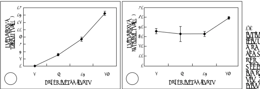
배양기간에 따른 분비세포와 섬모세포의 비율과 MUC8 발 현의 상관관계 정상 사람 코점막 상피세포는 레티노익산(retinoic acid) 이 있는 상태에서 배양되었는데 β-tubulin 항체에 반응하 는 섬모세포의 존재는 합류 후 2일째까지는 관찰되지 않았Fig. 1. Percentage of ciliated and
secretory cells according to the cul-ture duration in NHNE (Normal hu-man nasal epithelial) cells. The number of ciliated cells increased as a function of differentiation (A), but the number of secretory cells remain relatively constant (B). Th-ese figures are reprTh-esentative of three separate experiments. 16 14 12 10 08 06 04 02 00 2 7 14 28 Days after confluence
Per c e n ta ge of ci liat e d ce lls (% ) A A A A Per c e n ta ge of Se cr e to ry cel ls (% ) 60 50 40 30 20 10 00 2 7 14 28
Days after confluence B BB B 20 15 10 05 00 2 7 14 28
Days after confluence
No rmal ize d re la tiv e MU C 8 ge ne ex p res sion (fo ld ov er b a c a l)
Fig. 2. MUC8 mRNA expression level according to the culture
du-ration in NHNE (normal human nasal epithelial) cells using real-time quantitative PCR. MUC8 mRNA expression increased as a function of differentiation and its increasing pattern was similar to that of the ciliated cell number.
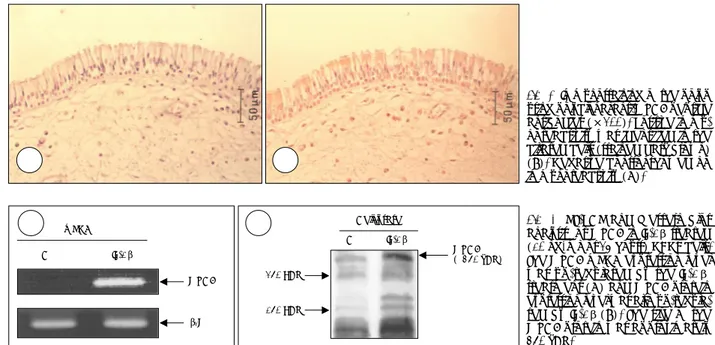
다. 그러나 합류 후 7일째 관찰된 섬모세포의 비율은 3.1± 0.2%였고, 14일째 섬모세포의 비율은 7.4±0.5%, 28일째 는 14.5±0.6%였다(Fig. 1A). 동시에, H6C5 항체에 반응 하는 분비세포의 비율은 합류 2일째 35.6±2.8%, 7일째 32.8±7.8%, 14일째는 32.8±2.5%였고 이때까지는 수치 상의 큰 차이는 없었다. 그러나 합류후 28일째 분비세포의 비율은 49.4±1.4%로 약간의 증가를 보였다(Fig. 1B). 그 리고 MUC8 유전자의 발현량은 레티노익산이 있는 경우 시 간에 따라 점차적으로 증가하였다(Fig. 2). 섬모세포의 수 와 MUC8 유전자의 발현량의 증가 형태는 매우 유사하였 다. 이는 MUC8이 코점막 상피세포에서 섬모세포의 분화 또는 기능과 관계가 있을 것으로 생각된다. 비용종에서 MUC8단백질의 분리 비용종에 대한 다클론 anti-MUC8 peptide 항체로 염색 하였을 때 염색이 되지 않은 음성 대조군(Fig. 2A)과 비교 하여 섬모세포들은 양성으로 염색되었다(Fig. 2B). 이로써 MUC8이 사람 비용종의 섬모세포에서 발현된다는 것을 알 게 되었다.
정상 코점막 상피세포에서 IL-1β에 의한 MUC8 mRNA 와 단백질의 증가
MUC8 mRNA의 발현량이 IL-1β에 의해 상승한다는 내 용이 이전에 여러 연구에서 보고 되었다.9)10) 이러한 내용은 본 연구에서도 같은 결과를 보였고(Fig. 3A), 새로 합성된 anti-MUC8 peptide를 이용한 Western blot 분석에서
IL-1β에 의해 단백질의 발현량도 상승 조절되는 것을 알 수 있었다(Fig. 3B). 본 연구의 결과에서 MUC8 cDNA의 크 기는 9.5 kb였고(data not shown), 결과적으로 MUC8 단 백질의 크기는 약 350 kDa으로 생각되었다. 명확하게 구별 되는 band가 350 kDa 근처에서 나타났고 이 band는 IL-1β에 의해 상승 되었다.
고 찰
점액과분비는 여러 염증성 호흡계 질환의 가장 흔한 병리 적 특징이며, 따라서 기도에서의 점액 유전자 발현과 조절 을 이해하는 것이 매우 중요하다. 점액 유전자 중에 MUC8 은 생체외 코상피세포 배양실험에서 염증 매개 인자들에 의 해 발현이 증가하고,9)10) 여러 염증인자에 의하여 자극받는 생체 비용종 상피에서 발현이 증가한다고 알려져 있다.8) 그 러나 cDNA의 일부 염기서열(323 amino acids)만 밝혀졌 기 때문에, cDNA의 역할은 아직 명확하게 밝혀지지 않았다. 본 연구의 목적은 첫째로 사람 코점막 상피세포의 분화 정 도와 MUC8 유전자의 발현 사이의 관계를 알아보는 것이 었다. 본 연구에서 섬모세포의 수는 분화정도에 따라 증가 하였다. 그러나 분비세포의 수는 상대적으로 일정하게 유지 되었으며, MUC8 mRNA 발현량의 증가는 섬모세포의 수 적 증가와 같은 양상을 보였다. 이는 MUC8 유전자의 발현 이 섬모세포의 분화와 관련이 있음을 나타낸다고 하겠다. 두 번째로 MUC8 단백질의 국재화를 알아보기 위하여 비 용종에서 다클론 anti-MUC8 peptide 항체를 이용하여 면 A AA A BBBBFig. 3. Immunostaining of the polyp
using polyclonal anti-MUC8 peptide antibodies (×200). Positive immu-noreactivity was identified in the ciliated cells (stained dark brown) (B). Negative control showed no immunoreactivity (A).
Fig. 4. RT-PCR and Western blot
analysis for MUC8 in IL-1β treated (10 ng/ml for 24 hours) NHNE cells. The MUC8 mRNA expression level was up-regulated by the IL-1β treatment (A) and MUC8 protein expression level was also up-regula-ted by IL-1β (B). The size of the MUC8 protein was approximately 350 kDa. mRNA C IL-1β β2M MUC8 A A A A Cell lysate 250 KDa 150 KDa MUC8 ( 350 kDa) C IL-1β B B B B
역염색을 시행하였으며, MUC8 단백질이 배세포에서가 아니 라, 섬모세포와 일부 염증세포에서 발현된다는 결과를 얻었다 (Fig. 3B). 그러나 Lopez-Ferrer A 등이 MUC8 단백질이 사람 기관지 상피(bronchial epithelium)의 배세포 및 섬모세 포 모두에서 발현된다고 보고하였으며,13) 이러한 차이는 본 연 구에서 사용된 항체들이 다양한 MUC8의 peptide 서열을 대 상으로 만들어 졌다는 점과 Lopez의 연구에서 사용된 기관지 점막 대신 코점막을 사용했다는 점에서 기인했다고 생각된다.
최근에 microarray와 2-D PAGE를 이용한 high-throu-ghput analysis로 인해 mRNA와 단백질 발현량의 사이에 큰 차이가 있음이 알려졌다.14)15) 이러한 차이를 가능하게 하 는 원인에는 전사후조절(post-transcriptional regulation) 이 대표적이다. 대부분의 조절유전자(regulated gene)는 그 단백질 양에 영향을 주는 것으로 생각되며 이를 전사조절 (transcriptional regulation)이라고 한다. 그러나 유전자발현 의 변화 없이 합성되는 단백질의 양만이 변화될 수 있는데, 이 는 전이조절과 같은 전사후조절의 가능성을 나타낸다. 따라서 본 연구는 MUC8 단백질이 IL-1β 처치 후 전사조절 또는 전사후조절에 의해 조절되는 지를 조사하였다. 본 연구에서 세 포들을 IL-1β로 처치했을 때 MUC8 mRNA의 발현량이 단 백량의 증가에 따라 같이 증가하였다. 이러한 결과는 MUC8 단백질 발현량이 전사조절에 의해 조절된다는 것을 보여준다.
최근에 IL-1β에 의한 MUC8 유전자 발현의 유도가 사 람 호흡 상피세포에서 일련의 ERK MAP kinase/RSK1/ CREB cascade pathway에 의해 매개된다는 연구가 보고 되었다.16) 그러나 MUC8 유전자의 cDNA와 promoter의 염 기 서열은 아직 완전하게 밝혀지지 않았고, cDNA와 pro-moter의 염기 서열이 완전히 밝혀질 때, 사람 호흡계에서 MUC8의 조절과 기능이 완전하게 밝혀질 수 있다고 생각된다. 본 연구를 통해 MUC8 단백질이 사람 코점막 상피세포 에서 특히 섬모세포에서 발현되고 IL-1β의 처치에 의해 증 가된다는 것을 알 수 있었다. 이러한 결과들은 MUC8 유전 자가 사람 코점막 상피세포에서 섬모세포의 분화 또는 그 기능과 관련이 있음을 유추하게 해준다. 결국, MUC8 유전 자와 단백질의 발현량은 사람 코점막 상피세포에서 섬모세 포의 표식자(marker)로서 사용될 수 있을 것으로 생각된다. 중심 단어:점액유전자 8・섬모세포・코 상피세포. 본 연구는 2003년도 범석학술장학재단 학술연구비에 의하여 이 루어졌음. REFERENCES
1) Basbaum C, Lemjabbar H, Longphre M, Li D, Gensch E, McNamara N. Control of mucin transcription by diverse injury-induced signaling
pathways. Am J Respir Crit Care Med 1999;160:S44-8.
2) Lapensee L, Paquette Y, Bleau G. Allelic polymorphism and
chromo-somal localization of the human oviductin gene (MUC9). Fertil Steril 1997;68:702-8.
3) Chen Y, Zhao YH, Kalaslavadi TB, Hamati E, Nehrke K, Le AD, et
al. Genome-wide search and identification of a novel gel-forming mu-cin MUC19/Muc19 in glandular tissues. Am J Respir Cell Mol Biol 2004;30:155-65.
4) Hovenberg HW, Davies JR, Carlstedt I. Different mucins are
produ-ced by the surface epithelium and the submucosa in human trachea: Identification of MUC5AC as a major mucin from the goblet cells. Biochem J 1996;318:319-24.
5) Thornton DJ, Howard M, Khan N, Sheehan JK. Identification of two
glycoforms of the MUC5B mucin in human respiratory mucus. J Biol Chem 1997;272:9561-6.
6) Buisine MP, Devisme L, Copin MC, Durand-Reville M, Gosselin B, Aubert JP, et al. Developmental mucin gene expression in the human
respiratory tract. Am J Respir Cell Mol Biol 1999;20:209-18. 7) Kim CH, Song KS, Kim HU, Seong JK, Yoon JH. Expression of
MUC5AC mRNA in the goblet cells of human nasal mucosa. Laryngo-scope 2000;110:2110-3.
8) Kim SS, Kim KS, Lee JG, Park IY, Koo JS, Yoon JH. Levels of
intra-cellular protein and mRNA of mucin and lysozyme in normal human nasal and polyp epithelium. Laryngoscope 2000;110:276-80. 9) Yoon JH, Kim KS, Kim HU, Linton JA, Lee JG. Effects of TNF-alpha
and IL-1beta on mucin, lysozyme, IL-6 and IL-8 in passage-2 normal human nasal epithelial cells. Acta Otolaryngol 1999;119:905-10. 10) Seong JK, Koo JS, Lee WJ, Kim HN, Park JY, Yoon JH, et al.
Upre-gulation of MUC8 and downreUpre-gulation of MUC5AC by inflammatory mediators in human nasal polyps and cultured nasal epithelium. Acta Otolaryngol 2002;122:401-7.
11) Yoon JH, Kim KS, Kim SS, Lee JG, Park IY. Secretory differentiation
of serially-passaged normal human nasal epithelial cells by retinoic acid: Expression of mucin and lysozyme. Ann Otol Rhinol Laryngol 2000;109:594-601.
12) Shankar V, Pichan P, Eddy RL Jr, Tonk V, Nowak N, Sait SN, et al.
Chromosomal localization of a human mucin gene (MUC8) and clo-ning of the cDNA corresponding to the carboxy terminus. Am J Respir Cell Mol Biol 1997;16:232-41.
13) Lopez-Ferrer A, Curull V, Barranco C, Garrido M, Lloreta J, Real FX,
et al. Mucins as differentiation markers in bronchial epithelium. Squa-mous cell carcinoma and adenocarcinoma display similar expression patterns. Am J Respir Cell Mol Biol 2001;24:22-9.
14) Gygi SP, Rochon Y, Franza BR, Aebersold R. Correlation between
pro-tein and mRNA abundance in yeast. Mol Cell Biol 1999;19:1720-30. 15) Juan HF, Lin JY, Chang WH, Wu CY, Pan TL, Khoo KH, et al.
Bio-mic study of human myeloid leukemia cells differentiation to macro-phages using DNA array, proteomic, and bioinformatic analytical methods. Electrophoresis 2002;23:2490-504.
16) Song KS, Lee WJ, Kim CH, Cho KN, Koo JS, Yoon JH, et al.
Induc-tion of MUC8 gene expression by Interleukin-1β is mediated by a sequential ERK MAP kinase/RSK1/CREB cascade pathway in human airway epithelial cells. J Biol Chem 2003;278:34890-6.