저작자표시-비영리-변경금지 2.0 대한민국 이용자는 아래의 조건을 따르는 경우에 한하여 자유롭게 l 이 저작물을 복제, 배포, 전송, 전시, 공연 및 방송할 수 있습니다. 다음과 같은 조건을 따라야 합니다: l 귀하는, 이 저작물의 재이용이나 배포의 경우, 이 저작물에 적용된 이용허락조건 을 명확하게 나타내어야 합니다. l 저작권자로부터 별도의 허가를 받으면 이러한 조건들은 적용되지 않습니다. 저작권법에 따른 이용자의 권리는 위의 내용에 의하여 영향을 받지 않습니다. 이것은 이용허락규약(Legal Code)을 이해하기 쉽게 요약한 것입니다. Disclaimer 저작자표시. 귀하는 원저작자를 표시하여야 합니다. 비영리. 귀하는 이 저작물을 영리 목적으로 이용할 수 없습니다. 변경금지. 귀하는 이 저작물을 개작, 변형 또는 가공할 수 없습니다.
Clinical significance of repetitive
compound muscle action potential in
myasthenia gravis patients
Hyo Eun Lee
Department of Medicine
Clinical significance of repetitive
compound muscle action potential in
myasthenia gravis patients
Directed by Professor Seung Min Kim
The Master's Thesis
submitted to the Department of Medicine,
the Graduate School of Yonsei University
in partial fulfillment of the requirements for the degree of
Master of Medical Science
This certifies that the Master's Thesis of
Hyo Eun Lee is approved.
Thesis Supervisor : Seung Min Kim
---Thesis Committee Member#1 : Bae Hwan Lee
---Thesis Committee Member#2 : Yeon-kyung Jung
The Graduate School
Yonsei University
ACKNOWLEDGEMENTS
I would like to express heartfelt thanks to Professor Seung Min
Kim for serving as my thesis advisor and committee chair. He has
provided me with the best opportunities to experience neurology.
Special thanks go to Professor Ha Young Shin for his concern and
careful guidance regarding completing this manuscript. I would
also like to thank Professor Bae Hwan Lee, Yeon-Kyung Jung for
serving on my committee, and providing invaluable advice.
Finally, I am deeply grateful to my parents and a congenial
colleague for their support and encouragement.
<TABLE OF CONTENTS>
ABSTRACT···1
I. INTRODUCTION ···3
II. MATERIALS AND METHODS ···5
III. RESULTS ···9
1. Clinical manifestations···9
2. Repetitive nerve stimulation test and neostigmine test ···9
3. Side effects of pyridostigmine bromide···10
IV. DISCUSSION ···16
V. CONCLUSION ···19
REFERENCES ···20
ABSTRACT (IN KOREAN) ···23
LIST OF FIGURES
Figure 1. RNS test on ADM stimulation by ulnar nerve
at the elbow ··· 8
LIST OF TABLES
Table 1. Clinical characteristics of myasthenia gravis
patients with and without R-CMAP ··· 12
Table 2. Antibody status in subgroup patients who checked
MuSK Ab··· 13
Table 3. Result of low frequency repetitive nerve stimulation
test and neostigmine test··· 14
Table 4. Response to oral pyridostigmine bromide ··· 15
ABSTRACT
Clinical significance of repetitive compound muscle action potential in
myasthenia gravis patients
Hyo Eun Lee
Department of Medicine
The Graduate School, Yonsei University
(Directed by Professor Seung Min Kim)
Repetitive compound muscle action potentials (R-CMAPs) are
multiple compound muscle action potentials (CMAPs) following an
initial CMAP induced by a single stimulation of a nerve. R-CMAPs
indicate cholinergic neuromuscular hyperactivity, and occur in several
conditions, such as congenital acetylcholinesterase (AChE) deficiency,
slow-channel congenital myasthenic syndrome, and organophosphate
poisoning. Although R-CMAPs may also be observed in myasthenia
gravis (MG) patients with AChE inhibitor overdose, these are seldom
seen in MG patients with the standard dose. The mechanism of R-CMAP
induction and its clinical significance in patients with MG is also unclear.
Therefore, we investigated the clinical characteristics of MG patients
with R-CMAPs who were given the standard diagnostic dose of
neostigmine. We retrospectively reviewed the medical records and
electrodiagnostic findings of patients who underwent electrodiagnostic
tests before and after the neostigmine test. Of the 71 MG patients
reviewed, 24 developed R-CMAPs after receiving the standard diagnostic
dose of neostigmine. There were no significant differences in age at
disease onset, myasthenia gravis activities of daily living score, disease
duration, and follow-up duration. Decremental response on the
baselinerepetitive nerve stimulation (RNS) test was lower in the
R-CMAP group. This group had a higher frequency of side effects and
intolerance to, and a lower maximal tolerable dose of pyridostigmine
bromide (PB). The appearance of R-CMAPs after a standard dose of
injected neostigmine may be a good indicator of the risk of PB side
effects. The detection of R-CMAPs after neostigmine injection may help
in creating a therapeutic plan for myasthenia gravis patients.
---Key words : myasthenia gravis, repetitive compound muscle action
potential, pyridostigmine bromide
Clinical significance of repetitive compound muscle action potential in
myasthenia gravis patients
Hyo Eun Lee
Department of Medicine
The Graduate School, Yonsei University
(Directed by Professor Seung Min Kim)
I. INTRODUCTION
Myasthenia gravis (MG), an autoimmune disorder affecting neuromuscular transmission, is characterized by weakness of voluntary muscles and fatigue. The identification of impaired neuromuscular transmission is an essential step for the diagnosis of MG. Therefore, electrodiagnostic studies including repetitive nerve stimulation (RNS) and single-fiber electromyography are used
to evaluate the electrophysiological status of the neuromuscular junction.1,2
Repetitive compound muscle action potentials (R-CMAPs) are multiple compound muscle action potentials (CMAPs) observed after an initial CMAP by a single stimulation of a nerve. The mechanism of R-CMAP production remains unclear. A generally accepted mechanism of R-CMAP is described below. The stimulation of presynaptic acetylcholine (ACh) receptors can lead to
backfiring of nerve action potentials.3,4 Excess Ach prolongs the activation of
the postsynaptic receptor, resulting in prolonged open channel time and
end-plate potentials.2,3,5 These abnormal synaptic events reactivate the nerve fiber,
leading to re-excitation of the muscle fiber of the motor unit.5 A single nerve
stimulation may elicit not only a single CMAP, but also multiple R-CMAPs following the initial CMAP in some neuromuscular junction disorders, such as organophosphate intoxication, congenital acetylcholinesterase deficiency, and
slow-channel congenital myasthenic syndrome.5-9These R-CMAPs result from
the repetitive discharge of muscle fibers after a single stimulation, and represent
the electrophysiological status of cholinergic neuromuscular hyperactivity.8
In patients with MG, R-CMAPs may be caused by acetylcholinesterase
(AChE) inhibitor overdose.7 However, R-CMAPs are rarely seen in patients
with MG taking a standard dose of AChE inhibitors.8,10In a previous study, MG
patients with nicotinic side effects of AChE inhibitors developed R-CMAP
more frequently than patients without the side effects.7 In addition, R-CMAPs
have been frequently observed in muscle-specific tyrosine kinase (MuSK) antibody-positive MG patients during a diagnostic neostigmine test. A substantial number of MuSK antibody-positive MG patients with R-CMAPs
cannot tolerate AChE inhibitors due to their cholinergic side effects.8 These
findings suggest that R-CMAPs may predict the risk of side effects and intolerance to oral AChE inhibitors by representing the cholinergic activity status at the neuromuscular junction. Despite the two aforementioned studies, little is known about the clinical significance of R-CMAP in patients with myasthenia gravis.
Therefore, we investigated the clinical characteristics of myasthenia gravis patients with R-CMAP in order to identify its clinical usefulness in therapeutic decision-making.
II. MATERIALS AND METHODS
We retrospectively reviewed the clinical records and eletrodiagnostic findings of MG patients who underwent electrodiagnostic studies (EDx) and diagnostic neostigmine tests (NT) from 2007 to 2015 at Severance Hospital. The diagnosis of MG was based on the symptoms and signs of muscle fatigue, decrement responses on low rate RNS, serum level of anti-AChR and MuSK antibodies, and the improvement of muscle fatigue after the intramuscular injection of neostigmine. Antibody analyses were performed through commercially available assays (anti-AChR-binding antibody: Seoul Clinical Laboratories, Seoul, Korea; anti-MuSK antibody: Athena Diagnostics, Worcester, MA, USA). The test for anti-AChR-binding antibody was considered negative if the value was ≤ 0.2 nmol/L. The result of anti-MuSK antibodies tests were expressed as negative (<10), borderline (≥10, <20), or positive (≥20). When patients with MG are evaluated in our electrodiagnostic laboratory, they usually undergo clinical evaluation, EDx, and NT. To confirm objective electrophysiology responsiveness to neostigmine methylsulfate, EDx may be repeated 30-45 minutes after neostigmine injection at the examiner's discretion.
EDx included baseline RNS and post-NT ulnar nerve stimulation recordings on the abductor digiti minimi (ADM). The RNS was performed according to previously described methods on the ADM, flexor carpi ulnaris (FCU) muscles with the ulnar nerve stimulation at the elbow, the orbicular oculi (OO), nasalis muscles with the facial nerve stimulation, and the trapezius muscle with spinal accessory nerve stimulation using the Neuroscreen system (Toennies, Bavaria,
Germany) or the Schwarzer topas EMG system (Natus, Bavaria, Germany)8. A
decrement of CMAP ≥ 10% was considered abnormal. In patients taking pyridostigmine bromide (PB), the EDx was performed at least 12 hours after the last dose. NT was performed after baseline clinical examination and EDx. The response was evaluated approximately 30 minutes after intramuscular injection
of 0.02 mg/kg neostigmine methylsulfate. The side effects of neostigmine were recorded.
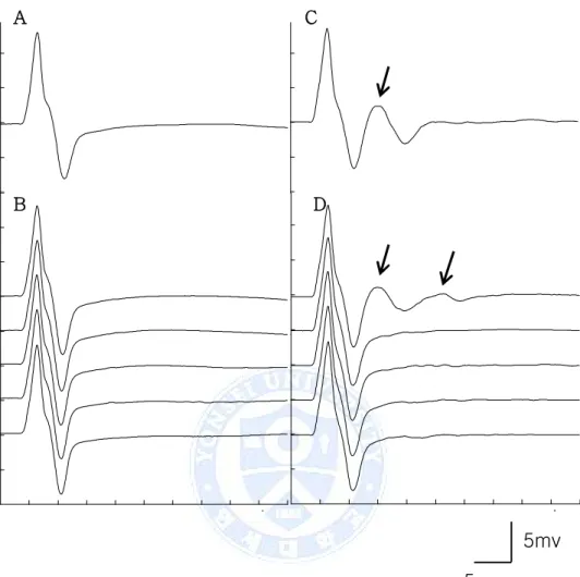
A total of 79 patients with MG underwent EDx before and after NT. Exclusion criteria were as follows: overdose of neostigmine methylsulfate, incomplete medical records, or less than twelve months of follow up. The patients were classified into two groups according to the development of R-CMAP after NT. R-CMAP was defined as a CMAP followed by repetitive discharges that did not exist before NT (Fig. 1).
Two neurologists (L.H.S and P.B.S) without clinical information about the patients reviewed the results of EDx. The two neurologists consensually determined the presence of R-CMAP on the ADM muscle at normal gain (5 mV/division).
We obtained patients’ background information from their medical records, including age of MG symptom onset, sex, myasthenia gravis foundation of America (MGFA) clinical classification at the first visit, the worst MGFA clinical classification during follow up, results of AChR-binding and anti-MuSK antibody tests, thymoma, follow-up duration, MGFA postintervention status, maximal tolerable dose of PB, and intolerance to PB. Intolerance to PB was defined as an inability to take PB due to cholinergic side effects such as severe muscle fasciculation, abdominal cramps, hypersalivation, and blurred vision. Age at EDx, disease duration, RNS results, myasthenia gravis activities of daily living (MG-ADL) score, baseline quantitative myasthenia gravis (QMG) score, result of NT, and side effects of neostigmine were obtained from EDx reports.
This study was approved by the institutional review board of Severance Hospital, Yonsei University Health System, and the requirement for informed consent from the study subjects was waived.
Statistical analysis
All data are expressed as the median (interquartile range) for skewed continuous variables. Comparisons of continuous variables for the two groups were performed using the Mann-Whitney U test. The Chi-square test (Pearson`s and Fisher`s exact test) was used for the comparison of the categorical variables between groups. All statistical analyses were performed using the IBM SPSS Statistics software for Windows Version 21.0 (IBM Corp., Armonk, NY, USA). A two-tailed probability value of p < 0.05 was considered statistically significant.
A C
B D
Figure 1. RNS test on ADM stimulation by ulnar nerve at the elbow. (A)
R-CMAP was not showed on single stimulation of nerve. (B) There was no decremental response by low frequency (3Hz) RNS test. (C, D) R-CMAP (arrow) was presented following initial CMAP after 30min of neostigmine methylsulfate injection.
5mv
5ms
III. RESULTS
Among the 79 patients who underwent EDx before and after NT, 71 patients were included in this study and eight were excluded. Seven patients had less than twelve months of follow up. One patient had incomplete medical records. None of these patients had R-CMAPs on baseline RNS tests.
1. Clinical manifestations
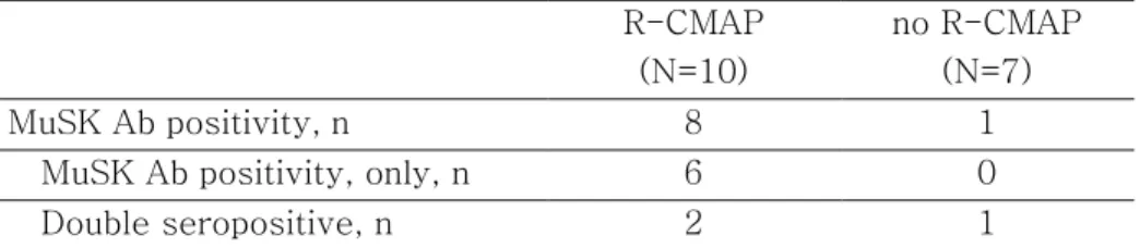
The clinical characteristics of the patients are summarized in Table 1. There were no significant differences in age at disease onset (39.5 vs. 34.0 years, p = 0.189), thymoma (16.7 vs. 19.1%, p = 1.000), and follow-up duration (36.5 vs. 34.0 months, p = 0.742) between the R-CMAP group and the no-R-CMAP group. The R-CMAP group showed ocular type (41.7 vs 14.9%, p = 0.012) and female (91.7 vs. 57.4%, p = 0.003) predominance. The Ach receptor antibody (Ab) seropositivity was lower in the R-CMAP group than in the no R-CMAP group (58.3 vs. 85.1%, p = 0.012). Although we did not perform MuSK-Ab detection for all patients, anti-Musk antibodies were found in 17 patients. Nine patients revealed MG with MuSK-Ab. Of these, eight patients were in the R-CMAP group (90 vs. 10%, p = 0.015), and one patient was in the no R-R-CMAP group. The antibody status of the subgroup of patients in which MuSK-Abs were checked is summarized in Table 2.
2. Repetitive nerve stimulation (RNS) test and neostigmine test (NT)
There were no significant differences in age at exam (44 vs. 39 years, p = 0.069), MG-ADL score at the RNS test (5.5 vs. 7.0, p = 0.390), and disease duration (60.0 vs. 63.0 months, p = 0.456) between the R-CMAP group and the no-R-CMAP group. The decremental response on the baseline RNS test was
lower in the R-CMAP group (62.5 vs. 91.5%, p = 0.007) (Table 3). After 30 minutes of neostigmine methylsulfate injection, R-CMAPs were found in the 24 patients after repeated ulnar nerve stimulation recording on the ADM (Fig. 1).
The R-CMAP group had fewer positive results in the neostigmine test, and lower differences between baseline QMG scores and post-neostigmine QMG scores. All patients in the R-CMAP group had neostigmine-related side effects (100 vs. 70.2%, p = 0.002). Twenty-three patients in the R-CMAP group had neostigmine-related muscarinic side effects; 23 patients had abdominal pain, one developed diarrhea. Thirty-two patients in the no-R-CMAP group had neostigmine-related muscarinic side effects: 29 had abdominal pain, five complained of diarrhea, one had nausea, and three had increased salivation. Nicotinic side effects of neostigmine were especially more frequent in the R-CMAP group (75.0 vs. 6.4%, p < 0.001). Eighteen patients in the R-R-CMAP group had nicotinic side effects; 17 had muscle fasciculation and two had squeezing sensation in the neck. Only three patients of the no R-CMAP group had nicotinic side effects: three had muscle fasciculation, and one had squeezing sensation in the neck.
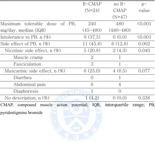
3. Side effects of pyridostigmine bromide (PB)
PB is one of the most commonly used AChE inhibitors for the treatment of myasthenia gravis in patients included in this study. Table 4 shows the side effects of PB. The R-CMAP group had a lower maximal tolerable dose of PB (240 vs. 480 mg/day, p < 0.001) and a higher frequency of PB intolerance (37.5 vs. 0%, p < 0.001). Nine patients in the R-CMAP group had intolerance to PB. The most commonly given reasons for intolerance were abdominal pain and muscle fasciculation. In the no-R-CMAP group, six patients showed discomfort about taking PB; complaints resolved after decreasing the dose of PB. The other
more frequent in the R-CMAP group (45.8 vs. 12.8%, p < 0.001). Nicotinic side effects of PB were especially more frequent in the R-CMAP group (20.8 vs. 4.3%, p = 0.040).
Table 1. Clinical characteristics of myasthenia gravis patients with and without R-CMAP R-CMAP (n=24) no R-CMAP (n=47) p-value Onset age, median (IQR),
years 39.5 (28.0-50.8) 34.0 (28.0-44.0) 0.189 Female, n (%) 22 (91.7) 27 (57.4) 0.003
Follow up duration, month, median (IQR) 57.5 (49.5-64.0) 56.0 (47.0-63.0) 0.316 AChR Ab seropostivity, n (%) 14 (58.3) 40 (85.1) 0.012
AChR Ab titier , median (IQR), nmol/L 1.23 (0.01-7.62) 8.15 (0.63-11.62) 0.011 Thymoma, n (%) 4 (16.7) 9 (19.1) 1.000
MGFA clinical classification at first visit I, n 10 7 0.012* IIa, n 1 19 IIb, n 10 3 IIIa, n 1 9 IIIb, n 1 7 IVa, n 0 1 IVb, n 1 1 MGFA worst I, n 4 7 IIa, n 6 11 IIb, n 6 1 IIIa, n 2 14 IIIb, n 1 2 IVa, n 1 3 IVb, n 3 4 V, n 1 5
* p-value; I vs. other MGFA
CMAP, compound muscle action potential; IQR, interquartile range;MG, myasthenia gravis; AChR Ab, acetylcholine receptor antibody; MGFA, Myasthenia Gravis
Table 2. Antibody status in subgroup patients who checked MuSK Ab
CMAP, compound muscle action potential; MuSK Ab, muscle specific tyrosine kinase antibody R-CMAP (N=10) no R-CMAP (N=7) MuSK Ab positivity, n 8 1
MuSK Ab positivity, only, n 6 0
Table 3. Result of low frequency repetitive nerve stimulation test and neostigmine test R-CMAP (N=24) no R-CMAP (N=47) p-value Age at examination, median
(IQR), years 44.0 (32.0-55.8) 39.0 (29.0-47.0) 0.069
Disease duration, median
(IQR), months 60.0 (54.5-76.3) 63.0 (52.0-107.0) 0.456 MG ADL score at RNST, median (IQR) 5.5 (3.3-8.8) 7.0 (3.0-10.0) 0.390 CMAP decremental on baseline
RNST of ≥ 10%, n (%) 15 (62.5) 43 (91.5) 0.007 ADM, n (%) 0 (0.0) 29 (61.7) <0.001 FCU, n (%) 4 (16.7) 34 (72.3) <0.001 OO, n (%) 12 (50.0) 35 (74.5) 0.039 Nasalis, n (%) 12(50.0) 41 (87.2) 0.001 Trapezius, n (%) 8 (33.3) 32 (68.1) 0.005
Result of neostigmine test
Positivity of neostigmine
test, n (%)
16 (66.7) 44 (93.6) 0.005
Baseline QMG score, median (range) 8.5 (7.3-13.3) 13.0 (8.0-18.0) 0.046 Change in QMG score, median (range) 3 (2.0-4.8) 6 (2.0-9.0) 0.017 Side effect of neostigmine, n
(%)
24 (100.0) 33 (70.2) 0.002
Nicotinic side effect, n (%) 18 (75.0) 3 (6.4) <0.001
Muscarinic side effect, n (%)
23 (95.8) 32 (68.1) 0.008
CMAP, compound muscle action potential; IQR, interquartile range; MG-ADL, myasthenia gravis activities of daily living; RNST, repetitive nerve stimulation test; ADM, abductor digiti minimi; FCU, flexor carpi ulnaris; OO, orbicularis oculi; QMG, quantitative myasthenia gravis
Table 4. Response to oral pyridostigmine bromide R-CMAP (N=24) no R-CMAP (N=47) p-value Maximum tolerable dose of PB,
mg/day, median (IQR)
240 (45-480) 480 (480-480) <0.001 Intolerance to PB, n (%) 9 (37.5) 0 (0.0) <0.001 Side effect of PB, n (%) 11 (45.8) 6 (12.8) 0.002
Nicotinic side effect, n (%) 5 (20.8) 2 (4.3) 0.040
Muscle cramp 2 1
Fasciculation 3 1
Muscarinic side effect, n (%) 6 (25.0) 4 (8.5) 0.077
Diarrhea 0 1
Abdominal pain 6 4
Diaphoresis 1 0
No description, n (%) 1 (4.2) 0 (0.0) 0.338
CMAP, compound muscle action potential; IQR, interquartile range; PB,
IV. DISCUSSION
AChE inhibitors facilitate neuromuscular transmission by inhibiting
acetylcholine breakdown at the neuromuscular junction.11 These drugs are the
first-line treatment for MG, and provide temporary relief of the symptoms.11-13
They are effective as an initial therapy in most patients with MG and may be
used as the sole therapeutic agent for long periods in mild forms of MG.14
Although AChE inhibitors are usually well tolerated at standard doses (for example, up to 60 mg of pyridostigmine bromide five times per day), some patients with MG show cholinergic hypersensitivity and are intolerant of even small doses of AChE inhibitors. In our study, nine patients with MG were intolerant to PB. All of them had R-CMAPs after NT. Despite these side effects, doctors prescribed PB, because it is usually well tolerated and the first-line
treatment for MG.2 Patients eventually discontinued PB and began
immunosuppressant therapies. Fortunately, they do not continue to experience the side effects of PB, and their MG symptoms are well controlled. The patients achieved MGFA postintervention status PR or MM at last visit. Therefore, therapeutic management with AChE inhibitors should be individualized. We suggest that R-CMAP after NT is a helpful tool to predict the side effects of PB.
Predicting the distinct responses to AChE inhibitors is very important to prevent unnecessary side effects of AChE inhibitors in MG patients. There are
no well-controlled studies to predict the side effects of PB.15 One method to
achieve this is by measuring AChE activities to predict side effects of PB.
Various methods are known to determine cholinesterase activities.16 Ellman's
assay is widely used to measure AChE activity but has technical-related
limitations.16,17 In some studies, AChE activities do not correlate with the
severity of PB side effects.18-20 In other studies, only older age correlated with
AChE gene.15,21-23 AChE activity measurement and genetic analysis are not
commonly used in the clinic. However, it is very easy to test for R-CMAP after NT in a clinical setting. Moreover, it requires no additional cost or equipment.
In our study, after neostigmine methylsulfate injection, 80% of the patients experienced side effects. All subjects in the R-CMAP group had side effects, and 70% of the patients in the no-R-CMAP group experienced side effects. However, the latter could tolerate PB well. Six patients (12.8%) had side effects to PB treatment. Despite the side effects observed after NT, most patients tolerated PB well. Therefore, side effects after NT are not a good indicator to base decisions regarding PB treatment on. Of the patients in the R-CMAP group who experienced side effects with neostigmine methylsulfate injection, 37.5% were intolerant to PB and 45.8% showed side effects after PB treatment.
MG is difficult to diagnose using MuSK-Ab due to negative results of
diagnostic tests such as RNS and NT.8,24,25 MuSK-Ab analysis is not easy to
perform in the clinic, mainly because it is too expensive. AChE inhibitors do not have a therapeutic effect or can worsen MG symptoms in patients with MuSK-Ab.8,11,15,24,26 In our study, we performed MuSK-Ab detection in 17 patients.
Nine patients tested positive. Eight out of the nine MuSK-Ab-positive MG patients were in the R-CMAP group (p = 0.015, Fisher`s exact test). Although we did not perform MuSK-Ab detection for all patients, we believe that an electrophysiological study could detect R-CMAPs after NT in MG patients with MuSK-Ab. Previous studies also suggest that the presence of cholinergic
hypersensitivity helps to diagnose MG with MuSK-Ab.8,10
This study had several limitations. First, we could not explain female predominance in the R-CMAP group. Second, this study had a retrospective design, a small number of patients and physicians made an arbitrary decision regarding patient selection. Therefore, a selection bias may exist. Third, previous studies applied grading systems using needle electrodes and surface
(5 mV/division). Thus, we might have missed small amplitude R-CMAPs. Further prospective studies are required to support our conclusions.
Our results showed that MG patients with R-CMAPs after NT had a higher frequency of side effects and intolerance to PB treatment. R-CMAP after standard doses of neostigmine may be a good indicator of side effects to PB. Testing for R-CMAPs after a neostigmine injection might help to diagnose MG and develop a therapeutic plan for MG treatment.
V. CONCLUSION
We investigated the clinical characteristics of myasthenia gravis patients with R-CMAP after NT. Patients in the R-CMAP group had several distinctive features compared with those in the no-R-CMAP group. The R-CMAP group showed a higher frequency of intolerance and side effects to PB. Therefore, we suggest that the presence of R-CMAPs after NT is a good indicator of the side effects of PB, and may help in therapeutic decision making.
REFERENCES
1. Berrih-Aknin S, Frenkian-Cuvelier M, Eymard B. Diagnostic and
clinical classification of autoimmune myasthenia gravis. J Autoimmun 2014;48-49:143-8.
2. Meriggioli MN, Sanders DB. Autoimmune myasthenia gravis:
emerging clinical and biological heterogeneity. Lancet Neurol 2009;8:475-90.
3. Besser R, Gutmann L, Dillmann U, Weilemann LS, Hopf HC.
End-plate dysfunction in acute organophosphate intoxication. Neurology 1989;39:561-7.
4. Maselli RA, Soliven BC. Analysis of the organophosphate-induced
electromyographic response to repetitive nerve stimulation: paradoxical response to edrophonium and D-tubocurarine. Muscle Nerve 1991;14:1182-8.
5. Kumar RS, Kuruvilla A. Repetitive compound muscle action
potentials in electrophysiological diagnosis of congenital myasthenic syndromes: a case report and review of literature. Ann Indian Acad Neurol 2010;13:139-41.
6. van Dijk JG, Lammers GJ, Wintzen AR, Molenaar PC. Repetitive
CMAPs: mechanisms of neural and synaptic genesis. Muscle Nerve 1996;19:1127-33.
7. Punga AR, Sawada M, Stalberg EV. Electrophysiological signs and
the prevalence of adverse effects of acetylcholinesterase inhibitors in patients with myasthenia gravis. Muscle Nerve 2008;37:300-7.
8. Shin HY, Park HJ, Lee HE, Choi YC, Kim SM. Clinical and
Electrophysiologic Responses to Acetylcholinesterase Inhibitors in MuSK-Antibody-Positive Myasthenia Gravis: Evidence for
Cholinergic Neuromuscular Hyperactivity. J Clin Neurol
2014;10:119-24.
10. Punga AR, Flink R, Askmark H, Stalberg EV. Cholinergic neuromuscular hyperactivity in patients with myasthenia gravis seropositive for MuSK antibody. Muscle Nerve 2006;34:111-5.
11. Skeie GO, Apostolski S, Evoli A, Gilhus NE, Illa I, Harms L, et al.
Guidelines for treatment of autoimmune neuromuscular
transmission disorders. Eur J Neurol 2010;17:893-902.
12. Aung T, Dowden AY. Successful desensitization protocol for
pyridostigmine hypersensitivity in a patient with myasthenia gravis. Ann Allergy Asthma Immunol 2013;110:308.
13. Mehndiratta MM, Pandey S, Kuntzer T. Acetylcholinesterase
inhibitor treatment for myasthenia gravis. Cochrane Database Syst Rev 2014;10:Cd006986.
14. Pohanka M. Acetylcholinesterase inhibitors: a patent review
(2008 - present). Expert Opin Ther Pat 2012;22:871-86.
15. Maggi L, Mantegazza R. Treatment of myasthenia gravis: focus on
pyridostigmine. Clin Drug Investig 2011;31:691-701.
16. Holas O, Musilek K, Pohanka M, Kuca K. The progress in the
cholinesterase quantification methods. Expert Opin Drug Discov 2012;7:1207-23.
17. Ellman GL, Courtney KD, Andres V, Jr., Feather-Stone RM. A
new and rapid colorimetric determination of acetylcholinesterase activity. Biochem Pharmacol 1961;7:88-95.
18. Cook MR, Gerkovich MM, Sastre A, Graham C. Side effects of
low-dose pyridostigmine bromide are not related to
cholinesterase inhibition. Aviat Space Environ Med
2001;72:1102-6.
19. Cook MR, Graham C, Sastre A, Gerkovich MM. Physiological and
performance effects of pyridostigmine bromide in healthy volunteers: a dose-response study. Psychopharmacology (Berl) 2002;162:186-92.
20. Sharabi Y, Danon YL, Berkenstadt H, Almog S, Mimouni-Bloch A,
Zisman A, et al. Survey of symptoms following intake of pyridostigmine during the Persian Gulf war. Isr J Med Sci
1991;27:656-8.
21. Mutero A, Camp S, Taylor P. Promoter elements of the mouse
acetylcholinesterase gene. Transcriptional regulation during muscle differentiation. J Biol Chem 1995;270:1866-72.
22. Shapira M, Tur-Kaspa I, Bosgraaf L, Livni N, Grant AD, Grisaru D,
et al. A transcription-activating polymorphism in the ACHE
promoter associated with acute sensitivity to
anti-acetylcholinesterases. Hum Mol Genet 2000;9:1273-81.
23. Loewenstein-Lichtenstein Y, Schwarz M, Glick D,
Norgaard-Pedersen B, Zakut H, Soreq H. Genetic predisposition to adverse consequences of anti-cholinesterases in 'atypical' BCHE carriers. Nat Med 1995;1:1082-5.
24. Evoli A, Tonali PA, Padua L, Monaco ML, Scuderi F, Batocchi AP,
et al. Clinical correlates with anti-MuSK antibodies in generalized seronegative myasthenia gravis. Brain 2003;126:2304-11.
25. Mori S, Kubo S, Akiyoshi T, Yamada S, Miyazaki T, Hotta H, et al.
Antibodies against muscle-specific kinase impair both
presynaptic and postsynaptic functions in a murine model of myasthenia gravis. Am J Pathol 2012;180:798-810.
26. Verschuuren JJ, Huijbers MG, Plomp JJ, Niks EH, Molenaar PC,
Martinez-Martinez P, et al. Pathophysiology of myasthenia gravis with antibodies to the acetylcholine receptor, muscle-specific kinase and low-density lipoprotein receptor-related protein 4. Autoimmun Rev 2013;12:918-23.
ABSTRACT(IN KOREAN)
중증 근무력증 환자에서 반복 복합 근 활동전위의 임상적 의의
<지도교수 김 승 민 >
연세대학교 대학원 의학과
이 효 은
반복 복합 근 활동전위는 신경을 자극 시 최초의 복합 근 활동전위 이후 연달아 검출되는 복합 근 활동전위들을 의미한다. 반복 복합 근 활동전위는 선천성 아세틸콜린 분해 효소 결핍, 선천성 느린 통로 중증 근무력 증후군, 유기인제 독성과 같은 질환에서 발견되며 이는 콜린성 신경근육 과민성 상태를 반영한다는 것을 의미한다. 중증 근무력증 환자에서도 아세틸콜린 분해 효소 억제제를 과량 사용하였을 때 반복 복합 근 활동 전위가 나타날 수 있다. 하지만 표준용량의 아세틸콜린 분해 효소 억제제를 중증 근무력증 환자에게 사용하였을 때 반복 복합 근 활동전위는 거의 발견되지 않는다. 반복 복합 근 활동전위의 발생기전은 확립된 바가 없으며 그 임상적 의의에 대해서도 연구된 바가 없다. 그러므로 우리는 표준용량의 네오스티그민주사 이후 반복자극 검사를 시행하였던 환자들에서 반복 복합 근 활동전위의 유무에 따른 임상적 특징에 대해 분석하여 그 임상적 의의에 대해 연구하였다. 총 71명의 환자가 포함되었으며 환자의 의무기록과 전기 생리학적 검사 결과를 검토하였다. 24명에서 표준용량의 네오스티그민주사 이후 반복 복합 근 활동전위가 관찰되었다. 네오스티그민주사 이후 반복 복합 근 활동전위가 발생한 군과 발생하지 않은 군 사이에 질환의 발생 나이, 중증 근무력증 일상생활 수행능력 점수, 추적기간은 차이가 없었다. 반복 복합 근활동전위가 발생한 군에서 피리도스티그민에 부작용 및 과민반응이 많았으며 최대 복용가능 용량도 적었다. 이 연구를 통해 반복 복합 근 활동전위가 아세틸 콜린 분해 효소 억제제의 부작용과 밀접한 관계가 있음을 확인하였으며 표준용량의 네오스티그민주사 이후 반복 신경자극검사를 한번 더 시행하여 반복 복합 근 활동전위의 유무를 살피는 것이 중증 근무력증 환자의 치료계획 수립에 도움이 될 것이다.
---핵심되는 말
: 중증 근무력증, 반복 복합 근 활동전위, 피리도스
티그민
PUBLICATION LIST
1. Shin HY, Park HJ, Lee HE, Choi YC, Kim SM. Clinical and
Electrophysiologic Responses to Acetylcholinesterase Inhibitors in MuSK-Antibody-Positive Myasthenia Gravis: Evidence for
Cholinergic Neuromuscular Hyperactivity. J Clin Neurol