Impact of Statin Treatment on Strut Coverage
after Drug-Eluting Stent Implantation
Yongsung Suh,
1Byeong-Keuk Kim,
1Dong-Ho Shin,
1Jung-Sun Kim,
1Young-Guk Ko,
1Donghoon Choi,
1Yangsoo Jang,
1,2and Myeong-Ki Hong
1,21Division of Cardiology, Severance Cardiovascular Hospital, Yonsei University College of Medicine, Seoul; 2Severance Biomedical Science Institute, Yonsei University College of Medicine, Seoul, Korea.
Received: January 29, 2014 Revised: March 10, 2014 Accepted: March 26, 2014
Corresponding author: Dr. Myeong-Ki Hong, Division of Cardiology,
Severance Cardiovascular Hospital, Yonsei University College of Medicine, 50-1 Yonsei-ro, Seodaemun-gu, Seoul 120-752, Korea.
Tel: 82-2-2228-8458, Fax: 82-2-393-2041 E-mail: mkhong61@ yuhs.ac
∙ The authors have no financial conflicts of interest.
© Copyright:
Yonsei University College of Medicine 2015
This is an Open Access article distributed under the terms of the Creative Commons Attribution Non-Commercial License (http://creativecommons.org/ licenses/by-nc/3.0) which permits unrestricted non-commercial use, distribution, and reproduction in any medium, provided the original work is properly cited.
Purpose: To evaluate the effect of statin treatment on strut coverage after drug-eluting stent (DES) implantation. Materials and Methods: In this study, 60 pa-tients were randomly assigned to undergo sirolimus-eluting stent (SES) or biolim-us-eluting stent (BES) implantation, after which patients were randomly treated with pitavastatin 2 mg or pravastatin 20 mg for 6 months. The degree of strut cover-age was assessed by 6-month follow-up optical coherence tomography, which was performed in 52 DES-implanted patients. Results: The percentages of uncovered struts were 19.4±14.7% in pitavastatin-treated patients (n=25) and 19.1±15.2% in pravastatin-treated patients (n=27; p=0.927). A lower percentage of uncovered struts was significantly correlated with a lower follow-up low-density lipoprotein (LDL) cholesterol level (r=0.486; p=0.009) and a greater decline of the LDL cho-lesterol level (r=-0.456; p=0.015) in SES-implanted patients, but not in BES-im-planted patients. In SES-imBES-im-planted patients, the percentage of uncovered struts was significantly lower among those with LDL cholesterol levels of less than 70 mg/dL after 6 months of follow-up (p=0.025), but no significant difference in this variable according to the follow-up LDL cholesterol level was noted among BES-implanted patients (p=0.971). Conclusion: Lower follow-up LDL cholesterol lev-els, especially those less than 70 mg/dL, might have a protective effect against de-layed strut coverage after DES implantation. This vascular healing effect of lower LDL cholesterol levels could differ according to the DES type.
Key Words: Stent, optical coherence tomography, statin
INTRODUCTION
Neointimal coverage over stent struts emerged as an important clinical issue after it was reported that incomplete strut coverage might be associated with the occur-rence of late stent thrombosis following drug-eluting stent (DES) implantation.1-4 Optical coherence tomography (OCT) is a useful tool that has excellent resolution for confirming neointimal stent strut coverage.5,6 Although several variables such as the implantation of sirolimus-eluting stents (SESs)7,8 and acute coronary syn-drome9,10 were reported as risk factors for delayed strut coverage in previous OCT
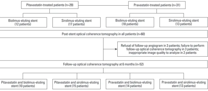
also randomly allocated. Thus, four groups were created as follows: pitavastatin-N-BES group (12 patients), pitavas-tatin-SES group (17 patients), pravastatin-N-BES group (18 patients), and pravastatin-SES group (13 patients). Post-in-tervention OCT examinations were performed for all tients immediately after DES implantation. Among 60 pa-tients, 6-month follow-up angiography was not performed for three patients, the OCT catheter could not be passed through the lesion due to severe angulation in three pa-tients, and there was poor image quality in two patients. Therefore, follow-up OCT evaluation was performed for 52 patients as follows: pitavastatin-N-BES group (10 pa-tients), pitavastatin-SES group (15 papa-tients), pravastatin-N-BES group (14 patients), and pravastatin-SES group (13 patients) (Fig. 1). Blood samples to evaluate lipid profiles were obtained at the time of stent implantation and follow-up angiography. All patients were clinically followed follow-up 1, 3, and 6 months after stent implantation.
Coronary intervention and quantitative coronary angiography analysis
All patients received at least 75 mg of aspirin and a loading dose of 300 mg of clopidogrel at least 12 h pre-interven-tion. Unfractionated heparin was administered to maintain the activated clotting time at >250 s. All percutaneous coro-nary interventions were performed according to current standard techniques. Post-intervention, dual antiplatelet therapy with aspirin 100 mg and clopidogrel 75 mg daily was prescribed for 12 months.
Quantitative coronary angiographic analysis was per-formed before and after stent implantation as well as at fol-studies, the relationship of specific medications (i.e., statins)
with strut coverage is not fully understood. Statins are the most widely used lipid-lowering agents in patients with coronary artery disease. These drugs reduce inflammation and enhance endothelial function via pleiotropic effects.11 Previous in vitro and animal studies reported that statins regulate the migration and proliferation of smooth muscle cells and control neointimal formation.12-15 However, there are no human data that assess the impact of statin treatment on strut coverage after DES implantation. Therefore, we conducted a randomized OCT study to evaluate the effect of statin treatment on DES strut coverage.
MATERIALS AND METHODS
Study design and patient enrollment
The present study consisted of subgroup analysis of a pre-vious randomized OCT study16 and evaluated the impact of statin treatment on strut coverage. The previous OCT study was performed to compare strut coverage 6 months after
Nobori biolimus-eluting stent (N-BES, Nobori®, Terumo
Corporation, Tokyo, Japan; n=60) or SES (CypherTM, Cor-dis Corp., Miami Lakes, FL, USA; n=60) implantation. The inclusion and exclusion criteria of the OCT study were provided in the previous report.16 Written informed consent was obtained from all participants, and the Institutional Re-view Boards of our institute approved this study. In total, 60 patients were enrolled in this study and randomly treated with pitavastatin 2 mg or pravastatin 20 mg, beginning on the day of stent implantation. Stenting (SES or N-BES) was
Fig. 1. A flowchart of the study is shown.
Refusal of follow-up angiogram in 3 patients; failure to perform follow-up optical coherence tomography in 3 patients;
inappropriate image quality to analyze in 2 patients
Pitavastatin and biolimus-eluting stent (10 patients) Biolimus-eluting stent
(12 patients)
Pravastatin and biolimus-eluting stent (14 patients) Biolimus-eluting stent
(18 patients)
Pitavastatin and sirolimus-eluting stent (15 patients) Sirolimus-eluting stent
(17 patients)
Pravastatin and sirolimus-eluting stent (13 patients) Sirolimus-eluting stent
(13 patients) Post-stent optical coherence tomography in all patients (n=60)
Follow-up optical coherence tomography at 6 months (n=52)
dent’s t-test, or the Mann-Whitney U test. To avoid prob-lems of sample size inflation and correlated data, only pa-tients with one target lesion were included in the study. Cross-section analysis or strut-level analysis may not be straightforward due to the congregation of struts within each lesion in an interindividual manner. For this analysis, we performed multilevel regression model analysis. Specifical-ly, the patient and lesion data were incorporated as random effect components using the lme4 package with R (http:// cran.r-project.org/web/packages/lme4/index.html).20 Pear-son’s correlation analysis was performed to evaluate the re-lationship between low-density lipoprotein (LDL) choles-terol levels and the percentage of uncovered struts. Values of p<0.05 denoted statistical significance.
RESULTS
Baseline clinical and angiographic characteristics were simi-lar between the pitavastatin-treated groups and pravastatin-treated groups (Table 1). Significant reductions of LDL cho-lesterol levels were observed at the 6-month follow-up time point (reduction: 24 mg/dL in the pitavastatin-treated group, p<0.001; 21 mg/dL in the pravastatin-treated group, p= 0.003). Follow-up LDL cholesterols level less than 70 mg/dL were achieved in 10 patients (34.5%) in the pitavastatin-treated group and 5 patients (16.1%) in the pravastatin-treated group (p=0.101). OCT findings were also similar between pitavastatin-treated patients and pravastatin-treated patients (Table 2). The percentages of uncovered struts at the 6-month follow-up OCT were 19.4±14.7% in pitavas-tain-treated patients (n=25) and 19.1±15.2% in pravastain-treated patients (n=27) (p=0.927); conversely, the values were 23.3±16.6% in SES-implanted patients (n=28) and 14.5±10.9% in BES-implanted patients (n=24) (p=0.026).
In all of 52 patients, follow-up cholesterol levels of less than 70 mg/dL were associated with smaller percentages of uncovered struts (less than 70 mg/dL vs. 70 mg/dL or more; 12.5±12.2% vs. 21.5±15.1%; p=0.058). Although the per-centage of uncovered struts among SES-implanted patients was significantly lower in patients with follow-up LDL cho-lesterol levels of less than 70 mg/dL (less than 70 mg/dL vs. 70 mg/dL or more; 10.1±12.4% vs. 26.9±15.6%, respective-ly; p=0.025), there were no significant differences in the percentage of uncovered struts among BES-implanted pa-tients between those with follow-up LDL cholesterol levels of less than 70 mg/dL (14.6±12.7%) and those with levels low-up, using an offline quantitative coronary angiographic
system (CASS system, Pie Medical Instruments, Maastricht, the Netherlands) in an independent core laboratory (Cardio-vascular Research Center, Seoul, Korea). Using the guiding catheter for magnification and calibration, reference vessel diameters and the minimal luminal diameter were measured from diastolic frames in a single, matched view showing the smallest minimal luminal diameter. Late loss was defined as the difference between the post-procedure and follow-up minimal luminal diameters.
OCT imaging and cross-sectional analysis
Immediately and 6 months after the intervention, OCT of the target lesion was performed using a frequency-domain OCT system (C7-XR OCT imaging system, LightLab Im-aging, Inc., St. Jude Medical, St. Paul, MN, USA). For this study, OCT cross-sectional images were generated at a ro-tational speed of 100 frames per second while the fiber was withdrawn at a speed of 20 mm/s within the stationary im-aging sheath. All OCT images were analyzed at a core lab-oratory (Cardiovascular Research Center, Seoul, Korea) by analysts who were blinded to patient and procedural infor-mation.
Cross-sectional OCT images were analyzed at 0.2-mm longitudinal intervals. Stent and luminal cross-sectional ar-eas (CSAs) were mar-easured, and the neointimal hyperplasia (NIH) CSA was calculated as the stent area minus the lumi-nal CSA. NIH thickness was measured as the distance be-tween the endoluminal surface of the neointima and the strut.8 An uncovered strut was categorized as an NIH thick-ness=0 μm.8 A malapposed strut was defined as a strut that had detached from the vessel wall by ≥130 μm (N-BES) or ≥160 μm (SES).17,18 The percentage of uncovered or malap-posed struts was calculated as the ratio of uncovered or malapposed struts to total struts in all OCT cross-sections. Intrastent thrombi were defined as irregular masses protrud-ing into the lumen by more than 250 μm at the thickest point.19
Statistical analysis
Statistical analysis was performed using Statistical Analysis System software (v. 9.1.3., SAS Institute, Cary, NC, USA) and R version 2.15.1 (R Development Core Team, Vienna, Austria, http://www.R-project.org). Categorical data were presented as numbers (%) and compared using the chi-square or Fisher’s exact test. Continuous data were presented as the mean±SD and compared using a paired t-test,
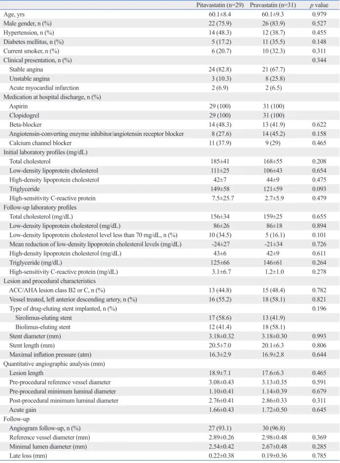
Stu-Table 1. Baseline Clinical and Angiographic Characteristics of the Pitavastatin-Treated Groups and Pravastatin-Treated Groups
Pitavastatin (n=29) Pravastatin (n=31) p value
Age, yrs 60.1±8.4 60.1±9.3 0.979 Male gender, n (%) 22 (75.9) 26 (83.9) 0.527 Hypertension, n (%) 14 (48.3) 12 (38.7) 0.455 Diabetes mellitus, n (%) 5 (17.2) 11 (35.5) 0.148 Current smoker, n (%) 6 (20.7) 10 (32.3) 0.311 Clinical presentation, n (%) 0.344 Stable angina 24 (82.8) 21 (67.7) Unstable angina 3 (10.3) 8 (25.8)
Acute myocardial infarction 2 (6.9) 2 (6.5)
Medication at hospital discharge, n (%)
Aspirin 29 (100) 31 (100)
Clopidogrel 29 (100) 31 (100)
Beta-blocker 14 (48.3) 13 (41.9) 0.622
Angiotensin-converting enzyme inhibitor/angiotensin receptor blocker 8 (27.6) 14 (45.2) 0.158
Calcium channel blocker 11 (37.9) 9 (29) 0.465
Initial laboratory profiles (mg/dL)
Total cholesterol 185±41 168±55 0.208
Low-density lipoprotein cholesterol 111±25 106±43 0.654
High-density lipoprotein cholesterol 42±7 44±9 0.475
Triglyceride 149±58 121±59 0.093
High-sensitivity C-reactive protein 7.5±25.7 2.7±5.9 0.479
Follow-up laboratory profiles
Total cholesterol (mg/dL) 156±34 159±25 0.655
Low-density lipoprotein cholesterol (mg/dL) 86±26 86±18 0.894
Low-density lipoprotein cholesterol level less than 70 mg/dL, n (%) 10 (34.5) 5 (16.1) 0.101
Mean reduction of low-density lipoprotein cholesterol levels (mg/dL) -24±27 -21±34 0.726
High-density lipoprotein cholesterol (mg/dL) 43±6 42±9 0.611
Triglyceride (mg/dL) 125±66 146±61 0.264
High-sensitivity C-reactive protein (mg/dL) 3.1±6.7 1.2±1.0 0.278
Lesion and procedural characteristics
ACC/AHA lesion class B2 or C, n (%) 13 (44.8) 15 (48.4) 0.782
Vessel treated, left anterior descending artery, n (%) 16 (55.2) 18 (58.1) 0.821
Type of drug-eluting stent implanted, n (%) 0.196
Sirolimus-eluting stent 17 (58.6) 13 (41.9)
Biolimus-eluting stent 12 (41.4) 18 (58.1)
Stent diameter (mm) 3.18±0.32 3.18±0.30 0.993
Stent length (mm) 20.5±7.0 20.1±6.3 0.806
Maximal inflation pressure (atm) 16.3±2.9 16.9±2.8 0.644
Quantitative angiographic analysis (mm)
Lesion length 18.9±7.1 17.6±6.3 0.465
Pre-procedural reference vessel diameter 3.08±0.43 3.13±0.35 0.591
Pre-procedural minimum luminal diameter 1.10±0.41 1.14±0.39 0.679
Post-procedural minimum luminal diameter 2.76±0.41 2.86±0.33 0.311
Acute gain 1.66±0.43 1.72±0.50 0.645
Follow-up
Angiogram follow-up, n (%) 27 (93.1) 30 (96.8)
Reference vessel diameter (mm) 2.89±0.26 2.98±0.48 0.369
Minimal lumen diameter (mm) 2.54±0.42 2.67±0.48 0.285
Late loss (mm) 0.22±0.38 0.19±0.36 0.785
ACC, American College of Cardiology; AHA, American Heart Association. Values are presented as n (%) or the mean±SD.
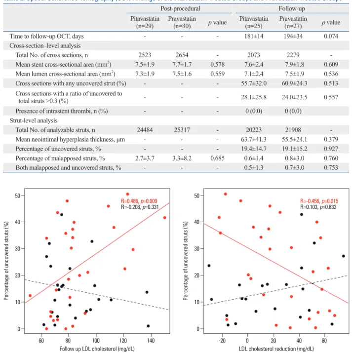
with the degree of LDL cholesterol reduction (r=-0.456; p=0.015) in SES-implanted patients but not in BES-implant-ed patients.
DISCUSSION
Previous studies identified several factors that were associat-ed with delayassociat-ed strut coverage, such as SES implantation,7,8 exceeding 70 mg/dL (14.4±10.5%; p=0.971). Fig. 2
pres-ents the relationship of follow-up LDL cholesterol levels with strut coverage according to DES type. The percentage of uncovered struts was significantly correlated with fol-low-up LDL cholesterol levels (r=0.486; p=0.009) in SES-implanted patients but not in BES-SES-implanted patients. Fig. 3 presents the relationship of the degree of LDL cholesterol re-duction with strut coverage according to DES type. The per-centage of uncovered struts was also significantly correlated
Table 2. Optical Coherence Tomography (OCT) Findings of the Pitavastatin-Treated Groups and Pravastatin-Treated Groups
Post-procedural Follow-up
Pitavastatin
(n=29) Pravastatin (n=30) p value Pitavastatin (n=25) Pravastatin (n=27) p value
Time to follow-up OCT, days - - - 181±14 194±34 0.074
Cross-section–level analysis
Total No. of cross sections, n 2523 2654 - 2073 2279
Mean stent cross-sectional area (mm2) 7.5±1.9 7.7±1.7 0.578 7.6±2.4 7.9±1.8 0.609
Mean lumen cross-sectional area (mm2) 7.3±1.9 7.5±1.6 0.559 7.1±2.4 7.5±1.9 0.536
Cross sections with any uncovered strut (%) - - - 55.7±32.0 60.9±24.3 0.513
Cross sections with a ratio of uncovered to
total struts >0.3 (%) - - - 28.1±25.8 24.0±23.5 0.557
Presence of intrastent thrombi, n (%) - - - 0 (0.0) 0 (0.0)
Strut-level analysis
Total No. of analyzable struts, n 24484 25317 - 20223 21908
Mean neointimal hyperplasia thickness, μm - - - 63.7±41.3 55.5±24.1 0.379
Percentage of uncovered struts, % - - - 19.4±14.7 19.1±15.2 0.927
Percentage of malapposed struts, % 2.7±3.7 3.3±8.2 0.685 0.6±1.4 0.8±3.0 0.760
Both malapposed and uncovered struts, % - - - 0.5±1.3 0.7±3.0 0.753
Fig. 2. The relationships of the percentage of uncovered struts with follow-up low-density lipoprotein (LDL) cholesterol levels are presented. Black and red dots represent biolimus-eluting stents and sirolimus-eluting stents, respectively.
Fig. 3. The relationships of the percentage of uncovered struts with the de-gree of low-density lipoprotein (LDL) cholesterol level reduction are pre-sented. Black and red dots represent biolimus-eluting stents and sirolimus-eluting stents, respectively.
Follow up LDL cholesterol (mg/dL) LDL cholesterol reduction (mg/dL)
0 0 10 10 20 20 30 30 40 40 50 50
Percentage of uncovered struts (%) Percentage of uncovered struts (%)
60 80 100 120 140 -20 0 20 40 60
R=0.486, p=0.009
for this finding is that the vascular healing response to statins or lower LDL cholesterol levels could be different ac-cording to the type of DES. Compared to SESs, BESs have several different characteristics, including a bioresorbable polymer carrier (poly-lactic acid), as well as coating only on the abluminal stent surface to allow the direct release of li-pophilic biolimus into the vessel wall.32,33 Differences in the polymer, drug, or drug-eluting period between BESs and SESs may influence the degree of strut coverage according to the LDL cholesterol-lowering effects of statins.
The present study has some limitations. First, although our study was designed as a randomized trial, the study pop-ulation included a relatively small number of patients. We feel that a future study with a larger sample size would be needed to confirm our findings. However, our main findings could provide a clue, suggesting the possible relationship of lowering LDL cholesterol or use of statin with the vascular healing process. Second, all control groups received statin therapy. Therefore, we could not evaluate the effect of statin therapy on strut coverage by making comparisons with pa-tients who did not receive statin treatment. However, a con-trol group with no statin treatment would not be ethically justified in current clinical practice for DES-implanted pa-tients with coronary artery disease.
In conclusion, this randomized study revealed a protec-tive effect of statins against delayed strut coverage in SES-implanted patients who achieved lower follow-up LDL cholesterol levels (especially less than 70 mg/dL). This vas-cular healing effect of lower LDL cholesterol levels in-duced by statins could be different according to the type of DES implanted.
ACKNOWLEDGEMENTS
This study was supported by a grant from the Korea Health-care Technology R&D Project, Ministry for Health, Welfare & Family Affairs, Republic of Korea (nos. A085012 and A102064), a grant from the Korea Health 21 R&D Project, Ministry of Health & Welfare, Republic of Korea (no. A085136), and the Cardiovascular Research Center, Seoul, Korea.
REFERENCES
1. Farb A, Burke AP, Kolodgie FD, Virmani R. Pathological
mecha-acute coronary syndrome,9,10 the time interval between DES implantation and follow-up OCT,21 baseline high-sensitivity C-reactive protein levels,22 the stent diameter,23 diabetes mel-litus,23 and American Heart Association/American College of Cardiology type B2/C lesions.23 Although several studies re-vealed that statins have beneficial effects on both mature en-dothelial cells and enen-dothelial progenitor cells,24-27 there are no human data associating the use of statins with an accelera-tion of strut coverage after DES implantaaccelera-tion. Wang, et al.14 demonstrated that atorvastatin pretreatment could accelerate both neointimal coverage and reendothelialization after SES implantation in a minipig model. Another animal study us-ing a wire-mediated vascular injury model in mice reported that fluvastatin has protective effects against impaired reen-dothelialization in sirolimus-treated arteries.24 The mecha-nism by which statins protect against delayed vascular heal-ing is linked to their ability to modulate smooth muscle cell proliferation and migration and increase circulating endo-thelial progenitor cell counts.14,24 Experimental animal stud-ies reported that each statin has different effects according to its water solubility.28,29
In this study, strut coverage was compared between pita-vastatin-treated lesions and prapita-vastatin-treated lesions, and we hypothesized that different statins may differentially af-fect OCT-based strut coverage in DES-implanted lesions. Compared to pravastatin (a hydrophilic statin), pitavastatin (a fully synthetic lipophilic statin) has powerful efficacy comparable with that of atorvastatin and rosuvastatin.30,31 In the present study, a significant difference in strut coverage was not observed between the pitavastatin-treated groups and pravastatin-treated groups; however, greater DES strut coverage was significantly related with lower follow-up LDL cholesterol levels and greater reductions of LDL cho-lesterol levels in SES-implanted lesions, but not in BES-implanted lesions.
These findings suggest that reducing LDL cholesterol lev-els alone has a role in strut coverage after first-generation DES implantation. However, as mentioned previously, the primary mechanism by which statins modify the vascular healing process is considered to involve a pleotropic effect rather than a direct LDL cholesterol-lowering effect. There-fore, this discrepancy might be explained by the fact that a lower LDL cholesterol level is one indicator of the intensity of the pleotropic effect of statins in the vascular healing pro-cess. Additionally, the relationship between follow-up LDL cholesterol levels and strut coverage was not observed for next-generation DESs (i.e., BESs). A possible explanation
cle and endothelial cell proliferation and reduces neointima on a drug-eluting stent platform. Cardiovasc Res 2005;68:483-92. 16. Kim BK, Ha J, Mintz GS, Kim JS, Shin DH, Ko YG, et al.
Ran-domised comparison of strut coverage between Nobori biolimus-eluting and sirolimus-biolimus-eluting stents: an optical coherence tomog-raphy analysis. EuroIntervention 2014;9:1389-97.
17. Tanigawa J, Barlis P, Dimopoulos K, Dalby M, Moore P, Di Ma-rio C. The influence of strut thickness and cell design on immedi-ate apposition of drug-eluting stents assessed by optical coherence tomography. Int J Cardiol 2009;134:180-8.
18. Davlouros PA, Mavronasiou E, Xanthopoulou I, Karantalis V, Tsigkas G, Hahalis G, et al. An optical coherence tomography study of two new generation stents with biodegradable polymer carrier, eluting paclitaxel vs. biolimus-A9. Int J Cardiol 2012;157: 341-6.
19. Kume T, Akasaka T, Kawamoto T, Ogasawara Y, Watanabe N, Toyota E, et al. Assessment of coronary arterial thrombus by opti-cal coherence tomography. Am J Cardiol 2006;97:1713-7. 20. Doran H, Bates D, Bliese P, Dowling M. Estimating the multilevel
Rasch model: with the lme4 package. J Stat Softw 2007;20:1-18. 21. Kim BK, Kim JS, Oh C, Ko YG, Choi D, Jang Y, et al. Major
de-terminants for the uncovered stent struts on optical coherence to-mography after drug-eluting stent implantation. Int J Cardiovasc Imaging 2012;28:705-14.
22. Kim BK, Kim JS, Oh C, Ko YG, Choi D, Jang Y, et al. Impact of preprocedural high-sensitivity C-reactive protein levels on uncov-ered stent struts: an optical coherence tomography study after drug-eluting stent implantation. Clin Cardiol 2011;34:97-101. 23. Ishigami K, Uemura S, Morikawa Y, Soeda T, Okayama S,
Nishi-da T, et al. Long-term follow-up of neointimal coverage of siroli-mus-eluting stents--evaluation with optical coherence tomography. Circ J 2009;73:2300-7.
24. Fukuda D, Enomoto S, Shirakawa I, Nagai R, Sata M. Fluvastatin accelerates re-endothelialization impaired by local sirolimus treat-ment. Eur J Pharmacol 2009;612:87-92.
25. Wolfrum S, Jensen KS, Liao JK. Endothelium-dependent effects of statins. Arterioscler Thromb Vasc Biol 2003;23:729-36. 26. Werner N, Priller J, Laufs U, Endres M, Böhm M, Dirnagl U, et
al. Bone marrow-derived progenitor cells modulate vascular reen-dothelialization and neointimal formation: effect of 3-hydroxy-3-methylglutaryl coenzyme a reductase inhibition. Arterioscler Thromb Vasc Biol 2002;22:1567-72.
27. Walter DH, Zeiher AM, Dimmeler S. Effects of statins on endo-thelium and their contribution to neovascularization by mobiliza-tion of endothelial progenitor cells. Coron Artery Dis 2004;15: 235-42.
28. Sakamoto T, Kojima S, Ogawa H, Shimomura H, Kimura K, Ogata Y, et al. Usefulness of hydrophilic vs lipophilic statins after acute myocardial infarction: subanalysis of MUSASHI-AMI. Circ J 2007;71:1348-53.
29. Satoh K, Ichihara K. Lipophilic HMG-CoA reductase inhibitors increase myocardial stunning in dogs. J Cardiovasc Pharmacol 2000;35:256-62.
30. Saku K, Zhang B, Noda K; PATROL Trial Investigators. Random-ized head-to-head comparison of pitavastatin, atorvastatin, and ro-suvastatin for safety and efficacy (quantity and quality of LDL): the PATROL trial. Circ J 2011;75:1493-505.
31. Hayashi T, Yokote K, Saito Y, Iguchi A. Pitavastatin: efficacy and safety in intensive lipid lowering. Expert Opin Pharmacother 2007;8:2315-27.
nisms of fatal late coronary stent thrombosis in humans. Circula-tion 2003;108:1701-6.
2. Finn AV, Joner M, Nakazawa G, Kolodgie F, Newell J, John MC, et al. Pathological correlates of late drug-eluting stent thrombosis: strut coverage as a marker of endothelialization. Circulation 2007;115:2435-41.
3. Guagliumi G, Sirbu V, Musumeci G, Gerber R, Biondi-Zoccai G, Ikejima H, et al. Examination of the in vivo mechanisms of late drug-eluting stent thrombosis: findings from optical coherence to-mography and intravascular ultrasound imaging. JACC Cardio-vasc Interv 2012;5:12-20.
4. Won H, Shin DH, Kim BK, Mintz GS, Kim JS, Ko YG, et al. Op-tical coherence tomography derived cut-off value of uncovered stent struts to predict adverse clinical outcomes after drug-eluting stent implantation. Int J Cardiovasc Imaging 2013;29:1255-63. 5. Prati F, Regar E, Mintz GS, Arbustini E, Di Mario C, Jang IK, et
al. Expert review document on methodology, terminology, and clinical applications of optical coherence tomography: physical principles, methodology of image acquisition, and clinical appli-cation for assessment of coronary arteries and atherosclerosis. Eur Heart J 2010;31:401-15.
6. Lee SY, Hong MK. Stent evaluation with optical coherence to-mography. Yonsei Med J 2013;54:1075-83.
7. Kim JS, Jang IK, Kim JS, Kim TH, Takano M, Kume T, et al. Op-tical coherence tomography evaluation of zotarolimus-eluting stents at 9-month follow-up: comparison with sirolimus-eluting stents. Heart 2009;95:1907-12.
8. Takano M, Inami S, Jang IK, Yamamoto M, Murakami D, Seimi-ya K, et al. Evaluation by optical coherence tomography of neo-intimal coverage of sirolimus-eluting stent three months after im-plantation. Am J Cardiol 2007;99:1033-8.
9. Kim JS, Fan C, Choi D, Jang IK, Lee JM, Kim TH, et al. Different patterns of neointimal coverage between acute coronary syndrome and stable angina after various types of drug-eluting stents implan-tation; 9-month follow-up optical coherence tomography study. Int J Cardiol 2011;146:341-6.
10. Kubo T, Imanishi T, Kitabata H, Kuroi A, Ueno S, Yamano T, et al. Comparison of vascular response after sirolimus-eluting stent implantation between patients with unstable and stable angina pectoris: a serial optical coherence tomography study. JACC Car-diovasc Imaging 2008;1:475-84.
11. Mihos CG, Salas MJ, Santana O. The pleiotropic effects of the hydroxy-methyl-glutaryl-CoA reductase inhibitors in cardiovascu-lar disease: a comprehensive review. Cardiol Rev 2010;18:298-304.
12. Indolfi C, Cioppa A, Stabile E, Di Lorenzo E, Esposito G, Pisani A, et al. Effects of hydroxymethylglutaryl coenzyme A reductase inhibitor simvastatin on smooth muscle cell proliferation in vitro and neointimal formation in vivo after vascular injury. J Am Coll Cardiol 2000;35:214-21.
13. Bellosta S, Bernini F, Ferri N, Quarato P, Canavesi M, Arnaboldi L, et al. Direct vascular effects of HMG-CoA reductase inhibitors. Atherosclerosis 1998;137 Suppl:S101-9.
14. Wang TJ, Yang YJ, Xu B, Zhang Q, Jin C, Tang Y, et al. Atorvas-tatin accelerates both neointimal coverage and re-endothelializa-tion after sirolimus-eluting stent implantare-endothelializa-tion in a porcine model: new findings from optical coherence tomography and pathology. Circ J 2012;76:2561-71.
15. Jaschke B, Michaelis C, Milz S, Vogeser M, Mund T, Hengst L, et al. Local statin therapy differentially interferes with smooth
mus-33. Ostojic M, Sagic D, Beleslin B, Jung R, Perisic Z, Jagic N, et al. First clinical comparison of Nobori -Biolimus A9 eluting stents with Cypher- Sirolimus eluting stents: Nobori Core nine months angiographic and one year clinical outcomes. EuroIntervention 2008;3:574-9.
32. Chevalier B, Silber S, Park SJ, Garcia E, Schuler G, Suryapranata H, et al. Randomized comparison of the Nobori Biolimus A9-elut-ing coronary stent with the Taxus Liberté paclitaxel-elutA9-elut-ing coro-nary stent in patients with stenosis in native corocoro-nary arteries: the NOBORI 1 trial--Phase 2. Circ Cardiovasc Interv 2009;2:188-95.