저작자표시-비영리-변경금지 2.0 대한민국 이용자는 아래의 조건을 따르는 경우에 한하여 자유롭게 l 이 저작물을 복제, 배포, 전송, 전시, 공연 및 방송할 수 있습니다. 다음과 같은 조건을 따라야 합니다: l 귀하는, 이 저작물의 재이용이나 배포의 경우, 이 저작물에 적용된 이용허락조건 을 명확하게 나타내어야 합니다. l 저작권자로부터 별도의 허가를 받으면 이러한 조건들은 적용되지 않습니다. 저작권법에 따른 이용자의 권리는 위의 내용에 의하여 영향을 받지 않습니다. 이것은 이용허락규약(Legal Code)을 이해하기 쉽게 요약한 것입니다. Disclaimer 저작자표시. 귀하는 원저작자를 표시하여야 합니다. 비영리. 귀하는 이 저작물을 영리 목적으로 이용할 수 없습니다. 변경금지. 귀하는 이 저작물을 개작, 변형 또는 가공할 수 없습니다.
CONTENTS
List of Abbreviation ---
II
List of Figures ---
III
List of Tables ---
IV
General Introduction ---
1
References ---
5
1 Abstract ---
9
2 Introduction ---
10
3 Materials and Methods ---
15
4 Results ---
22
5 Discussion ---
34
References ---
39
Abstract in Korean ---
46
List of Abbreviations
aa amino acid
ANOVA a one-way analysis of variance
BBB Basso, Beattie, and Bresnahan
CNS Central nervous system
DPI Day post-injury
ERK extracellular signal-regulated kinase
GAPDH glyceraldehyde-3-phosphate dehydrogenase
H&E Hematoxylin and eosin
HO-1 Heme oxygenase-1
IL-1β Interleukin-1 beta
JNK c-Jun N-terminal kinase
MAPK Mitogen-activated protein kinase
NIH National institutes of health
Nrf-2 Nuclear factor erythroid 2-related factor 2
PBS Phosphate-buffered saline
SCI Spinal cord injury
SEM Standard error of the mean
SOD Superoxide dismutase
List of Figures
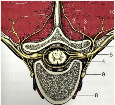
Figure 1. Schematic illustrations of cross-sections of spinal cord in
the vertebral column--- 1
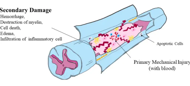
Figure 2. Schematic illustrations of both mechanical and
inflammatory response-induced damage of the spinal cord 2
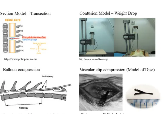
Figure 3. Schematic illustration of animal models of spinal cord injury 3
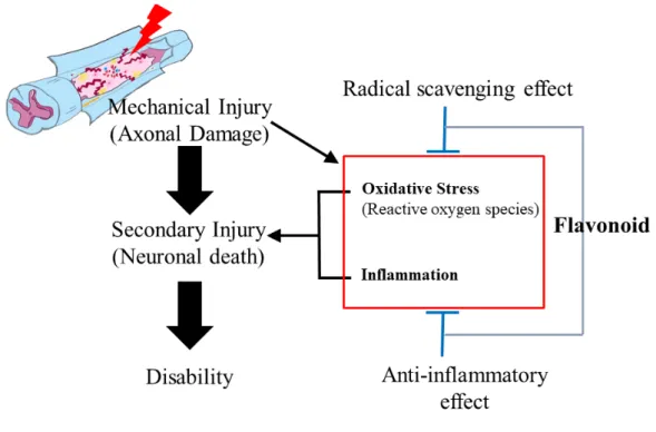
Figure 4. Schematic illustration of the neuropathogenesis of spinal cord
injury, including antiinflammatory and antioxidative effects---- 11
Figure 5. Schematic illustration of the purpose in the present study--- 14
Figure 6. Locomotor outcomes as evaluated by Basso, Beattie, and
Bresnahan (BBB) scoring (n = 5/daily)--- 23
Figure 7. Neuropathological changes of the spinal cords of sham
control (A and D), vehicle-treated (B and E), and hesperidin-treated groups (C and F) on 4 DPI--- 25
Figure 8. Western blotting to detect nuclear factor erythroid
2-related factor (Nrf)-2 and heme oxygenase (HO)-1 in the spinal cords of rats with SCI--- 27
Figure 9. Quantitative real time-PCR results in the spinal cords of
SCI rats--- 29 Figure 10. Quantitative real time-PCR results of superoxide
dismutase (SOD) and catalase in the spinal cords of SCI rats--- 31 Figure 11. Western blotting analysis of mitogen-activated protein
kinase (MAPK) in the spinal cord and chemical analysis of bilirubin and Fe2+ in the sera of SCI rats--- 33
Figure 12. Schematic illustration of neuro-protective mechanism in the spinal cords of hesperidin treatment--- 38
List of Tables
Table 1. Characterization of antibodies--- 18
General Introduction
Spinal cord anatomyThe spinal cord is connected to the brain. It is the part of the central nervous system (CNS) located within the vertebral column, and includes both sensory and motor nerves. It is also the main pathway for information between the brain and peripheral nervous system (Squire et al., 2013). The spinal cord carries sensory information from the body and part of the head to the CNS via afferent fibers, and performs the initial processing of this information (Figure 1).
Figure 1. Schematic illustrations of cross-sections of spinal cord in the
Axons of the motor neurons in the ventral horn project to the periphery to innervate skeletal muscle and smooth muscle, and mediate voluntary and involuntary reflexes. The spinal cord contains neurons, the descending axons of which mediate autonomic control of most visceral functions. The spinal cord is composed of the central gray matter and peripheral white matter (Figure 2). Two systems are responsible for the transmission of motor function: the upper and lower motor neuron systems (Olby et al., 1999).
Figure 2. Schematic illustrations of both mechanical and inflammatory
Spinal cord injury and animal model
Regardless of the primary injury type, secondary degeneration follows spinal cord injury (SCI) leading to severe damage (Figure 3) (Tator et al., 1991; Olby, 1999, 2010; Dumont et al., 2001). The neuropathological outcomes of SCI are characterized by edema, axonal degeneration, inflammatory cell infiltration, fibronectin exudation through the damaged blood –brain barrier, microglial activation, and reactive astrogliosis (Prewitt et al., 1997; Jung et al., 2003; Kim et al., 2003; Shin, 2013).
Many animal models have been developed to study the pathogenesis of SCI. Models based on mechanical impact include the New York University impact device injury, Ohio State University impact device injury, and clip-compression injury models (Kwon et al., 2002; Shin et al., 2013). Compression models include balloon compression (Tarlov, 1953) and lamina compression (Sun et al., 2017) models. Transection and partial transection have also been used to study SCI (Basso et al., 1996; Zeman et al., 1997). Basso et al. (1996) used a rat model of SCI in a preliminary study for the application of candidate therapeutic agents in dogs with SCI to study their therapeutic effects and mechanisms.
References
Basso DM, Beattie MS, Bresnahan JC. Graded histological and locomotor outcomes after spinal cord contusion using the NYU weight-drop device versus transection. Exp. Neurol., 1996; 139: 244-256.
Beattie MS. Inflammation and apoptosis: linked therapeutic targets in spinal cord injury. Trends Mol Med, 2004; 10(12): 580-583.
Dumont R, Okonkwo D, Verma S, et al. Acute spinal cord injury, partⅠ: pathophysiologic mechanisms. Clin Neuropharmcol, 2001; 24(5): 254-261.
Jung K, Min DS, Sim KB, Ahn M, Kim H, Cheong J, Shin T. Upregulation of phospholipase D1 in the spinal cords of rats with clip compression injury. Neurosci. Lett., 2003; 336: 126-130.
Kim DH, Heo SD, Ahn MJ, Sim KB, Shin TK. Activation of embryonic intermediate filaments contributes to glial scar formation after spinal cord injury in rats. J. Vet. Sci., 2003; 4: 109-112.
Kwon, BK, Oxland, TR, and Tetzlaff, W. Animal models used in spinal cord regeneration research. Spine (Phila Pa 1976), 2002; 27, 1504-1510.
Olby N. Current concepts in the management of acute spinal cord injury. J. Vet. Intern. Med., 1999; 13(5): 399-407.
Olby N. The pathogenesis and treatment of acute spinal cord injuries in dogs. Vet. Clin. North Am. Small Anim. Pract., 2010; 40(5): 791-807.
Prewitt CM, Niesman IR, Kane CJ, Houle JD. Activated macrophage/microglial cells can promote the regeneration of sensory axons into the injured spinal cord. Exp. Neurol., 1997; 148(2): 433-443.
Shin T, Ahn M, Moon C, Kim S, Sim KB. Alternatively activated macrophages in spinal cord injury and remission: another mechanism for repair? Mol. Neurobiol., 2013; 47: 1011-1019.
Squire, Larry Squire. Fundamental neuroscience (4th ed.). Amsterdam. Elsevier/Academic Press, 2013; p.628.
Sun GD, Chen Y, Zhou ZG, Yang SX, Zhong C, Li ZZ. A progressive compression model of thoracic spinal cord injury in mice: function assessment and pathological changes in spinal cord. Neural Regen Res, 2017; 12(8): 1365-1374.
Tarlov IM, Klinger H, Vitale S. Spinal cord compression studies. Ⅰ. Experimental techniques to produce acute and gradual compression. AMA Arch Neurol Psychiatry, 1953; 70: 813-819.
Tator CH, Fehlings MG. Review of the secondary injury theory of acute spinal cord trauma with emphasis on vascular mechanisms. J. Neurosurg., 1991; 75(1): 15-26.
Zeman RJ, Zhang Y, Etlinger JD. Clenbuterol, a beta2-adrenoceptor agonist, reduces scoliosis due to partial transection of rat spinal cord. Am. J. Physiol., 1997; 272(4 Pt 1): E712-E715.
Potential mechanism for attenuating inflammatory
oxidative stress of hesperidin in spinal cord injury
1. Abstract
S
pinal cord injury (SCI) is associated with inflammation along withconcurrent oxidative stress and glial activation. This study was performed to determine whether hesperidin, a representative flavonoid present in citrus fruits, ameliorates SCI-induced motor dysfunction and neuropathological degeneration in a rat model. Rats received hesperidin (100 mg/kg body weight daily, oral administration) from 7 days prior to SCI to 7 days after SCI. Behavioral tests were performed in rats until 6 weeks after SCI. To study inflammatory molecules, SCI rats treated with hesperidin were sacrificed on day 4 post-SCI, and their spinal cords were collected and subjected to histopathological examination. Behavioral tests indicated that pretreatment with hesperidin significantly ameliorated hindlimb paralysis in SCI rats beginning at day 21 post-SCI. Hesperidin pretreatment in SCI rats reduced the neuropathological changes (e.g., hemorrhage, inflammatory cell infiltration, and tissue loss) and levels of proinflammatory cytokines, including tumor necrosis factor-α and interleukin-1β. In addition, oxidative stress-related molecules, including superoxide dismutase, catalase, nuclear factor erythroid 2-related factor 2, and heme oxygenase-1, were also increased by hesperidin treatment. Furthermore,
Fe2+, bilirubin, and p38 mitogen-activated protein kinase, which are byproducts
of heme catabolism by heme oxygenase in serum and spinal cord tissue, were significantly increased in hesperidin-pretreated rats compared to vehicle-treated controls. Taken together, the findings of the present study suggest that hesperid in accelerated recovery of locomotor function and tissue repair of the damaged spinal cord, with concurrent upregulation of heme oxygenase-1 in the rat SCI model.
2. Introduction
Spinal cord injury (SCI) occurs due to mechanical injury, followed by secondary degeneration associated with the infiltration of inflammatory cells (Ahn et al., 2015; Jung et al., 2003; Shin et al., 2013). Clip compression-induced SCI is a useful animal model of spinal cord degeneration (Ahn et al., 2015; Shin et al., 2013). Regardless of the cause, SCI results in motor dysfunction depending on the severity of spinal cord tissues, where the neuropathological lesions are characterized by edema, inflammatory responses, and reactive gliosis, leading to permanent incomplete disability of the limbs. During blood–brain barrier leakage after SCI, an inflammatory response occurs in adjacent regions as well as in damaged lesions, where macrophages, neutrophils, and activated microglial cells generate free radicals that trigger neuronal death (Donnelly and Popovich, 2008). These observations suggest the involvement of oxidative stress from inflammatory cells and glial cells in neuroinflammation, followed by interruption of rewiring of axons in the spinal cord. Increased cellular antioxidant levels have been associated with the amelioration of a variety of inflammatory diseases, including SCI (Fatima et al., 2015). Therefore, antioxidant therapy represents an alternative choice for reducing neuroinflammation and subsequent motor disability in SCI patients (Hamann and Shi, 2009).
Figure 4. Schematic illustration of the neuropathogenesis of spinal cord injury,
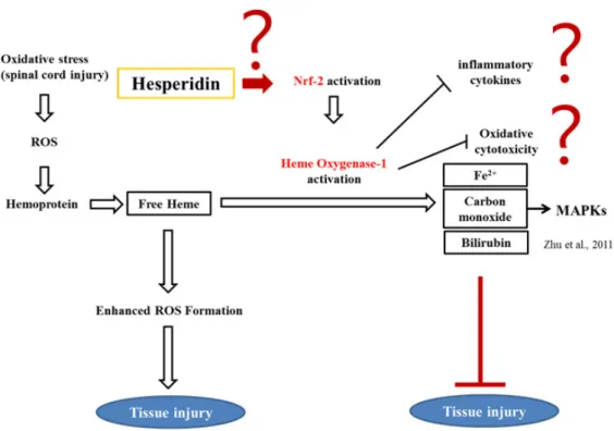
Hesperidin, a flavanoglycone abundantly present in citrus fruits, has been reported to have anti-inflammatory and antioxidant activity (Jain and Parmar, 2011;Parhiz et al., 2015;Umeno et al., 2016), radical scavenging activity (Iranshahi et al., 2015) and neuroprotective property (Justin-Thenmozhi et al., 2018;Justin Thenmozhi et al., 2015), implying that hesperidin is involved in the tissue protection through antioxidant, anti-inflammatory and neuroprotective roles which all are key elements in the course of neuro-inflammation caused by SCI. Hesperidin protects against chemically induced hepatocarcinogenesis by modulating heme oxygenase (HO)-1, an induced form of HO, and ameliorating oxidative stress and inflammation (Mahmoud et al., 2017). HO-1 is the rate-limiting enzyme in heme metabolism, a process that leads to the generation of equimolar quantities of
carbon monoxide, Fe2+, and biliverdin (Zhu et al., 2011). Carbon monoxide,
one of the three main byproducts of heme catabolism by HO, selectively inhibits expression of the proinflammatory cytokines, tumor necrosis factor (TNF)-α, interleukin (IL)-1β, and macrophage inflammatory protein-1β (Otterbein et al., 2000). In addition, carbon monoxide mediates these antiinflammatory effects specifically through the mitogen-activated protein kinases (MAPKs), in particular the MAPK kinase 3/p38 pathway (Otterbein et al., 2000). Therefore, HO-1 is commonly regarded as having potent antiinflammatory and antioxidant properties. The potential mechanism underlying the attenuation of locomotor disability by hesperidin in the rat spinal cord injury model remains unclear.
This study was performed to determine behaviorally whether hesperidin attenuates motor disability in SCI rats, and to examine which molecules, including HO-1, are involved in the mechanism of the amelioration of SCI by hesperidin (Figure 5).
3. Materials and methods
3.1. Animals and Surgical procedures
Female Sprague-Dawley rats (180–220 g, 6–8 weeks old) were obtained from Central Lab Inc. (Seocho-gu, Seoul, Korea) and were allowed to acclimatize to the laboratory environment for 1 week before use. Clip compression injury was performed as described previously (Ahn et al., 2012;Kim et al., 2017). Briefly, rats were anesthetized with Zoletil 50 (Virbac, Carros, France) and subjected to laminectomy at thoracic cord 9/10. Then, the thoracic spinal cord was compressed vertically using a vascular clip (Stoelting, Wood Dale, IL, USA) at an occlusion pressure of 15–20 g for 1 min. The wounds, including those in muscles and skin layers, were closed immediately following injury.
All experimental procedures were conducted in accordance with the Guidelines for the Care and Use of Laboratory Animals of Jeju National University (2018-0029). The animal protocols conformed to current international laws and the National Institutes of Health (NIH) Guide for the Care and Use of Laboratory Animals (NIH Publication No. 85-23, 1985, revised 1996).
3.2. Experimental groups with hesperidin treatment
To assess the effects of hesperidin on SCI, rats were divided into three groups (10 animals/group) as follows: Group A, sham operation control group; Group B, vehicle-treated control group; Group C, hesperidin-treated
(100 mg/kg body weight/day, H5254, Lot No. #019K1256; Sigma-Aldrich, St. Louis, MO, USA), hesperidin was administered to rats from 7 days prior to SCI to day 7 post-SCI (14 days in total). Vehicle solution alone was administered to rats in the same manner.
3.3. Behavioral test
Locomotor behavioral function after SCI was evaluated using the Basso, Beattie, and Bresnahan (BBB) rating scale during the experiment until 6 weeks, depending on the experimental groups. All evaluations were performed in a double-blind manner; average scores were calculated for each group and used to compare the severity of hindlimb paralysis.
After acknowledgement of behavioral improvement, further experiments were performed to assess spinal cord tissues. Briefly, SCI with 14-day hesperidin treatment was repeated because this schedule significantly ameliorated locomotor disability.
3.4. Tissue preparation and histological examination
For histopathological examination of spinal cord tissues, thoracic spinal cords (T8–T11) were collected at 4 days post-injury (DPI). Briefly, the spinal cords were fixed with 4% (v/v) paraformaldehyde in phosphate-buffered saline (PBS; pH 7.4) for 48 h. Subsequently, sagittal sections (5 µm thick) were cut for hematoxylin and eosin (H&E) staining and used for immunohistochemistry.
3.5. Serum chemistry
Serum samples prepared 4 DPI were subjected to biochemical analysis as described in our previous report (Kim et al., 2018). To evaluate the
contents of bilirubin and Fe2+, which are related to antioxidant activity (Klemz
et al., 2009), their serum levels were measured using a Roche-Hitachi Cobas 8000 C702 (Roche Diagnostic System, Basel, Switzerland).
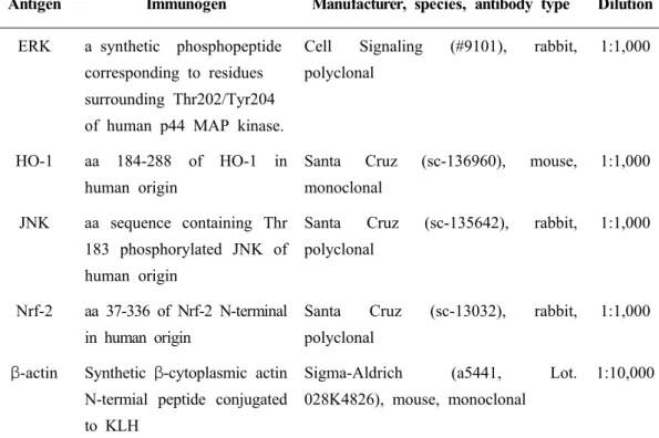
3.6. Antibody characterization
The characteristics of the antibodies used in the present study are summarized in Table 1.
Table 1 The characterization of antibodies used in this study.
Antigen Immunogen Manufacturer, species, antibody type Dilution
ERK a synthetic phosphopeptide corresponding to residues surrounding Thr202/Tyr204 of human p44 MAP kinase.
Cell Signaling (#9101), rabbit, polyclonal
1:1,000
HO-1 aa 184-288 of HO-1 in human origin
Santa Cruz (sc-136960), mouse, monoclonal
1:1,000 JNK aa sequence containing Thr
183 phosphorylated JNK of human origin
Santa Cruz (sc-135642), rabbit, polyclonal
1:1,000
Nrf-2 aa 37-336 of Nrf-2 N-terminal in human origin
Santa Cruz (sc-13032), rabbit, polyclonal
1:1,000 β-actin Synthetic β-cytoplasmic actin
N-termial peptide conjugated to KLH
Sigma-Aldrich (a5441, Lot. 028K4826), mouse, monoclonal
1:10,000
Secondary antibodies for Western blot analysis
Peroxidase anti-rabbit IgG (H+L) Vector Laboratories (BA-1000) 1:1,000 Peroxidase anti-mouse IgG (H+L) Vector Laboratories (PI-2000) 1:1,000 Abbreviatoins: aa, amino acid; ERK, extracellular signal-regulated kinase; HO-1,
hemeoxygenase-1; JNK, c-Jun N-terminal kinase; Nrf-2, nuclear factor erythroid 2-related factor-2; IgG, immunoglobulin.
3.7. Western blot analysis
We performed western blotting as described previously (Ahn et al., 2015). Spinal cords, including the core region (length, 1 cm), were homogenized, and then electrophoresed and immunoblotted onto nitrocellulose membranes (Schleicher and Schuell, Keene, NH, USA). Membranes were incubated with primary antibodies against nuclear factor erythroid-derived 2-related factor 2 (Nrf-2), HO-1, extracellular signal-regulated kinase (ERK), c-Jun N-terminal kinase (JNK), phospho-p38 (p-p38), and β-actin, and then reacted with the appropriate secondary antibody in accordance with the manufacturer’s protocol (Vector Laboratories, Burlingame, CA, USA) and visualized using a BS ECL Plus kit (W6002; Biosesang, Gyeonggi, Korea). Immunoblotting signals were detected using Fusion Solo 6X (Vilber Lourmat, Collegien, France) and analyzed using FUSION software. The expression levels were normalized relative to β-actin.
3.9. Quantitative real time-PCR
Total RNAs of spinal cords were extracted using TRIzol RNA Isolation Reagent (Life Technologies, Thermo Fisher Scientific, Waltham, MA, USA). Purified RNA was transcribed into cDNA using 5× First Strand cDNA Synthesis Master Mix (CellSafe, Gyeonggi-do, Korea) according to the manufacturer’s protocol.
Quantitative real-time PCR was performed using QuantiSpeed SYBR No-Rox Mix (PhileKorea Co., Ltd., Seoul, Korea) according to the manufacturer’s protocol. The primer sequences are presented in Table 2. PCR was performed using a Mic Real-Time PCR Cycler (Bio Molecular Systems,
Potts Point, NWS, Australia). The PCR profile consisted of 40 cycles of denaturation at 95°C for 15 s, followed by 60°C for 30 s to allow extension and amplification of the target sequence. The expression levels were normalized relative to that of glyceraldehyde-3-phosphate dehydrogenase
(GAPDH) using the 2−ᇞᇞCT method.
3.10. Statistical analysis
All measurements are shown as averages of three independent experiments. All values are presented as means ± standard error of the mean (SEM). BBB scores and western blotting data were analyzed by one-way analysis of variance (ANOVA) followed by Student–Newman–Keuls post hoc tests for multiple comparisons. In all analyses, P < 0.05 was taken
Table 2 Primer sequences used in the present study.
Primer Sequence
Catalase forward 5′-CCACGAGGGTCACGAACTGT-3′
Catalase reverse 5′-CTCCTATTGCCGTCCGATTC-3′
GAPDH forward 5′- CAGCGCATACCACTTCAGC-3′
GAPDH reverse 5′- ACCATCGAGCATCCCAAG-3′
IL-1β forward 5′- CCCTGCAGCTGGAGAGTGTG-3′
IL-1β reverse 5′-TGTGCTCTGCTTGAGAGGTG-3′
SOD1 forward 5′-GGCCACACCGTCCTTTCC-3′
SOD1 reverse 5′-CGGTCCAGCGGATGAAGA-3′
SOD2 forward 5′-TAAGCGTGCTCCCACACATC-3′
SOD2 reverse 5′-ATCAGGACCCACTGCAAGGA-3′
SOD3 forward 5′-TGCAGACTGCGTGCATCTC-3′
SOD3 reverse 5′-GCGACACGCACTCCAAAGA-3′
TNF-α forward 5′-CGTCGTAGCAAACCACCAAG-3′
TNF-α reverse 5′-CACAGAGCAATGACTCCAAA -3′
Abbreviatoins: GAPDH, glyceraldehyde-3-phosphate dehydrogenase; IL-1β, interleukin-1 beta; SOD, superoxide dismutase; TNF-α, tumor necrosis factor-alpha.
4. Result
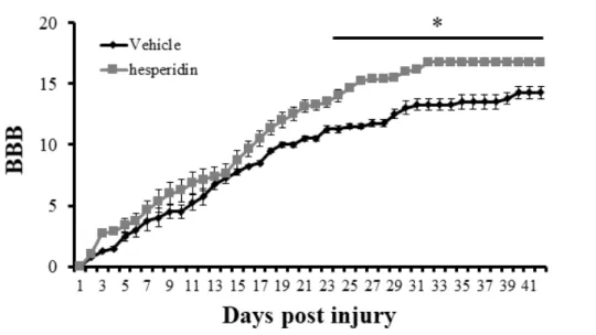
4.1. Hesperidin ameliorates locomotor movement in SCI rats
To evaluate locomotor function, we examined the BBB score after operation. Locomotor function recovered from 3 DPI, and the BBB score increased in both groups. BBB scores in the hesperidin-treated group were higher than those in the vehicle-treated control group during the experimental period. The BBB score was particularly significantly increased in the hesperidin-treated group (10.4 ± 0.52, P < 0.05), compared with the vehicle-treated controls at 21 DPI; this trend persisted throughout the experimental period (Figure 6).
Figure 6. Locomotor outcomes as evaluated by Basso, Beattie, and Bresnahan
(BBB) scoring (n = 5 per day). The BBB scores were very low in both groups until 3 days post-injury (DPI). The BBB score in the hesperidin-treated group was significantly higher than that in the vehicle-treated group at 21 DPI. This difference persisted until 42 DPI. * P < 0.05 vs. vehicle-treated group.
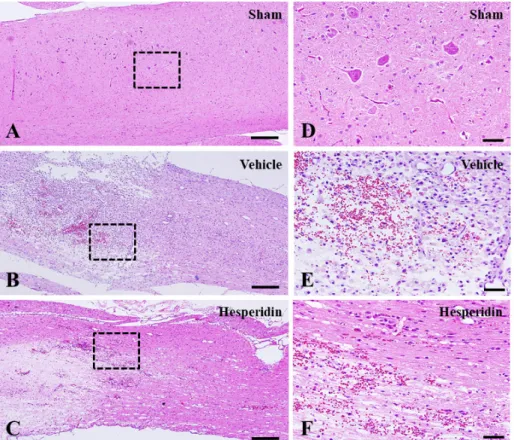
4.2. Neuropathological changes
In the sham operation control group, there were no neuropathological changes in the core region of the spinal cord (Figure 7A and 7D). The core region of the spinal cord in vehicle-treated rats showed severe edema, hemorrhage, and infiltration of inflammatory cells in longitudinal sections (Figure 7B and 7E). On the other hand, the neuropathological changes, including edema, hemorrhage, and inflammatory cell infiltration, were reduced in the hesperidin-treated group (Figure 7C and 7F).
Figure 7. Neuropathological changes in the spinal cords of sham-operated
control (A and D), vehicle-treated control (B and E), and hesperidin-treated groups (C and F) at 4 DPI. (A–C) Low magnification images of longitudinal sections. (D–F) High magnification images of the boxes in A–C, respectively. A–F, Hematoxylin and eosin staining. (A–C) scale bars = 100 μm; (D–F) scale bars = 50 μm.
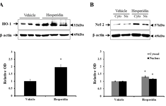
4.3. Hesperidin activated the Nrf-2/HO-1 signal pathway in the spinal cords
To evaluate activation of the Nrf-2/HO-1 signaling pathway, we performed western blotting analysis in the core region of the spinal cord at 4 DPI (n = 3 per group). Hesperidin treatment significantly increased the protein
levels of both cytoplasmic Nrf-2 (1.32 ± 0.04-fold, relative OD/mm2, P <
0.05) and nuclear Nrf-2 (1.16 ± 0.01-fold, P < 0.05), compared to the vehicle-treated control group (1.00 ± 0.03-fold in the cytoplasm and 1.00 ± 0.02-fold in the nucleus) (Figure 8A). Furthermore, the protein level of HO-1 in the hesperidin-treated group was significantly higher (1.96 ± 0.16-fold, P < 0.05) than that in the vehicle-treated control group (1.00 ± 0.12-fold, P < 0.05) (Figure 8B). These results indicated the upregulation of Nrf-2 expression and translocation into the nucleus in the spinal cord of injured rats following hesperidin treatment.
Figure 8. Western blotting to detect heme oxygenase (HO)-1 and nuclear
factor erythroid 2-related factor (Nrf)-2 in the spinal cords of SCI rats. (A and B) Representative immunoblots of HO-1 (~32 kDa), Nrf-2 (~57 kDa), and β-actin (~45 kDa). Bar graphs: the levels of both Nrf-2 and HO-1 were increased significantly and Nrf-2 was translocated from the cytoplasm to the nucleus in the spinal cords of hesperidin-treated rats. Normalization was achieved by reprobing the membranes with anti-β-actin antibody. Means ± SEM (n = 3 per group) are shown. * P < 0.05 vs. vehicle-treated control group.
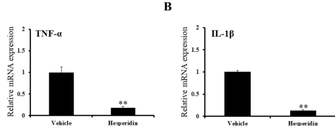
4.4. Hesperidin ameliorates the inflammation oxidative stress in the spinal cords of injured rats
To determine the antiinflammatory effects of hesperidin treatment in SCI rats, we evaluated the expression of proinflammatory cytokines, including TNF-α and IL-1β. Quantitative real-time PCR indicated that the mRNA expression levels of the representative proinflammatory cytokines, TNF-α (Figure 9A) and IL-1β (Figure 9B) were significantly lower in the hesperidin-treated SCI rats compared to vehicle-treated control rats (relative fold changes, P < 0.01 vs. vehicle-treated SCI rats).
Figure 9. Results of quantitative real-time PCR in the spinal cords of SCI rats
(n = 5 per group). Hesperidin treatment suppressed proinflammatory mediator-related gene expression in SCI rats. The mRNA expression levels of tumor necrosis factor-alpha (TNF-α) (A) and interleukin-1 beta (IL-1β) were significantly decreased in the hesperidin-treated group compared to vehicle-treated controls. * P < 0.05 vs. vehicle-treated control group.
We also analyzed superoxide dismutase (SOD) and catalase activities to evaluate the antioxidative effects of hesperidin treatment in SCI rats. The mRNA levels of SOD1 (Figure 10A), SOD2 (Figure 10B), SOD3 (Figure 10C), and catalase (Figure 10D) in the hesperidin-treated group were significantly higher than those in the vehicle-treated control group (Figure 10A –D).
Figure 10. Results of quantitative real-time PCR for superoxide dismutase
(SOD) and catalase in the spinal cords of SCI rats (n = 5 per group). The mRNA expression levels of SOD subtypes SOD1, SOD2, and SOD3, and catalase were significantly higher in the hesperidin-treated group than in the vehicle-treated controls. * P < 0.05 vs. vehicle-treated control group.
4.5. Hesperidin enhances p-p38 expression in the spinal cords of injured rats
ERK (1.07 ± 0.01-fold) and JNK (0.95 ± 0.02-fold) protein levels in the hesperidin-treated group were not significantly different from those in vehicle-treated controls (1.00 ± 0.01-fold and 1.00 ± 0.02-fold, respectively). However, the p-p38 protein level was significantly higher in the hesperidin-treated group than in the vehicle-treated control group (2.96 ± 0.17-fold vs. 1.00 ± 0.02-fold, respectively; P < 0.05) (Figure 11A–C).
4.6. Hesperidin enhances serum bilirubin and Fe2+ levels in the spinal cords
of injured rats
Serum chemistry analysis indicated that bilirubin (Figure 11D) and
Fe2+ (Figure 11E) levels were significantly increased in hesperidin-treated SCI
rats compared to those in vehicle-treated control rats (relative mRNA expression level, P < 0.05 vs. vehicle-treated SCI rats).
Figure 11. Western blotting analysis of mitogen-activated protein kinase
(MAPK) in the spinal cord and chemical analysis of bilirubin and Fe2+in the
sera of SCI rats. (A–E) Representative immunoblots of phosphorylated extracellular signal-regulated kinase (p-ERK; ~44 and 42 KDa) (A), c-Jun N-terminal kinase (p-JNK; ~54 and 46 KDa) (B), and p38 (p-p38; ~38 KDa) (C). Bar graphs: Gray bars represent the ratio of p-ERK-1/total ERK-1 and p-JNK-1/total JNK1 on densitometric analysis. Black bars represent the ratios of p-ERK-2/total ERK-2, p-JNK-2/total JNK2, and p-p38. There were no changes in p-ERK and p-JNK protein levels. On the other hand, p-p38 protein levels were significantly increased in the spinal cords of the hesperidin-treated group. Normalization was achieved by reprobing the membranes with anti-β-actin
antibody. Both bilirubin (D) and Fe2+ (E) concentrations in the hesperidin-treated
group were higher than those in vehicle-treated controls. Means ± SEM (n = 5 per group) are shown. * P < 0.05 vs. vehicle-treated control group.
5. Discussion
The results of the present study confirmed that hesperidin, a component of citrus fruits, exerts antiinflammatory effects in an animal model of SCI, which is characterized by edema, hemorrhage, inflammatory cell infiltration, and reactive gliosis in the affected spinal cord (Ahn et al., 2012;Kjell and Olson, 2016).
Hesperidin has been shown to exert antiinflammatory effects via its antioxidative and radical scavenging activities (Parhiz et al., 2015), which are effective in the amelioration of a variety of inflammation models, including rheumatoid arthritis (Li et al., 2010), dextran sulfate sodium-induced ulcerative colitis (Xu et al., 2009), and lipopolysaccharide-induced acute lung inflammation (Yeh et al., 2007). Hesperidin was confirmed to suppress the production of inflammatory cytokines, including TNF-α, IL-1β, and IL-6 (Parhiz et al., 2015; Xu et al., 2009; Yeh et al., 2007).
With regard to the involvement of flavonoids in neuroinflammation, it has been demonstrated that flavonoids mediate recovery of motor function through neuroprotective effects in SCI (Du et al., 2018;Jiang et al., 2016a;Ma et al., 2018;Ozdemir et al., 2016), as demonstrated in treatment with St. John’s wort (Hypericum perforatum) (Ozdemir et al., 2016), quercetin (Jiang et al., 2016a), eugenol (Ma et al., 2018), and oxyresveratrol (Du et al., 2018), possibly by reducing the cytotoxic effects of radicals. Based on these previous studies, we
The molecular mechanism underlying the effects of hesperidin in the SCI model remains to be determined. In the present study, we showed that the upregulation of HO-1 and Nrf-2 nuclear translocation in hesperidin-treated SCI rats played a role in the amelioration of hindlimb paralysis. The ameliorative effects of upregulated HO-1 and Nrf-2 on neuroinflammation in SCI models have been widely reported following treatment with beta carotene (Zhou et al., 2018), rosmarinic acid (Shang et al., 2017), asiatic acid (Jiang et al., 2016b), and lithium chloride (Kim et al., 2017). We postulated that upregulation of HO-1 and Nrf-2 protects neurons and axons in the same manner as antioxidative effects.
In contrast to the cytoprotective effect of HO-1 in tissues, HO-1 has been reported to mediate cell death with concurrent increases in reactive oxygen species levels (Bansal et al., 2014). Similarly, ferrous iron is known to react with oxygen to produce superoxide, which affects the surrounding cells (Yama et al., 2016). HO-1 is also involved in the generation of hydrogen peroxide, a source of hydroxyl radicals (Yama et al., 2016), which are neutralized by SOD and catalase, finally reducing the accumulation of reactive oxygen species. Furthermore, increasing SOD and catalase levels contributed to the tissue protective activity against oxidative stress in an SCI model (Wang et al., 2017). With regard to oxidative stress in SCI models, it was postulated that upregulation of radical scavenging enzymes, including SOD and catalase, in the hesperidin-treated group is closely associated with a reduction in SCI-induced hindlimb paralysis. Therefore, the upregulation of HO-1 in accordance with increased levels of SOD and catalase in the present study
ameliorates the damage after SCI.
Another mechanism of cytoprotection by HO-1 involves heme degradation products and their metabolic derivatives. Briefly, HO-1 has been shown to catalyze the degradation of heme to produce ferrous iron, carbon monoxide, and biliverdin, the latter of which is subsequently converted into bilirubin (Parhiz et al., 2015; Yama et al., 2016) (see schematic drawing in Fig. 7). Hesperidin enhances the potential antioxidant effects by increasing serum
levels of both bilirubin and Fe2+, the product of heme decomposition by HO,
in rats with SCI. Both bilirubin and Fe2+ are antioxidants with possible
physiological importance (Stocker et al., 1987; Zhu et al., 2011), and HO-1-derived bilirubin has been shown to ameliorate postischemic myocardial
dysfunction (Clark et al., 2000). The increased levels of bilirubin and Fe2+ in
serum observed in the present study may contribute to the scavenging of peroxyl radicals, leading to cytoprotection in SCI lesions.
With regard to the involvement of MAPK in inflammation, carbon monoxide has been shown to be one of the intermediate molecules in the cascade from HO-1 to antiinflammatory activity (Otterbein et al., 2000). In the present study, we found that p-p38, but not ERK and JNK, was significantly upregulated, suggesting that HO-1, through carbon monoxide, activated p-p38, leading to antiinflammatory effects. These findings were further supported by reports that the production of HO-1 through degradation of heme inhibited the
In conclusion, our findings suggest that hesperidin ameliorated paralysis caused by SCI in rats, and that the underlying molecular mechanism involved inhibition of inflammation in the spinal cord, with concurrent increases in nuclear translocation of Nrf-2 and subsequent upregulation of HO-1 (Figure 12).
Figure 12. Schematic illustration of the neuroprotective mechanism in the
spinal cord associated with hesperidin treatment. Hesperidin ameliorated rat hindlimb paralysis caused by SCI via antioxidative effects, suppression of inflammatory cytokines, and increased nuclear translocation of Nrf-2 with subsequent upregulation of HO-1.
References
Ahn, M, Lee, C, Jung, K, Kim, H, Moon, C, Sim, KB, Shin, T. Immunohistochemical study of arginase-1 in the spinal cords of rats with clip compression injury. Brain Res., 2012; 1445: 11-19.
Ahn, M, Moon, C, Park, C, Kim, J, Sim, KB, Shin, T. Transient activation of an adaptor protein, disabled-2, in rat spinal cord injury. Acta Histochem., 2015; 117: 56-61.
Bansal, S, Biswas, G, Avadhani, NG. Mitochondria-targeted heme oxygenase-1 induces oxidative stress and mitochondrial dysfunction in macrophages, kidney fibroblasts and in chronic alcohol hepatotoxicity. Redox Biol., 2013; 2: 273-283.
Clark, JE, Foresti, R, Sarathchandra, P, Kaur, H, Green, CJ, Motterlini, R. Heme oxygenase-1-derived bilirubin ameliorates postischemic myocardial dysfunction. Am. J. Physiol. Heart Circ. Physiol., 2000; 278(2): H643-651.
Donnelly, DJ, Popovich, PG, Inflammation and its role in neuroprotection, axonal regeneration and functional recovery after spinal cord injury. Exp Neurol., 2008; 209(2): 378-388.
inflammation and oxidative stress in rat model of spinal cord injury. Mol Med Rep, 2018; 17: 4067-4073.
Fatima, G, Sharma, VP, Das, SK, Mahdi, AA. Oxidative stress and antioxidative parameters in patients with spinal cord injury: implications in the pathogenesis of disease. Spinal Cord, 2015; 53: 3-6.
Hamann, K, Shi, R. Acrolein scavenging: a potential novel mechanism of attenuating oxidative stress following spinal cord injury. J. Neurochem., 2009; 111(6): 1348-1356.
Iranshahi, M, Rezaee, R, Parhiz, H, Roohbakhsh, A, Soltani, F. Protective effects of flavonoids against microbes and toxins: The cases of hesperidin and hesperetin. Life Sci., 2015; 137: 125-132.
Jain, M and Parmar, HS. Evaluation of antioxidative and anti-inflammatory potential of hesperidin and naringin on the rat air pouch model of inflammation. Inflamm. Res., 2011: 60(5); 483-491.
Jiang, W, Huang, Y, Han, N, He, F, Li, M, Bian, Z, Liu, J, Sun, T, Zhu, L. Quercetin suppresses NLRP3 inflammasome activation and attenuates histopathology in a rat model of spinal cord injury. Spinal Cord,
Neuroprotective effect of asiatic acid against spinal cord injury in rats. Life Sci., 2016b; 157: 45-51.
Jung, K, Min, DS, Sim, KB, Ahn, M, Kim, H, Cheong, J, Shin, T. Upregulation of phospholipase D1 in the spinal cords of rats with clip compression injury. Neurosci. Lett., 2003; 336(2): 126-130.
Justin-Thenmozhi, A, Dhivya Bharathi, M, Kiruthika, R, Manivasagam, T, Borah, A, Essa, MM. Attenuation of Aluminum Chloride-Induced Neuroinflammation and Caspase Activation Through the AKT/GSK-3beta Pathway by Hesperidin in Wistar Rats. Neurotox Res., 2018; 34(3): 463-476.
Justin Thenmozhi, A, Raja, TR, Janakiraman, U, Manivasagam, T. Neuroprotective effect of hesperidin on aluminium chloride induced Alzheimer's disease in Wistar rats. Neurochem. Res., 2015; 40(4): 767-776.
Kim, J, Bang, H, Ahn, M, Choi, Y, Kim, GO, Shin, T. Allyl isothiocyanate reduces liver fibrosis by regulating Kupffer cell activation in rats. J. Vet. Med. Sci., 2018; 80(6): 893-897.
Kim, Y, Kim, J, Ahn, M, Shin, T. Lithium ameliorates rat spinal cord injury by suppressing glycogen synthase kinase-3beta and activating heme oxygenase-1. Anat Cell Biol, 2017; 50(3): 207-213.
Kjell, J, Olson, L, Rat models of spinal cord injury: from pathology to potential therapies. Dis Model Mech, 2016; 9(10): 1125-1137.
Klemz, R, Mashreghi, MF, Spies, C, Volk, HD, Kotsch, K. Amplifying the fluorescence of bilirubin enables the real-time detection of heme oxygenase activity. Free Radic. Biol. Med., 2009; 46(2): 305-11.
Li, R, Cai, L, Xie, XF, Yang, F, Li, J. Hesperidin suppresses adjuvant arthritis in rats by inhibiting synoviocyte activity. Phytother Res, 2010; 24: Suppl 1, S71-76.
Ma, L, Mu, Y, Zhang, Z, Sun, Q. Eugenol promotes functional recovery and alleviates inflammation, oxidative stress, and neural apoptosis in a rat model of spinal cord injury. Restor. Neurol. Neurosci., 2018; 36(5): 659-668.
Mahmoud, AM, Mohammed, HM, Khadrawy, SM, Galaly, SR. Hesperidin protects against chemically induced hepatocarcinogenesis via modulation of Nrf2/ARE/HO-1, PPARgamma and TGF-beta1/Smad3 signaling, and amelioration of oxidative stress and inflammation. Chem. Biol. Interact., 2017; 277: 146-158.
effects involving the mitogen-activated protein kinase pathway. Nat. Med., 2000; 6(4): 422-428.
Ozdemir, US, Naziroglu, M, Senol, N, Ghazizadeh, V. Hypericum perforatum Attenuates Spinal Cord Injury-Induced Oxidative Stress and Apoptosis in the Dorsal Root Ganglion of Rats: Involvement of TRPM2 and TRPV1 Channels. Mol. Neurobiol., 2016; 53(6): 3540-3551.
Parhiz, H, Roohbakhsh, A, Soltani, F, Rezaee, R, Iranshahi, M. Antioxidant and anti-inflammatory properties of the citrus flavonoids hesperidin and hesperetin: an updated review of their molecular mechanisms and experimental models. Phytother Res, 2015; 29(3): 323-331.
Shang, AJ, Yang, Y, Wang, HY, Tao, BZ, Wang, J, Wang, ZF, Zhou, DB. Spinal cord injury effectively ameliorated by neuroprotective effects of rosmarinic acid. Nutr Neurosci, 2017; 20(3): 172-179.
Shin, T, Ahn, M, Moon, C, Kim, S, Sim, KB. Alternatively activated macrophages in spinal cord injury and remission: another mechanism for repair? Mol. Neurobiol. 2013; 47(3): 1011-1019.
Stocker, R, Yamamoto, Y, McDonagh, AF, Glazer, AN, Ames, BN. Bilirubin is an antioxidant of possible physiological importance. Science, 1987; 235(4792): 1043-1046.
Umeno, A, Horie, M, Murotomi, K, Nakajima, Y, Yoshida, Y. Antioxidative and Antidiabetic Effects of Natural Polyphenols and Isoflavones. Molecules, 2016; 21(6): E708.
Wang, X, Wu, X, Liu, Q, Kong, G, Zhou, J, Jiang, J, Wu, X, Huang, Z, Su, W, Zhu, Q. Ketogenic Metabolism Inhibits Histone Deacetylase (HDAC) and Reduces Oxidative Stress After Spinal Cord Injury in Rats. Neuroscience, 2017; 366: 36-43.
Xu, L, Yang, ZL, Li, P, Zhou, YQ. Modulating effect of Hesperidin on experimental murine colitis induced by dextran sulfate sodium. Phytomedicine, 2009; 16(10): 989-995.
Yama, K, Sato, K, Murao, Y, Tatsunami, R, Tampo, Y. Epalrestat Upregulates Heme Oxygenase-1, Superoxide Dismutase, and Catalase in Cells of the Nervous System. Biol. Pharm. Bull., 2016; 39(9): 1523-1530.
Yeh, CC, Kao, SJ, Lin, CC, Wang, SD, Liu, CJ, Kao, ST. The immunomodulation of endotoxin-induced acute lung injury by hesperidin in vivo and in vitro. Life Sci., 2007; 80(20): 1821-1831.
a rat model of spinal cord injury. Int. Immunopharmacol., 2018; 61: 92-99.
Zhu, X, Fan, WG, Li, DP, Kung, H, Lin, MC. Heme oxygenase-1 system and gastrointestinal inflammation: a short review. World J. Gastroenterol., 2011; 17(38): 4283-4288.
Abstract in Korean
척수손상모델에서 헤스페리딘의 치료 효과 및
그 기전에 대한 연구
(지도교수 : 신 태 균) 허 승 담 제주대학교 일반대학원 수의학과척수손상(Spinal Cord Injury)은 일차적 기계적 손상과 이후 발생하는 이차적 조직 손상을 특징으로 하며, 조직병리학적 검사에서 부종, 축삭 변성, 염증세포침윤, Fibronectin의 삼출 등의 특징이 있다. 감귤 추출물의 주요 성분인 헤스페리딘(Hesperidin)은 최근 여러 연구에서 신경 염증 및 항산화 손상 실험을 통해 항산화 및 항염증 작용을 갖는 것으로 알려졌다. 본 연구에서는 강력한 항산화제의 하나인 헤스페리딘을 이용하여 랫트의 압박성 척수손상모델에서 효능을 평가하였다. 그 기전연구로 산화적 손상을 억제시켜 세포를 보호하는 것으로 알려져 있는 nuclear factor erythroid 2-related factor-2 (Nrf-2) 및 그 표적 단백질인 heme oxygenase-1 (HO-1)와 관련 기전을 확인하였다. 랫트 흉추 10번 수준에서 추궁 절제술을 시행하여 척수를 노출시킨 후, 혈관 클립으로 1분간 척수를 압박하여 척수손상 모델을 제작하였고,
행동학적 평가에서는 Basso, Beattie, Bresnahan (BBB) score와 foot printing을 이용한 좌골 기능 검사 방법을 변형하여 보행정도를 평가하였고, 이를 통해 손상 후 21일부터 대조군에 비해 헤스페리딘 투여군이 유의성 있게 행동학적 차이를 보였으며, 손상 후 42일까지 유지되는 것을 확인하였다(p<0.05). 좌골기능 검사에서 뒷다리 보행은 손상 직후에 발등을 끄는 형태를 나타내다가 헤스페리딘 투여 후 14일째부터 발바닥 전체 보행에서 발가락 보행으로 변하는 것을 확인할 수 있었다. 조직학적 평가에서는 척수손상 후, 척수 조직 내 출혈, 염증세포의 침윤 및 조직의 손실이 확인되었으나, 헤스페리딘 투여 그룹에서 이러한 증상이 개선되었음을 확인하였다. 척수 조직 내 염증을 확인하기 위해 염증 매개 사이토카인을 real time-PCR로 확인한 결과, 헤스페리딘을 투여한 경우에 TNF-alpha와 IL-1 beta의 수치가 감소되었음을 확인하였다. 또한 헤스페리딘은 척수 조직에서 Nrf-2의 세포질/핵내 발현을 증가시켰고, 표적단백질은 HO-1의 발현도 증가시켰다. 산화적 손상의 지표 마커인 Superoxide dismutase와 Catalase는 헤스페리딘 투여 그룹에서 유의성 있게 증가하였고, 혈청내 빌리루빈과 철2가이온이 헤스페리딘 그룹에서 증가하였음을 확인하였다. 본 연구는 헤스페리딘이 척수손상이 유도된 랫트의 마비를 개선시켰고, 척수손상에 의한 염증세포의 침윤 및 조직 내 출혈, 조직의 손실을 완화시켰으며, Nrf-2의 핵내 전이 증가로 인한 표적 단백질인 HO-1의 활성화를 통해 신경세포를 산화적 손상으로부터 보호하는 효과가 있음을 확인하였다. 이 결과를 바탕으로 헤스페리딘은 척수손상의 치료에 보조적인 역할을 할 것으로 기대된다. 주요어: 척수손상, 헤스페리딘, 행동장애, 헴산화효소.
감사의 글
박사학위 논문을 마무리하면서 생각해보면 동물병원 생활과, 대학원 수업, 그리고 논문 진행에 힘들고 어려운 일이 많았지만 많은 분들의 격려와 도움 덕분에 졸업을 앞두게 되었습니다. 저를 아껴주시고 도와주신 모든 분들께 진심으로 감사의 마음을 전합니다. 2002년 대학졸업을 앞두고 동물병원 인턴을 할까 대학원 진학을 할까 고민하고 망설이고 있을 때 대학원에 진학할 수 있도록 기회를 주신 신태균 교수님, 이런 인연으로 저의 석사와 박사과정의 지도교수님이 되어주셨고 오늘이 있기까지 너무나도 부족했던 저에게 마지막 순간까지 어머니처럼 따뜻하게 챙겨주시고 도와주시고 이끌어주신 교수님께 머리 숙여 감사드립니다. 앞으로도 교수님과의 인연과 은혜를 가슴에 간직하고 살아가도록 하겠습니다. 소중한 시간을 내어 논문심사 해주시고 따뜻한 격려와 조언을 주신 지영흔 교수님, 강태영 교수님께도 깊은 감사드립니다. 먼 거리를 마다하지 않고 기꺼이 와주신 농림축산검역본부 김황용 사무관님, 실험실 연구로 바쁜 와중에도 밝은 웃음으로 유익한 조언을 해주신 안미정 교수님께 감사드립니다. 지난 박사과정동안 물심양면으로 옆에서 도와주고 심사기간 내내 부족한 부분을 꼼꼼히 조언해준 실험실의 듬직한 김정태 박사님, 내년부터 석박사 과정의 새로운 삶을 시작하는 유나, 멀고먼 한국으로 유학 와서마지막으로 제가 가장 사랑하는 가족들에게 감사의 말씀을 드립니다. 홀로 3남매를 부족함 없이 보살펴주신 어머니와 하늘에서 보고 계실 아버지께 감사를 올립니다. 항상 저를 아껴주시는 장인어른과 장모님께도 감사를 드리며, 오래도록 건강하길 기원합니다. 끝으로 오늘이 있기까지 저를 다독이고 힘이 되어준 영원한 동반자이며 사랑하는 아내 순주와 딸 예빈, 정원에게 고마움을 전하며 좋은 남편과 훌륭한 아버지가 되도록 노력하겠습니다. 2018. 12. 3. 허 승 담