INTRODUCTION
AD is a progressive neurodegenerative disorder which is recognized as the most common form of dementia among the elderly characterized by progressive dysfunction of cognition and memory (Scheltens et al., 2016). It affects the millions of the elderly and the number of AD patients has dramati-cally increased. In the worldwide, approximately 35.6 mil-lion people are currently affected from AD, but it is expected that the number of people living with AD will reach 135 mil-lion people in 2050 (Prince et al., 2013; Kim et al., 2015a). Although the cause of AD has not yet been fully understood, but several studies investigated that Aβ deposition is one of the major causes of AD pathology in the early onset familial AD (Hardy and Orr, 2006). Senile plaques formed by extra-cellular Aβ accumulation are one of the AD pathologic hall-marks which believed to not only interfere the synapses
neu-ron communication but also lead to cell death (Karran and De Strooper, 2016). The Aβ is known to be produced from APP (Gu et al., 2018). The shedding of APP generates sAPPβ and CTFβ through β-secretase, and consequential CTFβ cleavage produces Aβ40 as well as Aβ42 by γ-secretase (Park, 2010; Prince et al., 2013). However, α-secretase cleaves APP and then generates sAPPα and CTFα precluding the Aβ formation (Park, 2010). Eventually, the Aβ production will directly be re-lated with the sAPPβ generation and contrary correre-lated with the proportion of sAPPα (Kim et al., 2015b). Considering such previous findings, reduction of Aβ or delaying the Aβ deposi-tion in the brain would be the possible therapeutic targets in the treatment or prevention of AD (Hardy and Orr, 2006; Kim et al., 2015b; Karran and De Strooper, 2016). Furthermore, various secretases would be the key factors to modulate to deliver such beneficial effects. The ADAM family are known as α-secretases such as ADAM9, ADAM10, and ADAM17, Extracellular beta amyloid (Aβ) plaques are the neuropathological hallmarks of Alzheimer’s disease (AD). Accordingly, reducing Aβ levels is considered a promising strategy for AD prevention. 3’-O-acetyl-24-epi-7,8-didehydrocimigenol-3-O-β-D-xylopryranoside significantly decreased the Aβ production and this effect was accompanied with reduced sAPPβ production known as a sol-uble ectodomain APP fragment through β-secretases in HeLa cells overexpressing amyloid precursor proteins (APPs). This compound also increased the level of sAPPα, which is a proteolytic fragment of APP by α-secretases. In addition, 3’-O-acetyl-24-epi-7,8-didehydrocimigenol-3-O-β-D-xylopryranoside decreased the protein level of β-secretases, but the protein levels of A disintegrin and metalloproteinase (ADAM) family, especially ADAM10 and ADAM17, are increased. Thus, 3’-O-acetyl-24-epi-7,8-didehydrocimigenol-3-O-β-D-xylopryranoside could be useful in the development of AD treatment in the aspect of amyloid pathology.
Key Words: Alzheimer’s disease, Anti-amyloidogenic effect, Secretases
Abstract
*
Corresponding Author E-mail: [email protected]Tel: +82-2-3408-1959, Fax: +82-2-3408-4336
†The first two authors contributed equally to this work.
Received Nov 2, 2020 Revised Dec 16, 2020 Accepted Dec 17, 2020
Published Online Feb 23, 2021
Copyright © 2021 The Korean Society of Applied Pharmacology
https://doi.org/10.4062/biomolther.2020.195 Open Access
This is an Open Access article distributed under the terms of the Creative Com-mons Attribution Non-Commercial License (http://creativecomCom-mons.org/licens- (http://creativecommons.org/licens-es/by-nc/4.0/) which permits unrestricted non-commercial use, distribution, and reproduction in any medium, provided the original work is properly cited.
www.biomolther.org
3’-O-Acetyl-24-Epi-7,8-Didehydrocimigenol-3-O-β-D-Xylopryranoside Decreases Amyloid Beta Production in Amyloid
Precursor Protein-Transfected HeLa Cells
Sang-Bin Lee
1,2,†, Ansun Park
2,3,†, Chi Thanh Ma
4, Young Ho Kim
5and Hyun Ok Yang
1,2,3,*
1Department of Integrative Biological Sciences and Industry, Sejong University, Seoul 05006,2Natural Product Research Center, Korea Institute of Science and Technology, Gangneung 25451,
3Division of Bio-Medical Science & Technology, KIST School, Korea University of Science and Technology, Seoul 02792, Republic of Korea
4Department of Pharmacognosy, University of Medicine and Pharmacy at Ho Chi Minh City, Ho Chi Minh City 700000, Vietnam 5College of Pharmacy, Chungnam National University, Daejeon 34134, Republic of Korea
which catalyze the APP ectodomain shedding. ADAM10 is the major ADAM family member which is responsible for the con-stitutive activity, while other ADAM family such as ADAM9 and ADAM17 are accountable for the cleavage regulation (Vassar et al., 2009; Kuhn et al., 2010; Kim et al., 2015a).
Several drugs are used for slowing down the progression of AD, but they do not significantly delay symptom develop-ment or cure the disease. However naturally derived thera-peutics showed advantages in slowing down AD development and delaying the onset of symptoms (Kim et al., 2015b; Gu et al., 2018; Chen et al., 2019; Lee et al., 2019). Considering the facts that natural products usually show less side effects compared to the synthetic chemicals and exhibit diversified beneficial effects, natural resources are gaining much atten-tions in the field of neurodegenerative diseases. Cimicifuga dahurica (Turcz.) Maxim. (C. dahurica) is traditionally used as an antipyretic and analgesic in East Asia region such as Korea, China, Japan, and Russia. Several studies have been conducted to determine the activity of specific flavonoid com-pounds isolated from C. dahurica. Distinguishable comcom-pounds such as cycloartane-type triterpenoids, indolinone alkaloids, phenolics contained in this medicinal plant are believed to have antioxidative effects, anti-cancer, and anti-inflammatory (Tian et al., 2007; Qin et al., 2016; Zhang et al., 2016; Lv et al., 2017; Nguyen Phuong Thao et al., 2018). However, the Aβ inhibition effects of compounds isolated from C. dahurica have not been studied. In this study, we hypothesized that a com-pound isolated from C. dahurica roots might exhibit anti-Aβ effects in APP overexpressing Hela cells. To test this hypothe-sis, we examined the effect of the compound on Aβ production and its underlying mechanisms by investigating the formation of sAPPα and sAPPβ as well as the activities of both α- and β-secretases.
MATERIALS AND METHODS
Chemicals and reagents
Rabbit anti-APP antibodies to detection the C-terminal of APP were purchased from Sigma-Aldrich Co (St. Louis, MO, USA). FBS was purchased from ATCC Company (Manassas, VA, USA). DMEM, penicillin/streptomycin, G418, and 0.25% trypsin-EDTA were purchased from GIBCO–BRL Company (Carlsbad, CA, USA). Zeocin were purchased from Invitrogen Company (Carlsbad, CA, USA). Rabbit anti-GAPDH, anti-rab-bit horseradish peroxidase linked IgG, anti-ADAM9 antibodies and lysis buffer were obtained from Cell Signaling Technol-ogy (Danvers, MA, USA). Anti-BACE1 antibody, and anti-APP antibody to detection both mAPP and imAPP were obtained from Abcam Company (Cambridge, UK). Anti-ADAM10 anti-body was obtained from Calbiochem Company (San Diego, CA, USA). Anti-TACE and anti-ADAM17 antibodies were obtained from Chemicon Company (Billerica, MA, USA). All other chemicals were of analytical grade obtained from Sig-ma-Aldrich Co.
Plant materials
The roots of C. dahurica were obtained from Naemome Dah (Ulsan, Korea) which is the herbal resource company on February 2016. Prof. Young Ho Kim from Chungnam National University (Daejeon, Korea) identified and the herbarium of the College of Pharmacy, Chungnam National University
de-posited a voucher specimen (CNU-16003).
Plant extraction and isolation of compounds
The 95% ethanol extract of C. dahurica (65.3 g) was pre-pared by extracting dried roots of C. dahurica (2.5 kg) with ethanol and concentrating. Then, the extract was successively partitioned with n-hexane, CH2Cl2, and H2O. From the CH2Cl2
fraction and water layer, 52 compounds were isolated by sev-eral rounds of column chromatography and their structures were elucidated by spectroscopic methods. These results have been previously reported elsewhere (Thao et al., 2017a, 2017b, 2017c). Among the 52 compounds isolated from C. dahurica, 3’-O-acetyl-24-epi-7,8-didehydrocimigenol-3-O-β-D-xylopryranoside (Comp 27) was selected for further studies on its beneficial effects against the cytotoxicity of amyloido-genic Hela cell line.
Cell culture and viability assay
Hela cells stably transfected with an APP carrying Swedish mutation (APPsw) using BioT (Bioland Scientific LLC, Para-mount, CA, USA) according to the manufacturer’s instructions. APPsw transfected HeLa cells were grown in DMEM supple-mented with 10% FBS, 1% penicillin/streptomycin, 260 µg/mL Zeocin and 400 µg/mL G418. The cells were maintained 37°C under a humidified atmosphere of 5% CO2 : 95% air. The stock
sample solution was dissolved in DMSO and kept at −20°C, and diluted to the final concentration in fresh media before each experiment. The final DMSO concentration of the sample solutions did not exceed 0.5% in all experiments.
For measuring the cell viability, we used the EZ-Cytox kit according to the manufacturer’s instruction obtained from DAEILLAB Co (Cheongwon, Korea). Hela cells were seeded in 96-well plates for 24 h and then various concentrations of comp 27 (0-10 µM) were treated for 12 h. The microplate reader (BIO-TEK® Dower-Wave; BioTek, Winooski, VT, USA)
used to measured absorbance at 450 nm. The results were expressed as the percentage of MTT reduction, assigning the 100% value to the absorbance of the control group.
Western blot analysis
APPsw-transfected HeLa cells were collected after they were treated with the indicated concentrations of samples and chemicals for the indicated times. After harvested the cells us-ing PBS (pH 7.2), the proteins from cell pellets were lysed in a cold lysis buffer containing protease inhibitor cocktail ob-tained from Sigma-Aldrich Co. The lysates were centrifuged at 13,000 rpm for 20 min at 4°C. The protein content of the supernatant was determined by the Bradford assay obtained from Bio-Rad Laboratories (Hercules, CA, USA) and used in the subsequent experiments. Protein from each sample was separated by sodium dodecyl sulfate-polyacrylamide gel electrophoresis (SDS-PAGE) and then transferred to polyvi-nylidene fluoride membranes. After blocked membranes with 5% non-fat milk for 1 h at room temperature, the membranes incubated overnight at 4°C with primary antibodies includ-ing anti-APP, ADAM9, ADAM10, ADAM17, and BACE1. The membranes were washed three times using tris-buffered sa-line buffer with tween 20 (TBST). The membranes were incu-bated with horseradish peroxidase-conjugate anti-rabbit IgG antibodies for 1 h. Immunoreactive bounds were visualized using enhanced chemiluminescence (ECL) Advance western blotting detection reagents (34095, Bio-Rad Laboratories).
Lu-minescent Image Analyzer (LAS-4000, Fujifilm, Minato City, Tokyo, Japan) performed for the Imaging and quantitative densitometry analyses. All the protein levels were normalized to that of GAPDH.
Aβ and sAPPα peptide assay
APPsw-transfected HeLa cells were cultured with the com-pound or DMSO in DMEM for 8 h and then the medium was harvested for subsequent analyses. For the secreted Aβ de-tection, the kits for Aβ42 (KHB3442) and Aβ40 (KHB3482) were obtained from Invitrogen Company and used according to the supplier’s instructions. For sAPPα detection, sAPPα (27734) ELISA kit obtained from IBL Company was used in this study according to the supplier’s instructions as well.
Statistical analysis
Data were analyzed with Prism 7.0 software (GraphPad Software Inc., San Diego, CA, USA) using one-way analysis of variance (ANOVA) followed by the Tukey multiple compari-son test. Statistical significance was set at p<0.05 and the re-sults are expressed as the mean ± SEM.
RESULTS
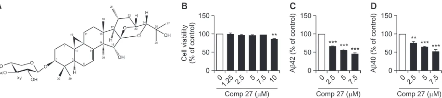
The structure of comp 27 isolated from C. dahurica is shown in Fig. 1A. To test the effect of comp 27 on cell viability, HeLa cells transfected with APPsw were treated with comp 27 (1.25, 2.5, 5, 7.5, and 10 µΜ) for 8 h. It did not affect cell vi-ability except for 10 µM concentration (Fig. 1B). Therefore, we
used these concentrations except 10 µM in this study. Then, we examined the effect of comp 27 on Aβ secretion. Cells were incubated with 2.5, 5, and 7.5 µM comp 27 for 8 h, and we measured the levels of Aβ42 and Aβ40 used specific ELISA kits from the conditioned media. The production of both were decreased in a dose-dependent manner. The Aβ42 level was reduced by 66.7%, 56.1%, and 46.2% at 2.5, 5, and 7.5 µM of comp 27, respectively (Fig. 1C). The Aβ40 level was also reduced by 75.7%, 64.5%, 52.5% at 2.5, 5, and 7.5 µM of comp 27, respectively (Fig. 1D).
β-Secretase and γ-secretase generated Aβ through se-quential cleavage of APP. On the other hand, α-secretase and γ-secretase generated precluding Aβ by cleavage within the Aβ domain. Thus, we further tested the effects of comp 27 on the production of APP proteolytic fragments, sAPPβ and sAPPα, as well as the APP expressions to investigate the two pathways. The secreted level of sAPPα was increased by 116.2%, 121.1%, and 131.2% at 2.5, 5, and 7.5 µM of comp 27, respectively (Fig. 2A). In addition, treatment of 7.5 µM comp 27 significantly decreased the level of sAPPβ to 50.6% (Fig. 2B, 2C). On the other hand, comp 27 did not change the levels of both mature and immature APP (mAPP and imAPP) (Fig. 2C, 2D).
Comp 27 increased sAPPα secretion and decreased the secretion of Aβ and sAPPβ. However, it did not affect total APP expression. Therefore, we expected that comp 27 may affect either ADAM family or BACE1 which are respectively acting as α- and β-secretases.
Next, we investigated whether comp 27 affect ADAM fam-ily expressions and activities. Cells were incubated with 2.5,
0 150 100 50 Cell viability (% of control) Comp 27 ( M) 0 150 100 50 A 4 2 (% o f control) Comp 27 ( M) 0 1.25 2.5 5 7.5 10 0 2.5 5 7.5 HO AcO OH O O H OH O O OH H H H Xyl 30 29 3 5 1 10 8 6 19 11 13 14 15 18 17 21 22 23 24 25 27 26 28 ** *** ****** 150 100 50 A 4 0 (% o f control) Comp 27 ( M) 0 0 2.5 5 7.5 *** *** **
A
B
C
D
Fig. 1.
Effect of comp 27 on cell viability and Aβ secretion. (A) Chemical structure of 3’-O-acetyl-24-epi-7,8-didehydrocimigenol-3-O-β-D-xylopyranoside (comp 27). (B) The effect of comp 27 on cell viability. (C, D) ELISA result of Aβ42 and Aβ40 secretion after comp 27 treat-ment. **p<0.01, ***p<0.001 compared to the control group.Mature APP Immature APP sAPP GAPDH 0 2.5 5 7.5 Comp 27 ( M)
B
150 100 50 Relative protein expression (% of control) Comp 27 ( M) 0 0 2.5 5 7.5 0 2.5 5 7.5D
150 100 50 Relative protein expression (% of control) Comp 27 ( M) 0 0 2.5 5 7.5C
* 150 100 50 sAPP (% of control) Comp 27 ( M) 0 0 2.5 5 7.5A
*** *** *** Mature APP Immature APP sAPPFig. 2.
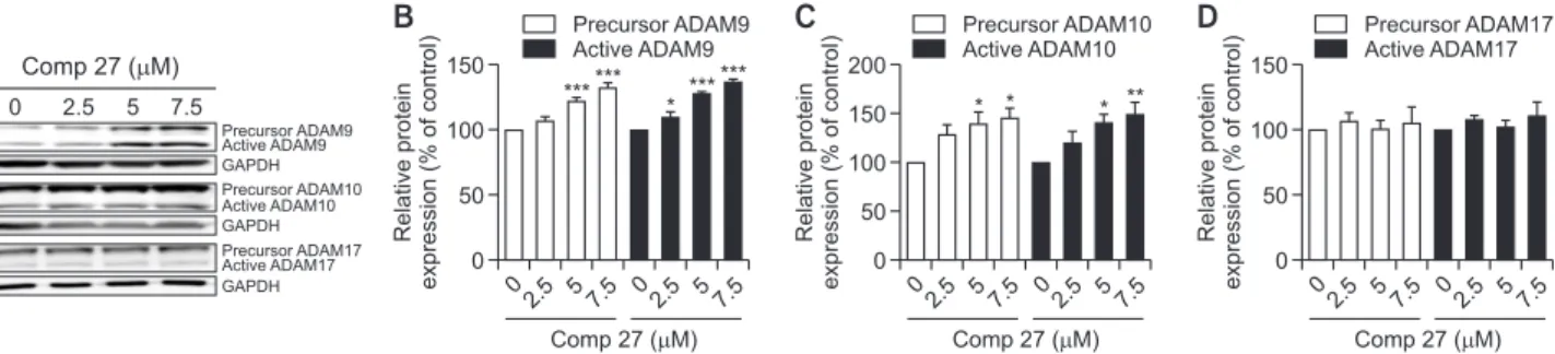
Effect of comp 27 on APP processing. (A) ELISA result of sAPPα after comp 27 treatment. (B) Western blot analysis of sAPPβ, ma-ture APP and immama-ture APP after comp 27 treatment. (C) Comp 27 dose-dependently decreased the level of sAPPβ. (D) Comp 27 did not change the levels of both mature and immature forms of APP. *p<0.05, ***p<0.001 compared to the control group.5, and 7.5 µM of comp 27 for 8 h, and the levels of ADAMs in lysates were measured using Western blot analysis. ADAM9 and ADAM10 exist as a pro-enzyme state, which are con-verted to mature form to cleavage of the APP (Lammich et al., 1999). We found that both precursor and active ADAM9 and ADAM10 were increased in a dose-dependent manner (Fig. 3A). Treatment with 7.5 µΜ of the compound increased ADAM9 levels to 136.5%, and ADAM10 levels to 150.3%, re-spectively (Fig. 3B, 3C). On the other hand, comp 27 did not change the expression of any forms of ADAM17, precursor and active ADAM17 (Fig. 3D).
We tried to determine whether comp 27 influences BACE1 protein expression. As we expected, compound 27 dose-de-pendently decreased BACE1 expression. The level of BACE1 was decreased by 77.2%, 68.5%, and 55.5% at 2.5, 5, and 7.5 µM comp 27, respectively (Fig. 4).
DISCUSSION
Cimicifuga dahurica (Turcz.) Maxim. is commonly called ‘shengma’. It is one of the ancient herbal medicines that has been subject of extensive studies. It is distributed widely in Northeast Asia and Russia, and traditionally used as an
anti-pyretic and analgesic agent (Thao et al., 2018). In this study, we first showed that comp 27, one of the isolated compounds from C. dahurica, significantly decreased both Aβ42 and Aβ40 secretion in HeLa cells overexpressing mutant APPs within the range of no cytotoxicity. We further investigated the pos-sible underlying mechanism of comp 27. Comp 27 increases sAPPα secretion which may be attributed to the expression of α-secretases. As expected, comp 27 increased ADAM9 and ADAM10 expression without affecting ADAM 17 activity. This result indicates that the comp 27-induced Aβ reduction occurs through increasing the level of α-secretase, especially ADAM9 and ADAM10. In addition, we found that comp 27 decreases sAPPβ formation concomitantly. Reduction of secreted sAPPβ might be due to either decreasing APP expression itself or in-hibiting the expression level of BACE1, which is a β-secretase responsible for the cleavage generating the Aβ peptides in the amyloidogenic pathology. According to our observation, comp 27 did not affect the expressions of both mature and immature APPs. Instead, we detected that comp 27 decreased BACE1 expression which can be the main cause of reduced expres-sion of sAPPβ. BACE1 expresexpres-sion, however, can be regulated at the amount of transcription, translation, or protein degrada-tion. So, further studies need to be performed to investigate the specific mechanisms underlying the comp 27 in such pro-cesses.
Aβ oligomers stimulate the kind of biological signaling pathway involving oxidative stress and neuroinflammation (Agostinho et al., 2010). This process leads not only a neuro-nal synapses and dendrites impairment but also disintegration of the neural circuits and neuronal loss eventually (Vargas et al., 2018). Accordingly, reducing the Aβ generation using any substance is considered to likely a good approach for treat-ment or prevention of AD. To date, it was not successful to de-velop an effective drug to stop or modify the progression of AD. Natural products could be an excellent source to reveal a hint for the therapeutic candidates against chronic and complexed disorders including neurodegenerative diseases. Since APP is cleaved within its extracellular domain by α-secretase or β-secretase, the promoting effect of comp 27 on α-secretase expression could decrease amyloidogenic process of APP by β-secretase. It also suggests that inhibitory effect of comp 27 on β-secretase expression could result in the same effect. Thus, our data suggest that comp 27 decreases Aβ produc-tion in vitro via modulaproduc-tion of two kinds of enzymes directly in-volved in APP cleavage. Confirmation of such significant
anti-A
Precursor ADAM9 Active ADAM9 GAPDH Precursor ADAM10 Active ADAM10 GAPDH Precursor ADAM17 Active ADAM17 GAPDH 0 2.5 5 7.5 Comp 27 ( M) 150 100 50 Relative protein expression (% of control) Comp 27 ( M) 0 0 2.5 5 7.5B
Precursor ADAM9 Active ADAM9 0 2.5 5 7.5 200 100 50 Relative protein expression (% of control) Comp 27 ( M) 0 0 2.5 57.5C
Precursor ADAM10 Active ADAM10 0 2.5 57.5 150 100 50 Relative protein expression (% of control) Comp 27 ( M) 0 0 2.5 57.5D
Precursor ADAM17 Active ADAM17 0 2.5 57.5 150 * ****** ****** * * * **Fig. 3.
Effect of comp 27 on the Expression of ADAM family. (A) Western blot analysis of ADAM9, 10 and 17 after comp 27 treatment. (B) Comp 27 dose-dependently increased the levels of precursor and active ADAM9. (C) Comp 27 dose-dependently increased the levels of precursor and active ADAM10. (D) Comp 27 did not change the levels of both precursor and active ADAM17. *p<0.05, **p<0.01, and ***p<0.001 compared to the control group.0 150 100 50 7.5 Relative protein expression (% of control) Comp 27 ( M) 0 2.5 5 BACE1 GAPDH BACE1 *** *** *
Fig. 4.
Effect of comp 27 on BACE1 Expression. comp 27 dose-dependently reduced the expression of BACE1. *p<0.05 and ***p<0.001 compared to the control group.Aβ effects in the future animal studies would promote comp 27 to be a possible therapeutic candidate for the AD pathology.
CONFLICT OF INTEREST
The authors declare no competing financial interest.
ACKNOWLEDGMENTS
This work was funded by the National Research Founda-tion of the Ministry of Science, InformaFounda-tion and Communica-tions Technology (ICT) & Future Planning of Republic of Korea (NRF-2015M3A9A5030735) and the Bio-Synergy Research Project (NRF-2012M3A9C4048793).
REFERENCES
Agostinho, P., Cunha, R. A. and Oliveira, C. (2010) Neuroinflamma-tion, oxidative stress and the pathogenesis of Alzheimer’s dis-ease. Curr. Pharm. Des. 16, 2766-2778.
Chen, X., Xu, B., Nie, L., He, K., Zhou, L., Huang, X., Spencer, P., Yang, X. and Liu, J. (2019) Flavanol-rich lychee fruit extract sub-stantially reduces progressive cognitive and molecular deficits in a triple-transgenic animal model of Alzheimer disease. Nutr.
Neuro-sci. doi: 10.1080/1028415X.2019.1673527 [Online ahead of print].
Gu, M. Y., Chun, Y. S., Zhao, D., Ryu, S. Y. and Yang, H. O. (2018) Glycyrrhiza uralensis and semilicoisoflavone B reduce Aβ secre-tion by increasing PPARγ expression and inhibiting STAT3 phos-phorylation to inhibit BACE1 expression. Mol. Nutr. Food. Res. 62,
e1700633.
Hardy, J. and Orr, H. (2006) The genetics of neurodegenerative dis-eases. J. Neurochem. 97, 1690-1699.
Karran, E. and De Strooper, B. (2016) The amyloid cascade hypoth-esis: are we poised for success or failure? J. Neurochem. 139 Suppl 2, 237-252.
Kim, J., Park, Y., Chun, Y. S., Cha, J. W., Kwon, H. C., Oh, M. S., Chung, S. and Yang, H. O. (2015a) Effect of lycoris chejuensis and its active components on experimental models of Alzheimer’s dis-ease. J. Agric. Food Chem. 63, 6979-6988.
Kim, J. M., Hwang, K. W., Joo, H. B. and Park, S. Y. (2015b) Anti-amyloidogenic properties of dryopteris crassirhizoma roots in Al-zheimer’s disease cellular model. J. Food Biochem. 39, 478-484.
Kuhn, P. H., Wang, H., Dislich, B., Colombo, A., Zeitschel, U., Ellwart, J. W., Kremmer, E., Rossner, S. and Lichtenthaler, S. F. (2010) ADAM10 is the physiologically relevant, constitutive alpha-secre-tase of the amyloid precursor protein in primary neurons. EMBO
J. 29, 3020-3032.
Lammich, S., Kojro, E., Postina, R., Gilbert, S., Pfeiffer, R., Jasion-owski, M., Haass, C. and Fahrenholz, F. (1999) Constitutive and regulated alpha-secretase cleavage of Alzheimer’s amyloid precur-sor protein by a disintegrin metalloprotease. Proc. Natl. Acad. Sci.
U.S.A. 96, 3922-3927.
Lee, J., Cho, E., Kwon, H., Jeon, J., Jung, C. J., Moon, M., Jun, M., Lee, Y. C., Kim, D. H. and Jung, J. W. (2019) The fruit of Crataegus pinnatifida ameliorates memory deficits in beta-amyloid protein-induced Alzheimer’s disease mouse model. J. Ethnopharmacol.
243, 112107.
Lv, C., Yang, F., Qin, R., Qi, Z., Zhou, W. and Lu, J. (2017) Bioactivity-guided isolation of chemical constituents against H2O2-induced neurotoxicity on PC12 from Cimicifuga dahurica (Turcz.) Max-im. Bioorg. Med. Chem. Lett. 27, 3305-3309.
Park, S. Y. (2010) Potential therapeutic agents against Alzheimer’s disease from natural sources. Arch. Pharm. Res. 33, 1589-1609.
Prince, M., Bryce, R., Albanese, E., Wimo, A., Ribeiro, W. and Ferri, C. P. (2013) The global prevalence of dementia: a systematic review and metaanalysis. Alzheimers Dement. 9, 63-75.e2.
Qin, R., Zhao, Y., Zhao, Y., Zhou, W., Lv, C. and Lu, J. (2016) Polyphe-nolic compounds with antioxidant potential and neuro-protective effect from Cimicifuga dahurica (Turcz.) Maxim. Fitoterapia 115,
52-56.
Scheltens, P., Blennow, K., Breteler, M. M., de Strooper, B., Frisoni, G. B., Salloway, S. and Van der Flier, W. M. (2016) Alzheimer’s disease. Lancet 388, 505-517.
Thao, N. P., Kim, J. H., Thuy Luyen, B. T., Dat, N. T. and Kim, Y. H. (2017a) In silico investigation of cycloartane triterpene derivatives from Cimicifuga dahurica (Turcz.) Maxim. roots for the develop-ment of potent soluble epoxide hydrolase inhibitors. Int. J. Biol.
Macromol. 98, 526-534.
Thao, N. P., Lee, Y. S., Luyen, B. T. T., Oanh, H. V., Ali, I., Arooj, M., Koh, Y. S., Yang, S. Y. and Kim, Y. H. (2018) Chemicals from Cimi-cifuga dahurica and their inhibitory effects on pro-inflammatory cytokine production by LPS-stimulated bone marrow-derived den-dritic cells. Nat. Prod. Sci. 24, 194-198.
Thao, N. P., Luyen, B. T., Lee, J. S., Kim, J. H. and Kim, Y. H. (2017b) Soluble epoxide hydrolase inhibitors of indolinone alkaloids and phenolic derivatives from Cimicifuga dahurica (Turcz.) Max-im. Bioorg. Med. Chem. Lett. 27, 1874-1879.
Thao, N. P., Luyen, B. T. T., Lee, J. S., Kim, J. H., Dat, N. T. and Kim, Y. H. (2017c) Inhibition potential of cycloartane-type glycosides from the roots of cimicifuga dahurica against soluble epoxide hydro-lase. J. Nat. Prod. 80, 1867-1875.
Tian, Z., Si, J., Chang, Q., Zhou, L., Chen, S., Xiao, P. and Wu, E. (2007) Antitumor activity and mechanisms of action of total gly-cosides from aerial part of Cimicifuga dahurica targeted against hepatoma. BMC Cancer 7, 237.
Vargas, L. M., Cerpa, W., Munoz, F. J., Zanlungo, S. and Alvarez, A. R. (2018) Amyloid-beta oligomers synaptotoxicity: the emerging role of EphA4/c-Abl signaling in Alzheimer’s disease. Biochim.
Bio-phys. Acta Mol. Basis Dis. 1864, 1148-1159.
Vassar, R., Kovacs, D. M., Yan, R. and Wong, P. C. (2009) The beta-secretase enzyme BACE in health and Alzheimer’s disease: regu-lation, cell biology, function, and therapeutic potential. J. Neurosci.
29, 12787-12794.
Zhang, L. L., Si, J. Y., Zhang, L. J., Xiao-Wei, H., Lin, L., Li, R. Y., Chen, D. and Cao, L. (2016) Synergistic anti-tumor activity and mecha-nisms of total glycosides from Cimicifuga dahurica in combination with cisplatin. Chin. J. Integr. Med. doi: 10.1007/s11655-015-2108-3 [Online ahead of print].