Korean J Gastroenterol Vol. 59 No. 4, 321-323 http://dx.doi.org/10.4166/kjg.2012.59.4.321
IMAGE OF THE MONTH
Korean J Gastroenterol, Vol. 59 No. 4, April 2012 www.kjg.or.kr
내시경 점막절제술로 제거된 Epstein-Barr Virus 음성 위 수질암
1예
권영일, 이오영, 백승삼
1한양대학교 의과대학 내과학교실, 병리학교실1
A Case of Epstein-Barr Virus Negative Gastric Medullary Carcinoma Excised by Endoscopic
Mucosal Resection
Young Il Kwon, Oh Young Lee and Seung Sam Paik1
Departments of Internal Medicine and Pathology1, Hanyang University College of Medicine, Seoul, Korea
CC This is an open access article distributed under the terms of the Creative Commons Attribution Non-Commercial License (http://creativecommons.org/licenses/ by-nc/3.0) which permits unrestricted non-commercial use, distribution, and reproduction in any medium, provided the original work is properly cited.
교신저자: 이오영, 133-792, 서울시 성동구 왕십리로 222, 한양대학교병원 소화기내시경실
Correspondence to: Oh Young Lee, Department of Gastroenterology, Hanyang University Seoul Hospital, 222 Wangsimni-ro, Sungdong-gu, Seoul 133-792, Korea. Tel: +82-2-2290-8354, Fax: +82-2-2298-9183, E-mail: [email protected]
Financial support: None. Conflict of interest: None.
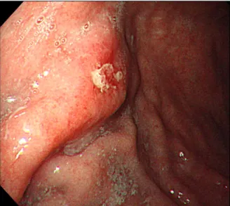
Fig. 1. Endoscopic finding. Elevated submucosal lesion with apical ulceration at the anterior wall of the antrum was observed.
Fig. 2. Endoscopic ultrasonography. It showed about 1.1 cm-sized homogenous hypoechoic mass in the submucosal layer.
증례: 70세 남자가 건강검진을 위해 본원에 내원하였다. 환
자는 특이 병력은 없었으며 특별한 증상을 호소하지도 않았
다. 내원 당시 혈압은 110/80 mmHg, 맥박은 68회/분, 호흡
수는 20회/분, 체온은 36.8
oC였다. 흉부에서 폐음 및 심음은
정상이었고, 복부청진에서 장음은 정상이었다. 촉진에서 간과
비장종대는 없었으며 만져지는 림프절도 없었다. 내원 당시
시행한 말초혈액검사는 백혈구 8,400/mm
3, 혈색소 13.4
g/dL, 헤마로크릿 41.7%, 혈소판 278,000/mm
3이었다. 혈청
322
권영일 등. 내시경 점막절제술로 제거된 Epstein-Barr Virus 음성 위 수질암 1예The Korean Journal of Gastroenterology
Fig. 3. (A) Endoscopic mucosal resection (EMR) site. EMR was done to obtain more specimen of submucosal mass. (B) Excised mass. 1.3×1.2×0.5 cm sized well demarcated oval, grayish mass was excised by EMR.
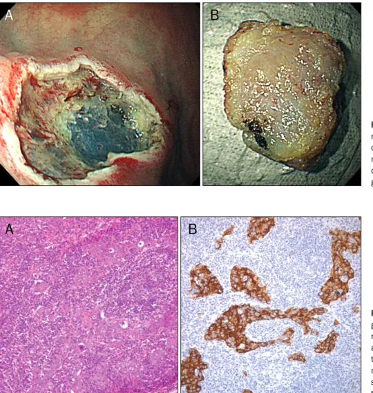
Fig. 4. (A) Pathologic study revealed gastric lymphoepithelioma-like carci-noma, with syncytial growth pattern of abundant lymphoid cell infiltrations in the stroma (H&E, ×200). (B) Immu-nohistochemical stain for cytokeratin showed strong positive on the epi-thelial tumor nests (×200).
생화학검사에서 total protein/albumin 6.9/4.5 g/dL,
AST/ALT 18/16 IU/L, total bilirubin 0.3 mg/dL, BUN 18
mg/dL, creatinine 0.9 mg/dL로 특이소견 없었다. 그러나 상
부위장관 내시경에서 하체부 전벽측에 얕은 중심부 궤양을 동
반한 점막 융기성 병변이 관찰되었고(Fig. 1), 병변에서 조직
검사를 시행하였으나 만성염증 소견만이 나왔다. 점막하 병변
의 침윤 정도를 파악하기 위해 내시경 초음파를 시행하였고
점막하층에 국한된 약 1.1 cm의 균질한 저에코 병변이 관찰
되었다(Fig. 2). 복부컴퓨터단층촬영(CT)에서는 특이 소견 없
었다.
정확한 진단을 위해 진단목적의 내시경 점막절제술을 시행
하였고(Fig. 3A), 경계가 비교적 명확하며 회색빛을 띄는
1.3×1.2×0.5 cm 크기의 점막하종양이 절제되었다(Fig. 3B).
조직검사 결과 임파구 침윤이 풍부한 간질에 종양세포가 융합
하여 군집을 이루는 양상으로 관찰되었고(Fig. 4A), 면역조직
화학 염색검사에서 종양세포에 cytoketatin 양성을 보여(Fig.
4B) 위 수질암으로 진단되었다. EBV 감염 여부를 알기 위해
시행한 EBV 면역조직화학 염색에는 음성을 보였다. 시술 전
시행한 PET CT에서는 특이소견 관찰되지 않았으나 조직검사
결과 일부에서 주변 경계와의 침범 여부가 명확하지 않아 위
아전 절제술과 림프절 절제술을 시행하였다. 그러나 수술 후
검체에서는 잔여 암조직은 발견되지 않아 추가적인 치료 없이
현재 외래에서 추적관찰 중이며, 현재까지 1년 이상의 추적관
찰 기간 동안 재발이나 새로운 병변은 발견되지 않고 있다.
진단: 위 수질암(gastric medullary carcinoma)
위 수질암은 위암의 약 3%를 차지하는 드문 암으로, 현재
까지 발표된 연구들에 의하면
1-3주로 50대, 남성이 많고 다른
위암에 비해 좋은 예후를 보인다. 대개 진행된 상태에서 진단
되며 조기암의 경우 IIa+IIc 형태가, 진행암의 경우
Borr-mann type II 형태가 많지만 드물게는 용종처럼 관강 내로
돌출된 형태나 점막하종양의 형태로도 발견된다. 복막 전이보
다는 간 전이가 흔하며 주된 사망원인이다. 주로 상체부에 많
으며 종양의 단면은 단단하나 부드럽게 탄력이 있으며 황색이
Kwon YI, et al. A Case of Gastric Medullary Carcinoma Excised by Endoscopic Mucosal Resection
323
Vol. 59 No. 4, April 2012
나 회색을 띈다.
위 수질암의 원인에 대해서는 명확히 밝혀지지 않았지만
EBV 감염, Fas ligand, interleukin-10 등과 연관이 있다는
보고가 있고,
4-7특히 EBV 감염의 경우 위 수질암의
77.8-100%에서 양성이었다는 보고가 있어 EBV 감염이 중요
한 원인일 것이라 생각된다. 또한 위 수질암에서 EBV 감염과
높은 MSI는 좋은 예후와도 연관이 있었다.
8,9우리나라에서 보고된 위 수질암의 경우 대부분 조기에 점
막하종양의 형태로 발견되었고 진행된 궤양 형태로 발견된 경
우는 드물어,
10-15기존의 외국 보고와 차이를 보였다. 이는 아
마도 건강검진 목적의 내시경 시행 증가와 내시경검사를 받기
용이한 의료환경 등에 의해 조기발견 기회가 증가되었기 때문
일 것이다. 또한 EBV 검사를 시행하지 않은 경우를 제외하고
는 모두 EBV 양성이었다.
이 증례는 고령의 무증상 환자에서 우연히 조기 진단되어
치료된 경우로, 비록 EMR 이후 수술치료를 받았지만 수술
후 검체에서 잔여암 조직이 발견되지 않아 조기 발견시 내시
경적으로도 치료가 가능함을 시사한다. 또한 저자들이 아는
바로는 국내 보고된 위 수질암 중 EBV 음성인 첫 증례로, 다
른 원인에 대한 추가적인 검사가 이루어지지 못한 점은 아쉬
운 부분이다. 향후의 위 수질암에서는 EBV 외에 MSI 등에
대한 검사도 필요하며 이는 향후 치료방침의 결정에도 도움이
될 것이다.
REFERENCES
1. Herath CH, Chetty R. Epstein-Barr virus-associated lymphoepi-thelioma-like gastric carcinoma. Arch Pathol Lab Med 2008; 132:706-709.
2. Minamoto T, Mai M, Watanabe K, et al. Medullary carcinoma with lymphocytic infiltration of the stomach. Clinicopathologic study of 27 cases and immunohistochemical analysis of the subpopulations of infiltrating lymphocytes in the tumor. Cancer 1990;66:945-952.
3. Watanabe H, Enjoji M, Imai T. Gastric carcinoma with lymphoid stroma. Its morphologic characteristics and prognostic corre-lations. Cancer 1976;38:232-243.
4. Oda K, Tamaru J, Takenouchi T, et al. Association of Epstein-Barr virus with gastric carcinoma with lymphoid stroma. Am J Pathol 1993;143:1063-1071.
5. Nakamura S, Ueki T, Yao T, Ueyama T, Tsuneyoshi M. Epstein- Barr virus in gastric carcinoma with lymphoid stroma. Special reference to its detection by the polymerase chain reaction and in situ hybridization in 99 tumors, including a morphologic analysis. Cancer 1994;73:2239-2249.
6. Kume T, Oshima K, Yamashita Y, Shirakusa T, Kikuchi M. Relationship between Fas-ligand expression on carcinoma cell and cytotoxic T-lymphocyte response in lymphoepithelioma-like cancer of the stomach. Int J Cancer 1999;84:339-343. 7. Hsu DH, de Waal Malefyt R, Fiorentino DF, et al. Expression of
in-terleukin-10 activity by Epstein-Barr virus protein BCRF1. Science 1990;250:830-832.
8. Lü BJ, Lai M, Cheng L, Xu JY, Huang Q. Gastric medullary carcino-ma, a distinct entity associated with microsatellite instability-H, prominent intraepithelial lymphocytes and improved prognosis. Histopathology 2004;45:485-492.
9. Grogg KL, Lohse CM, Pankratz VS, Halling KC, Smyrk TC. Lymphocyte-rich gastric cancer: associations with Epstein-Barr virus, microsatellite instability, histology, and survival. Mod Pathol 2003;16:641-651.
10. Lee SH, Jang BI, Eun JR, Kim KO, Lee KH, Kim TN. A case of sub-mucosal gastric lymphoepithelioma-like carcinoma. Korean J Med 2009;76(Suppl 1):S35-S39.
11. Cho JH, Lee WS, Lee KR, et al. Gastric lymphoepithelioma-like carcinoma diagnosed and treated by endoscopic submucosal dissection: review of the literature. Korean J Gastrointest Endosc 2010;40:256-260.
12. Goh PG, Kim ES, Kim YJ, et al. A case of gastric lymphoepithelio-ma-like carcinoma presenting as panperitonitis by perforation of stomach. Korean J Gastroenterol 2011;58:208-211. 13. Park WI, Kim HW, Park JH, et al. A case of gastric
lymphoepithe-lioma-like carcinoma presenting as a submucosal tumor. Korean J Gastrointest Endosc 2004;28:123-126.
14. Woo H, Shin SJ, Kim YB, et al. A case of endoscopic enucleation for gastric lymphoepithelioma-like carcinoma. Korean J Gastro-intest Endosc 2010;41:163-167.
15. Moon HS, Kang SH, Seong JK, Jeong HY, Song KS. Lymphoepi-thelioma-like gastric carcinoma resected by endoscopic sub-mucosal dissection (ESD). Endoscopy 2010;42(Suppl 2):E73- E74.