plSSN: 1976-8257 eISSN: 2234-2753 Toxicol. Res. Vol. 35, No. 4, pp. 353-359 (2019)
https://doi.org/10.5487/TR.2019.35.4.353 Open Access
353
Alleviation of Ultraviolet B-Induced Photoaging by 7-MEGA
TM500 in Hairless Mouse Skin
Kyo-Hyun Park1
, JiYeon Kim1
, Suryun Jung1
, Kyung-hwa Sung2
, Yeon-Kyoung Son3
, Jung Min Bae4
and Bae-Hwan Kim1
1Department of Public Health, Keimyung University, Daegu, Korea 2
Environmental Health Services Center, Daegu Catholic University, Daegu, Korea
3R&D Team, Food & Supplement Health Claims, Vitech, Jeonju, Korea
4Department of Technical Assistance, Agency for Korea National Food Cluster (AnFC), Iksan, Korea
Abstract
The purpose of this study was to investigate the effect of 7-MEGATM 500 on the improvement of skin aging in an UVB-induced photo-aging model of hairless mice. The dorsal skin of hairless mice was exposed to UVB three times a week for 12 weeks to induce skin wrinkle. After inducing the wrinkle, 7-MEGATM 500 was orally admin-istered once a day for 4 weeks. Skin thickness, skin barrier function, and wrinkle indicators were improved by treatment with 7-MEGATM 500. Both gene and protein expression levels of MMP-3 and c-Jun in skin were signifi-cantly decreased by 7-MEGATM 500. Therefore, the intake of 7-MEGATM 500 is thought to have a positive effect on the improvement of skin aging, although further studies are needed.
Key words: 7-MEGATM 500, Photoaging, Hairless mice, c-Jun, MMP-3
INTRODUCTION
Ultraviolet (UV) is abundant in the environment. It is the most important risk factor for skin cancer. It not only increases epidermal thickness, but also increases wrinkle formation (1). UV irradiation also induces the synthesis of matrix metalloproteinases (MMPs) and decrease of colla-gen synthesis (2).
Collagen degradation is closely related to the presence of MMPs that are mainly secreted by epidermal keratino-cytes and dermal fibroblasts. MMPs are usually expressed at low levels in unstimulated cells or normal skin tissues. However, they can be induced by various extracellular stimuli, including cytokines, growth factors, and UV radi-ation. Up-regulation of MMPs can also be induced by
even a minimal dose of UV (3). After chronical exposure to UV-irradiation, mouse skin shows epidermal hyperpla-sia, skin wrinkles, and significant increase of several MMPs, including stromelysin-1 3), metalloelastase (MMP-12), and collagenase (MMP-1) (4).
It has been shown that omega polyunsaturated fatty acids (PUFAs) possess anti-oxidative (5), anti-inflamma-tory (6), neuroprotective (7), and chemopreventive (8) effects. Omega-6 and -9 have been linked to obesity pre-vention (9) and anti-inflammation (10). Recent studies have shown that PUFAs can defend a wide range of dis-eases characterized by increased MMPs activity (11). They can also suppress UV-induced expression of proin-flammatory cytokines and MMPs in skin cells in vitro or skin tissues in vivo (1,4). Moreover, it has been reported tht palmitoleic acid (omega-7) and gamma-linolenic acid (omega-6) can affect skin regeneration and repair (12). However, there have been few reports on omega-7 com-pared to other omega fatty acids. Omega-7, also known as palmitoleic acid (16:1, Cis-9-hexadecenoic acid), is a monounsaturated fatty acid that is found in fish and plants such as macadamias, cold water fish, and sea buckthorn berries (13).
It has been previously shown that omega-3 and omega-6 Correspondence to: Bae-Hwan Kim, Department of Public Health,
Keimyung University, 1095 Dalgubeoldaero, Dalseo-gu, Daegu 42601, Korea
E-mail: [email protected]
This is an Open-Access article distributed under the terms of the Creative Commons Attribution Non-Commercial License (http:// creativecommons.org/licenses/by-nc/3.0) which permits unre-stricted non-commercial use, distribution, and reproduction in any medium, provided the original work is properly cited.
4 MED until 12 weeks. After skin photoaging induction (1-8 weeks), the test material or vehicle was orally admin-istered (10 mL/kg, once a day) for 4 weeks.
Determination of wrinkle grade. To determine the severity of wrinkling, each hairless mouse was anesthe-tized and the UVB exposed dorsal skin (wrinkle formation area) was photographed. The severity of wrinkling was measured using Bissett’s visual wrinkle scale. Skin impres-sions (replicas) were prepared by applying Repliflo Car-tridge Kit (CuDerm Corp., Dallas, TX, USA) to dorsal skin of each mousee. Replicas were analyzed using a skin visi-oline VL650 (CK Electronics GmbH, Cologne, Germany). Measurement of skin barrier function. Transepi-dermal water loss (TEWL) and stratum corneum (SC) water content were assessed under standardized condi-tions (external temperature 23 ± 3oC and 50 ± 10% RH) using a Tewameter (Courage-Khazaka Electronic GmbH, Cologne, Germany) and a Corneometer (Courage-Khaz-aka Electronic GmbH) apparatus, respectively.
Measurement of skin thickness. Skin thickness was measured using a digimatic micrometer (Mitutoyo Co. Ltd., Tokyo, Japan) once a week. The dorsal skin of each mouse was pulled up from the neck to the bottom of the body by hand and the skin fold thickness was measured between the neck and hips.
Histological observation. Dorsal skin from autopsy was fixed in 10% formalin for 24 hr and embedded in par-affin with common process. Embedded tissue was cut into 4μm-thick sections and stained with hematoxylin and eosin (H&E). Changes of skin tissue such as epidermal thickness and inflammatory cell infiltration were observed under an optical microscope.
RNA isolation and RT-PCR. Total RNA was isolated from dorsal skin of mouse using TRIzol reagent (Life Technologies Inc., Rockville, MD, USA) according to the manufacturer’s protocol. Total RNA was used to synthe-size cDNA with an iScript cDNA Synthesis Kit (Bio-Rad Laboratories, Hercules, CA, USA). PCR amplification of cDNA (4μL) was performed with PCR premix (SolGent, Daejeon, Korea) and primer pairs (Bionics, Seoul, Korea; act as inhibitors of MMPs (11), and 7-MEGATM 500 (more
than 50% of palmitoleic acid containing fish oil, omega-7) can show the effects of anti-oxidant and anti-inflamma-tion in vitro (14). However, in vivo informaanti-inflamma-tion on its effects on skin has been insufficient. Therefore, the aim of this study was to investigate the effect of 7-MEGATM 500 by observing expression levels of MMP-3 and c-Jun on skin of mouse.
MATERIALS AND METHODS
Preparation of 7-MEGATM
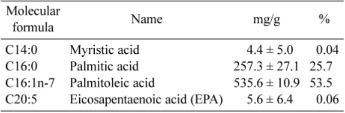
500. 7-MEGATM 500 was obtained from Organic Technologies (OH, USA). Pollock was collected from Alaskan Bering Sea and 7-MEGATM 500 containing palmitoleic acid (> 500 mg/g) was pre-pared (Table 1). 7-MEGATM 500 was administered by vol-ume of 10 mL/kg after dissolving a defined concentration of each group in 30% EtOH.
Animals. Male HR-1 hairless mice (5-week old, 18-20 g) were purchased from Orient Bio (Seongnam, Korea). After acclimation for one week, they were randomly assigned to five groups (Table 2). The animal room was maintained at a temperature of 22 ± 3oC with relative humidity of 50 ± 10% and 12-hr light/12-hr dark cycle per day. They were provided free access to feed (Purina, Korea) and water ad libitum during the experiment period. All experimental protocols were approved by the Institu-tional Animal Care and Use Committee (IACUC) of Keimyung University, South Korea (permit number: KM-2017-005).
Design of skin photoaging model. The dorsal skin of hairless mice was exposed to UVB three times a week. The irradiation dose was increased weekly by 1 MED (1 MED = 130 mJ/cm2) to 4 MED. It was then maintained at
Table 1. The main ingredients of 7-MEGATM
500 Molecular formula Name mg/g % C14:0 C16:0 C16:1n-7 C20:5 Myristic acid Palmitic acid Palmitoleic acid
Eicosapentaenoic acid (EPA)
04.4 ± 5.0 257.3 ± 27.1 535.6 ± 10.9 05.6 ± 6.4 00.04 25.70 53.50 00.06
Table 2. Experimental groups
Groups Induction of skin photoaging Test compound No. of mice
Normal Control (NC) Vehicle Control (VC) Experimental 1 (E1) Experimental 2 (E2) Experimental 3 (E3) − + + + + Distilled water (DW) 30% EtOH 7-MEGATM 500 (200 mg/kg) 7-MEGATM 500 (100 mg/kg) 7-MEGATM 500 (50 mg/kg) 7 7 7 7 7
plSSN: 1976-8257 eISSN: 2234-2753 Table 3). Before PCR amplification, the PCR mixture was
denatured at 95oC for 2 min. Amplification consisted of 35 cycles of denaturation at 95oC for 20 sec, annealing at 57oC for 40 sec, and extension at 72oC for 1 min, fol-lowed by a final extension at 72oC for 5 min. PCR prod-ucts were separated by 1% agarose gel electrophoresis and visualized with 6X loading dye and UV illumination.
Western blot analysis. Mouse dorsal skin sections were homogenized in radioimmunoprecipitation assay buffer (Sigma, USA) containing 1% protease inhibitor cocktail and phosphatase inhibitor cocktail. The homoge-nate was centrifuged at 14,000 rpm for 10 min at 4oC. The
supernatant was collected and protein concentration was estimated by the Bradford protein assay. A 30μL aliquot of protein was separated by 10% sodium dodecyl sulfate-polyacrylamide gel electrophoresis (SDS-PAGE). Result-ing separated proteins were transferred onto nitrocellulose membranes. Membranes were blocked with 5% skim milk in TBS-T (Tris-Buffered Saline plus 0.05% Tween 20). The following primary antibodies were used for western blotting: c-Jun, MMP-3, and β-actin (Santa Cruz Biotech-nology, Santa Cruz, CA, USA). Immunoreactive bands were visualized using enhanced chemiluminescence (ECL) detec-tion reagents (Amersham Biosciences, Amersham, UK). Band intensities were measured using ImageJ software
Table 3. Primer sequences for RT-PCR
Primer sequences1) Amplicon size (bp)2)
GAPDH Sense 5'-AACTTTGGCATTGTGGAAGG-3' 223
Antisense 5'-ACACATTGGGGGTAGGAACA-3'
c-Jun Sense 5'-TCCCCTATCGACATGGAGTC-3' 146
Antisense 5'-TTTTGCGCTTTCAAGGTTTT-3'
MMP-3 Sense 5'-CAGGTGTGGTGTTCCTGATG-3' 317
Antisense 5'-GCCTTGGCTGAGTGGTAGAG-3'
1)
Primer sequences (Bionics, Seoul, Korea).
2)
bp, basepair.
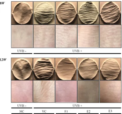
Fig. 1. Replica production and visual wrinkle patterns of skin. 8w: Before oral administration, 12w: Four weeks after oral
adminis-tration. NC: Normal control, VC: Vehicle control, E1: 7-MEGATM 500 (200 mg/kg), E2: 7-MEGATM 500 (100 mg/kg), E3: 7-MEGATM 500 (50 mg/kg).
were made at 8 weeks (before oral administration) and 12 weeks (before autopsy). The prepared replica was ana-lyzed with a wrinkle analyzer (VisioLine, VL650, Cour-age-Khazaka Electronic GmbH, Cologne, Germany). We found that UVB-irradiated skin showed wrinkles. How-ever, 7-MEGATM 500 blocked wrinkle formation (Fig. 1, Table 4). The experimental group showed a dose-depen-dent recovery pattern. At 12 weeks, wrinkles were weak-ening and skin surface was soft with elasticity. Wrinkles from in the test substance group were significantly reduced compared to those in the vehicle control (VC) group.
Measurement of skin barrier function. Results of moisture content and TEWL after treatment with 7-MEGATM 500 are shown in Table 5. In both UVB irradi-ated groups, TEWL gradually increased while water con-tent gradually decreased (1 to 8 weeks). From the first week after treatment with the test substance, moisture content of the skin increased dose-dependently in all groups while TEWL tended to decrease. These results strongly suggest that 7-MEGATM 500 can help protect against or restore UVB irradiation-induced skin barrier (National Institutes of Health, Bethesda, MD, USA).
Statistical analysis. SPSS 17.0 software was used for all statistical analyses. Skin thickness data a and mRNA or protein expression levels were analyzed by one-way anal-ysis of variance (ANOVA) followed by Duncan’s test. Skin wrinkles and skin barrier function data were ana-lyzed by two-way ANOVA followed by Duncan’s multi-ple range test when appropriate. Data are expressed as mean ± standard error (SE). Value of p < 0.05 was consid-ered significant for all comparisons made.
RESULTS
Determination of wrinkle grade. This in vivo study demonstrated that oral administeration of 7-MEGATM 500 alleviated the photoaging effect of UVB-radiation on skin. To investigate the effect of 7-MEGATM 500 on UVB-induced wrinkle formation, we UVB-induced skin photoaging by repeatedly exposing the skin of hairless mice to UVB for 8 weeks. The 7-MEGATM 500 was then orally adminis-tered once a day for 4 weeks. Visual analysis and replica
Table 4. Evaluation of wrinkles through replica analysis of hairless mouse before autopsy
NC VC E1 E2 E3 Wrinkle area (mm2) 08w 19.0 ± 10.5** 050.0 ± 7.5 041.5 ± 5.8 054.0 ± 31.1 057.8 ± 42.1 12w 17.8 ± 5.9** 076.6 ± 26.2 019.9 ± 9.9** 033.2 ± 10.9** 035.2 ± 43.6* No. of wrinkles 08w 52.0 ± 14.2 089.0 ± 40.1 095.0 ± 18.4 101.5 ± 23.3 091.0 ± 7.1 12w 46.7 ± 25.4* 083.0 ± 28.2 052.0±2.8* 068.5 ± 6.4 067.5 ± 12.0 Total length (mm) 08w 38.5 ± 3.2** 097.5 ± 8.0 092.6 ± 24.2 092.5 ± 29.92 081.2 ± 24.1 12w 35.5 ± 7.4** 094.9 ± 37.3 030.2 ± 10.3** 038.6 ± 4.1** 049.18 ± 8.06* Mean length (mm) 08w 00.6 ± 0.1* 000.9 ± 0.3 001.1 ± 0.2 000.9 ± 0.1 000.9 ± 0.2 12w 00.4 ± 0.1** 001.1 ± 0.1 000.5 ± 0.1** 000.7 ± 0.2** 000.8 ± 0.2** Mean depth (μm) 08w 90.6 ± 2.8** 116.3 ± 13.1 103.7 ± 8.6 103.6 ± 13.4 102.4 ± 7.0 12w 84.2 ± 7.1* 120.7 ± 29.0 069.7 ± 14.5* 090.1 ± 5.1 091.7 ± 4.1 8w: Just before oral administration, 12w: Four weeks after oral administration. NC: Normal control, VC: Vehicle control, E1: 7-MEGATM
500 (200 mg/kg), E2: 7-MEGATM
500 (100 mg/kg), E3: 7-MEGATM
500 (50 mg/kg). Values are means ± SE (n = 7). *p < 0.05, **p < 0.01 as com-pared to the VC group by ANOVA and Duncan’s multiple range test.
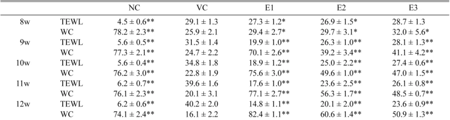
Table 5. Changes of trans-epidermal water loss (TEWL) and skin water content (WC) by time and group
NC VC E1 E2 E3 08w TEWL 04.5 ± 0.6** 29.1 ± 1.3 27.3 ± 1.2* 26.9 ± 1.5* 28.7 ± 1.3 WC 78.2 ± 2.3** 25.9 ± 2.1 29.4 ± 2.7* 29.7 ± 3.1* 32.0 ± 5.6* 09w TEWL 05.6 ± 0.5** 31.5 ± 1.4 19.9 ± 1.0** 26.3 ± 1.0** 28.1 ± 1.3** WC 77.3 ± 2.1** 24.7 ± 2.2 70.1 ± 2.6** 39.2 ± 3.4** 41.1 ± 4.2** 10w TEWL 05.6 ± 0.4** 34.8 ± 1.8 18.9 ± 1.2** 25.0 ± 2.2** 27.4 ± 0.6** WC 76.2 ± 3.0** 22.8 ± 1.9 75.6 ± 3.0** 49.6 ± 1.0** 47.0 ± 1.5** 11w TEWL 06.2 ± 0.7** 39.6 ± 1.6 17.6 ± 1.0** 23.6 ± 2.5** 26.1 ± 0.8** WC 76.1 ± 2.3** 20.1 ± 3.1 77.1 ± 2.7** 56.3 ± 1.7** 48.5 ± 0.7** 12w TEWL 06.2 ± 0.6** 40.2 ± 2.0 14.8 ± 1.1** 20.1 ± 2.0** 23.6 ± 0.9** WC 74.1 ± 2.4** 16.1 ± 2.2 82.4 ± 1.1** 60.6 ± 1.4** 50.9 ± 1.3** Unit: TEWL (g/h/m2
), WC (AU). 8w: Just before oral administration, 9w to 12w: From one week to four weeks after oral administration. NC: Normal control, VC: Vehicle control, E1: 7-MEGATM
500 (200 mg/kg), E2: 7-MEGATM
500 (100 mg/kg), E3: 7-MEGATM
500 (50 mg/kg). Values are means ± SE (n = 7). *p < 0.05, **p < 0.01 as compared to the VC group by ANOVA and Duncan’s multiple range test.
plSSN: 1976-8257 eISSN: 2234-2753 dysfunction.
Measurement of skin thickness. Skin thickness of hairless mice gradually increased after 8 weeks of UVB irradiation. However, skin thickness significantly decreased from the first week after oral administration of 7-MEGATM 500. Such decrease was proportional to the duration of administration. These results suggest that 7-MEGATM 500 can improve skin thickness thickened by ultraviolet light (Fig. 2).
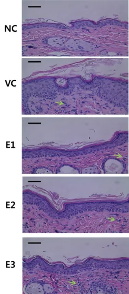
Histological observation. To investigate the effect of 7-MEGATM 500 on UV-induced photoaging in hairless mice, the thickness of epidermis was observed by H & E staining. Epidermal thickness was measured with a ruler at 400 magnification of microscopy. The epidermal thick-ness in the VC group (31.5 ± 0.5μm) was significantly increased compared to that in the NC group (11 ± 0.2μm) (p < 0.05). However, 4 weeks of intake of 7-MEGATM 500 significantly reduced the thickness of the epidermis. Such decrease was dependent on the dose of the concentration of 7-MEGATM 500 (E1 < E2 < E3, 15 ± 0.3 < 21 ± 0.1 < 23 ± 0.2μm, Fig. 3). These results suggest that 7-MEGATM 500 treatment can significantly improve the epidermal thickness of hairless mice thickened by UV irradiation.
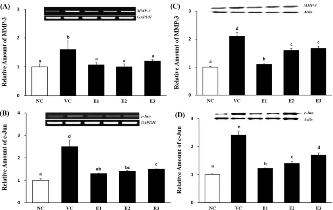
MMP-3 and c-Jun expression. MMPs and c-Jun can be used as photo-aging markers. Our results revealed that 12 weeks of UVB irradiation significantly increased mRNA and protein levels of MMP-3 and c-Jun in dorsal skin. However, 4 weeks of 7-MEGATM 500 treatment sig-nificantly reduced MMP-1 and c-Jun expression levels in the dose-dependent manner (Fig. 4).
DISCUSSION
UV irradiation is known as a skin photoaging factor. It causes wrinkle, roughness, relaxation, and pigmentation of skin (2). Especially, UVB irradiation causes photo-aging determined by expression of c-Jun and MMPs that can degrade the extracellular matrix (ECM). Developing c-Jun and MMPs inhibitor can be a promising strategy for photo-aging therapy (15). This study suggests that 7-MEGATM 500 might be useful as a functional ingredient by observing changes in expression levels of c-Jun and MMP-3 on skin.
Effect of 7-MEGATM 500 on UVB-induced photo-aging was evaluated by determining changes in clinical sign and biomolecular markers. In photo-aging symptoms, wrinkle
Fig. 2. Effects of the 7-MEGATM
500 on skin thickness in chronic UVB-irradiated hairless mice. NC: Normal control, VC: Vehicle control, E1: 7-MEGATM
500 (200 mg/kg), E2: 7-MEGATM
500 (100 mg/kg), E3: 7-MEGATM 500 (50 mg/kg). Values represent the mean ± SE (n = 7). *Significantly different from NC group (p < 0.05). **Significantly different from other groups (p < 0.05).
Fig. 3. Histopathological evaluation of 7-MEGATM
500 treat-ment on skin thickness in UVB-irradiated hairless mice. NC: Normal control, VC: Vehicle control, E1: 7-MEGATM
500 (200 mg/kg), E2: 7-MEGATM
500 (100 mg/kg), E3: 7-MEGATM
500 (50 mg/kg). Arrow: infiltration of inflammatory cells. bar = 20μm.
is exacerbated by increased TEWL. In this study, TEWL and wrinkle were also increased in UVB-induced photo-aging model. However, 7-MEGATM 500 treatment decreased both TEWL and wrinkles compared to VC. Water content in the group treated with 7-MEGATM 500 was increased compared to that in the VC group. Fine lines are geneally made by alteration in the surface of the skin or the epidermis whereas deep wrinkles are formed by changes in the dermis (16). Treatment with 7-MEGATM 500 visibly reduced formation of both fine lines and deep wrinkles in hairless mice.
UVB can induce photo-aging by damaging skin and inducing epidermal hyperplasia (16). Thus, measuring the thickness of irritated skin is a reliable indicator of photo-aging (17,18). Previous studies have reported that topical application of polyunsaturated fatty acids can attenuate UV-induced epidermal and dermal thickness in hairless mice (19), consistent with results of this study. Results of H&E staining showed that epidermis thickness and inflammatory cell infiltration were significantly decreased in 7-MEGATM 500 treated groups, similarly to those in the NC group. Another previous study (20) evaluated the effect of cis-pal-mitoleic acid supplementation on inflammatory activity and the expression of genes HNF4γ, HNF4α and IL6 in the colonic mucosa of patients with ulcerative colitis (UC). Cis-palmitoleic acid as co-adjuvant therapy for 8 weeks seemed
to decrease the inflammatory activity through the increased expression of HNF4α and HNF4γ in patients with UC.
In this study, high levels of mRNA expression of c-Jun and MMP-3 were observed in the photo-aging model induced by UVB. c-Jun and MMP-3 mRNA expression levels were significantly lower in E1, E2, and E3 groups treated with 7-MEGATM 500 than those in the VC group. Protein expression levels of c-Jun and MMP-3 using west-ern blot showed the same results. MMPs expression is generally low in unstimulated skin cells or normal skin tis-sue. However, it is significantly increased concomitant with symptoms such as epidermal hyperplasia and skin wrinkles in hairless mice chronically exposed to ultravio-let (21). In addition, c-Jun is a nuclear protein that is not expressed or expressed very low in normal skin tissue. However, it is rapidly increased by UV stimulation (22). Previous studies have shown that overexpression of c-Jun can reduce the expression of type I collagen (23).
Results of this study suggest that 7-MEGATM 500 could be effective in preventing and treating photo-aging by altering various indices related to photo-aging induced by UVB. However, further studies are needed to understand which components of 7-MEGATM 500 are directly respon-sible for the improvement of photo-aging. In conclusion, results of this study indicate that 7-MEGATM 500 could
Fig. 4. Effect of the 7-MEGATM
500 on the expression of MMP-3 and c-Jun induced by UVB in hairless mice skin. (a) MMP-3 mRNA expression level. (b) c-Jun mRNA expression level. (c) MMP-3 protein level. (d) c-Jun protein level. NC: Normal control, VC: Vehicle control, E1: 7-MEGATM
500 (200 mg/kg), E2: 7-MEGATM
500 (100 mg/kg), E3: 7-MEGATM
500 (50 mg/kg). Data are expressed as the mean ± SE (n = 7). Values with different letters are significantly different from each other (p < 0.05).
plSSN: 1976-8257 eISSN: 2234-2753 help recover photo-aging induced by UVB. Thus,
7-MEGATM 500 might be useful as a functional raw mate-rial to improve skin photo-aging.
ACKNOWLEDGMENTS
This research was supported by the collaborative R&BD program (2017) of Agency for Korea National Food Clus-ter (AnFC).
CONFLICT OF INTEREST
The authors have no conflict of interest to disclose. Received March 4, 2019; Revised April 1, 2019; Accepted April 25, 2019
REFERENCES
1. Kim, H.H., Cho, S., Lee, S., Kim, K.H., Cho, K.H., Eun, H.C. and Chung, J.H. (2006) Photoprotective and anti-skin-aging effects of eicosapentaenoic acid in human skin in vivo. J. Lipid Res., 47, 921-930.
2. Kim, H.H., Lee, M.J., Lee, S.R., Kim, K.H., Cho, K.H., Eun, H.C. and Chung, J.H. (2005) Augmentation of UV-induced skin wrinkling by infrared irradiation in hairless mice. Mech. Ageing Dev., 126, 1170-1177.
3. Storey, A., McArdle, F., Friedmann, P.S., Jackson, M.J. and Rhodes, L.E. (2005) Eicosapentaenoic acid and docosahex-aenoic acid reduce UVB- and TNF-alpha-induced IL-8 secretion in keratinocytes and UVB-induced IL-8 in fibro-blasts. J. Invest. Dermatol., 124, 248-255.
4. Jo, W.S., Yang, K.M., Park, H.S., Kim, G.Y., Nam, B.H., Jeong, M.H. and Choi, Y.J. (2012) Effect of microalgal extracts of tetraselmissuecica against UVB-induced photoa-ging in human skin fibroblast. Toxicol. Res., 28, 241-248. 5. Otton, R., Marin, D.P., Bolin, A.P., Macedo, R.D.C.S.,
Cam-poio, T.R., Fineto, C., Jr., Guerra, B.A., Leite, J.R., Barros, M.P. and Mattei, R. (2012) Combined fish oil and astaxan-thin supplementation modulates rat lymphocyte function. Eur. J. Nutr., 51, 707-718.
6. Calder, P.C. (2008) Polyunsaturated fatty acids, inflamma-tory processes and inflammainflamma-tory bowel diseases. Mol. Nutr. Food Res., 52, 885-897.
7. Bazan, N.G. (2007) Omega-3 fatty acids, pro-inflammatory signaling and neuroprotection. Curr. Opin. Clin. Nutr. Metab. Care, 10, 136-141.
8. Park, J.M., Kwon, S.H., Han, Y.M., Hahm, K.B. and Kim, E.H. (2013) Omega-3 polyunsaturated Fatty acids as poten-tial chemopreventive agent for gastrointestinal cancer. J. Cancer Prev., 18, 201-208.
9. Whigham, L.D., Watras, A.C. and Schoeller, D.A. (2007) Efficacy of conjugated linoleic acid for reducing fat mass: a meta-analysis in humans. Am. J. Clin. Nutr., 85, 1203-1211. 10. Finucane, O.M., Lyons, C.L., Murphy, A.M., Reynolds, C.M.,
Klinger, R., Healy, N.P., Cooke, A.A., Coll, R.C., McAllan, L., Nilaweera, K.N., O’Reilly, M.E., Tierney, A.C., Morine,
M.J., Alcala-Diaz, J.F., Lopez-Miranda, J., O’Connor, D.P., O’Neill, L.A., McGillicuddy, F.C. and Roche, H.M. (2015) Monounsaturated fatty acid-enriched high-fat diets impede adipose NLRP3 inflammasome-mediated IL-1β secretion and insulin resistance despite obesity. Diabetes, 64, 2116-2128. 11. Nicolai, E., Sinibaldi, F., Sannino, G., Laganà, G., Basoli, F.,
Licoccia, S., Cozza, P., Santucci, R. and Piro, M.C. (2017) Omega-3 and Omega-6 fatty acids act as inhibitors of the matrix metalloproteinase-2 and matrix metalloproteinase-9 activity. Protein J., 36, 278-285.
12. Zielińska, A. and Nowak, I. (2017) Abundance of active ingredients in sea-buckthorn oil. Lipids Health Dis., 16, 95. 13. Maguire, L.S., O’Sullivan, S.M., Galvin, K., O’Connor, T.P.
and O’Brien, N.M. (2004) Fatty acid profile, tocopherol, squalene and phytosterol content of walnuts, almonds, pea-nuts, hazelnuts and the macadamia nut. Int. J. Food Sci. Nutr., 55, 171-178.
14. Song, I.B., Gu, H., Han, H.J., Lee, N.Y., Cha, J.Y., Son, Y.K. and Kwon, J. (2018) Effects of 7-MEGATM 500 on oxida-tive stress, inflammation, and skin regeneration in H2O2
-treated skin cells. Toxicol. Res., 34, 103-110.
15. Moon, H.J., Lee, S.R., Shim, S.N., Jeong, S.H., Stonik, V.A., Rasskazov, V.A., Zvyagintseva, T. and Lee, Y.H. (2008) Fucoidan inhibits UVB-induced MMP-1 expression in human skin fibroblasts. Biol. Pharm. Bull., 31, 284-289.
16. Sirerol, J.A., Feddi, F., Mena, S., Rodriguez, M.L., Sirera, P., Aupí, M., Perez, S., Asensi, M., Ortega, A. and Estrela, J.M. (2015) Topical treatment with pterostilbene, a natural phyto-alexin, effectively protects hairless mice against UVB radia-tion-induced skin damage and carcinogenesis. Free Radic. Biol. Med., 85, 1-11.
17. Saw, C.L., Huang, M.T., Liu, Y., Khor, T.O., Conney, A.H. and Kong, A.N. (2011) Impact of Nrf2 on UVB-induced skin inflammation/photoprotection and photoprotective effect of sulforaphane. Mol. Carcinog., 50, 479-486.
18. Huang, M.T. (2006) Inhibitory effects of black tea theafla-vin derivatives on 12-O-tetradecanoylphorbol-13-acetate-induced inflammation and arachidonic acid metabolism in mouse ears. Mol. Nutr. Food Res., 50, 115-122.
19. Jin, X.J., Kim, E.J., Oh, I.K., Kim, Y.K., Park, C.H. and Chung, J.H. (2010) Prevention of UV-induced skin damages by 11,14,17- eicosatrienoic acid in hairless mice in vivo. J. Korean Med. Sci., 25, 930-937.
20. Bueno-Hernández, N., Sixtos-Alonso, M.S., MilkeGarcía, M.D.P. and Yamamoto-Furusho, J.K. (2017) Effect of Cis-palmitoleic acid supplementation on inflammation and expres-sion of HNF4γ, HNF4α and IL6 in patients with ulcerative colitis. Minerva Gastroenterol. Dietol., 63, 257-263. 21. Hunt, D.P., Jaholda, C. and Chandran, S. (2009) Multipotent
skin-derived precursors: from biology to clinical translation. Curr. Opin. Biotechnol., 20, 522-530.
22. Quan, T.H., Qin, Z.P., Xu, Y.R., He, T., Kang, S., Voorhees, J.J. and Fisher, G.J. (2010) Ultraviolet irradiation induces CYR61/CCN1, a mediator of collagen homeostasis, through activation of transcription factor AP-1 in human skin fibro-blasts. J. Invest. Dermatol., 130, 1697-1706.
23. Guo, B.R., Liu, P. and Ma, C.C. (2009) The expression of c-jun, c-fos in light aging disease and significance. Chin. Skin Venereol. Mag., 23, 791-793.