심에는 암줄기세포가 있으며, 최근 암 극복과 완치를 위한 치료 표적 으로서 암줄기세포를 표적으로 하는 많은 연구가 이루어지고 있다.
정상 장줄기세포의 항상성 유지 및 표지자
정상 장선와의 항상성 조절 기전에 대한 이해는 줄기세포 표지자 발굴과 함께 대장암의 발생과 진행에 대한 연구에 많은 기여를 하고 있다. 장점막은 상피세포의 증식, 분화와 사멸이 선와-융모 축(crypt-villus axis)을 따라 순차적으로 일어나는 독특한 구조를 가지고 있으 며, 세포의 순환이 빠르고 일정하며 증식 부위와 분화 부위가 분리되 어 있으므로 상피세포의 줄기세포 연구에 좋은 모델이 되고 있다. 소 장 상피의 융모 부위는 충분히 분화되어 더 이상 증식할 수 없는 세포 로 구성되어 있으며, 영양소 흡수 및 물리적 방어벽으로서 역할을 한 다. 선와에 해당하는 증식부위는 빠르게 증식하는 미분화 세포로 구 성되어 세포 순환을 조절하고 줄기세포의 niche와 상호관계를 유지 하고 있으며, 주요 증식세포는 선와의 하부에 위치하게 된다. 소장에서 줄기세포는 흡수장세포(absorptive enterocyte), 점액분Stem Cells in Colorectal Cancer: New Potential
Therapeutic Target
Tae Il Kim
Department of Internal Medicine and Institute of Gastroenterology, Yonsei University College of Medicine, Seoul, Korea
Received March 11, 2013. Revised March 14, 2013. Accepted March 17, 2013.
Correspondence to Tae Il Kim, Institute of Gastroenterology, Department of Internal Medicine, Yonsei University College of Medicine, 50 Yonsei-ro, Seodaemun-gu, Seoul 120-749, Korea. Tel: +82-2-2228-1965, Fax: +82-2-393-6884, E-mail: [email protected]
서 론
난치성 질환의 치료 가능성을 제시하면서 줄기세포에 대한 연구가 많은 주목을 받고 있으며, 재생의학(regenerative medicine) 분야 뿐 만 아니라 암질환의 병태생리의 이해에서부터 치료에 이르기까지 여 러 분야에 기여하고 있다. 모든 장기에 줄기세포와 그로부터 분화되 어 최종기능을 수행하는 분화세포에 이르는 구성단위가 있듯이, 장점 막도 장선와(intestinal crypt)의 구성단위로 이루어져 있다. 장선와 내 줄기세포와 분화세포, 그리고 주위 미세환경의 상호작용에 의한 장 상피세포의 항상성은 매우 잘 조절되지만, 1주 정도의 주기로 빠르게 세포의 이동, 분화, 사멸, 재생의 과정이 반복되는 장선와에서 이러한 조절의 이상은 암 발생의 위험과 높은 연관성을 가진다. 정상 장선와 와 같이 대장암에서도 구성세포 간의 계층적 분화과정이 있고 그 중Within the crypts of the intestinal mucosa, intestinal epithelium is a permanently renewing tissue, the architecture of which is
maintained by the ability of the intestinal stem cells to self-renew and to generate a hierarchy of proliferative and differentiated
cells. In the hierarchical structure of intestinal epithelia, the balance between proliferation and cell death is important for
ho-meostasis. This unique structure of intestinal mucosa, crypt axis, is supported by micro-environmental factors, and the
disrup-tion of the homeostasis of the crypt axis can develop colorectal neoplasia. Recent evidence suggests that colorectal cancer may
arise from mutated colorectal stem or progenitor cells termed colorectal cancer stem cells (CSC) or initiating cells because of
their exclusive ability to sustain tumor formation. Colorectal CSC have been identified based on the expression of cell surface
markers such as CD133, CD44 and CD166, and these cells have stem/progenitor cell properties, the ability to self-renew,
differ-entiate, and proliferate indefinitely to drive continuous expansion of the malignant cell population. The CSCs, in limited
num-ber within the bulk of the tumor, may account for their capability of escaping conventional therapies, thus leading to disease
relapse and metastasis. To overcome these malignant features of cancer, the researchers emphasize the importance of better
characterizing CSC to target the CSC.
(Intest Res 2013;11:85-91)
Key Words: Stem cells; Colorectal neoplasms; Therapy
ISSN1598-9100(Print)
• ISSN2288-1956(Online)
http://dx.doi.org/10.5217/ir.2013.11.2.85
Intest Res 2013;11(2):85-91
비 술잔세포(goblet cell), 장내분비(enteroendocrine) 세포, 그리고 Paneth 세포의 4가지 상피세포로 분화한다. 장세포, 술잔세포, 장내 분비 세포는 융모의 위쪽 방향으로 이동할수록 분화하며, 융모 끝에 이르게 되면 떨어져 나가면서 세포사멸에 이르게 된다. Paneth 세포 는 줄기세포 위치로부터 선와의 기저부 쪽으로, 즉 다른 세포들의 분 화 방향과는 반대인 아래쪽으로 이동함에 따라 분화하게 되며 여기 에는 EphB3 발현이 관여한다고 알려져 있다.1,2 대장점막에서는 융모 와 Paneth 세포가 없다는 점이 다르고 그 외 장선와의 구조는 비슷하 다(Fig. 1). 장줄기세포 표지자가 알려져 있지 않았던 시기에 장줄기세포의 상 대적 위치는 bromo-deoxyuridine 및 3H-thymidine labeling을 이용 한 실험에서 표지된 한쪽 DNA 가닥을 계속 유지하는 줄기세포의 특 성을 통해 알려졌고, 이 세포는 장선와에서 선와 기저부로부터 4번째 세포층에 위치하여 +4 labeling retaining cell (LRC)로 알려져 있다. 그리고 선와 형태형성(crypt morphogenesis) 동안 각 선와에서 하나 의 줄기세포가 선택되어 해당 장상피 선와를 채우게 되며, 이 같은 사 실은 다클론 배아선와(polyclonal embryonic crypt)가 단클론 성인선 와(monoclonal adult crypt)로 바뀌는 것으로 확인되었다.3
줄기세포 연구에서 가장 중요한 조건이 특이적인 줄기세포 표지 자인데, 최근 장선와의 기저부에 존재하는 장줄기세포 표지자에 대 해 lineage-tracing 마우스 실험을 이용한 몇 가지 주요한 발견이 있 었다. Leucine-rich repeat containing G protein-coupled receptor 5 (LGR5)는 장선와 기저부의 Paneth 세포의 사이에 위치한 crypt base columnar 세포(CBC cell)를 표지하는 WNT 신호전달의 표적 분자이 고,4 B lymphoma Mo-MLV insertion region 1 (BMI1)과 telomerase reverse transcriptase (TERT)는 장선와의 +4 LRC 세포에서 발현되
는 줄기세포 표지자이다.5-7 이들 장줄기세포 표지자들로 대표되는 장 줄기세포들은 위치 및 특성이 서로 다르므로 장줄기세포도 어느 한가 지 표지자로 설명할 수 없고 여러 다른 특성의 줄기세포가 혼재되어 있다고 볼 수 있다. 여러 특성 상 +4 LRC 세포들이 비활성 상태의 줄 기세포, CBC 세포가 활성 상태의 줄기세포로 생각되지만 같은 +4 LRC 세포에서도 BMI1 발현세포와 TERT 발현세포는 또 다른 특성 을 보이고 LGR5 세포와의 상하 관계도 다르게 나타나므로 이들 다른 특성의 장줄기세포 간의 상호작용, 상하 관계, 그리고 단클론성 장선 와를 형성하게 되는 기전 등에 대해서는 아직 논란이 많다.8,9 그 외에
도 musashi 1 (Msi-1), OLFM4, ASCL2 등이 오래 전부터 장줄기세 포의 다른 표지자들로 알려져 있고,10 최근 CD133 (Prominin-1), al-dehyde dehydrogenase-1 (ALDH-1), doublecortin and CAM kinase-like 1 (DCAMKL-1), Ephrin B2 (EphB2), leucine-rich repeats and immunoglobulin-like domain1 (Lrig1) 등도 주요 장줄기세포 표지자 로 보고되고 있다.11-14
대장암 암화과정의 암줄기세포 및 표지자와 치료 적용
대장암의 암화과정을 대표하는 adenoma-carcinoma sequence는 대장암의 발생과 진행에 있어 여러 주요 유전자들의 변이가 축적되면 서 암이 발생하고 진행된다는 잘 정립된 내용이지만, 그 외 미세환경 의 영향, 암 종양 내에 세포간 특성의 다양성 등과 함께 암줄기세포 의 개념이 같이 추가되었다. 즉 종양 내 암세포의 다양성에 대한 많은 보고와 함께, 이들 중 소수의 특정 세포군 만이 원래 종양과 동일 특 성의 종양을 형성할 수 있는 능력이 있다는 내용으로 암줄기세포의 개념이 시작되었다.15,16최근 BMI1, CD133, 그리고 LGR5의 Cre recomninase 마우스를 이용한 연구에서 장줄기세포에서 유도된 adenomatous polyposis coli (APC) 유전자 결손은 마우스 장샘종의 발생을 빠르게 유도하였고, 줄기세포 단계를 지나 분화된 세포에 발생한 APC 결손은 일시적인 변형 소견이나 느린 샘종 발생의 소견을 보였다.7,11,17 이러한 소견은 분화단계의 세포가 아닌 줄기세포에서의 주요 암관련 유전자 변이가 암발생의 궁극적 세포표적이며, 암세포의 기원이 되는 세포로서 진정 한 암줄기세포가 됨을 의미한다. 그러나 최근 보고에 의하면 미세환
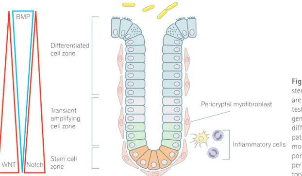
Fig. 1. Homeostasis of colon crypt and stem cell-regulating factors. Colon crypts are maintained by the ability of the in-testinal stem cells to self-renew and to generate a hierarchy of proliferative and differentiated cells via complex signaling pathway, such as WNT, Notch and bone morphogenetic protein (BMP), and sup-ported by microenvironments, including pericryptal myofibroblast and inflamma-tory cells.
경에서 유래한 hepatocyte growth factor (HGF), 또는 염증성 전사인 자인 NFĸB 등의 주요인자가 이미 줄기세포가 아닌 암세포를 다시 암 줄기세포로 역분화키는 작용을 할 수 있다.18,19 이런 의미에서 대장암 의 여러 다른 특성들은 암 발생의 기원세포의 차이, 그리고 해당 기원 세포에 축적되는 유전적 변이의 차이가 주요 원인일 수 있고, 이와 더 불어 주위 미세환경에 의한 영향도 포함되어야 할 주요인자이다. 그러나 아직 암줄기세포의 존재 및 중요성에 대해 여러 이견이 있 으며, 이것은 아직 뚜렷한 암줄기세포의 공통 표지자가 부족하다는 점에도 원인이 있다. 현재 CD133, CD166, CD44, CD24, ALDH-1, LGR5, epithelial cell adhesion molecule (EpCAM), BMI1, DCAM-KL-1 등의 암줄기세포 표지자가 보고되었고,12,13,15,16,20-22 이들 표지 자를 이용한 암줄기세포의 특성이 검증되고 있다. 이런 암줄기세포의 개념을 중요하게 여기는 이유는 여러 치료에 저 항성을 보이는 대표적인 세포군이라는 점과23-25 재발암 및 암전이의 주요 원인이 될 수 있다는 점이 있다(Fig. 2).18,23 여러 연구에서 일반 적인 항암치료 후 전체 종양크기는 감소하지만 종양 내 암줄기세포 의 분획은 오히려 증가하는 소견을 보여 치료 저항성과 재발암의 주 요 원인이 될 수 있음을 보고하고 있고, 암전이에 중요한 역할을 하는 epithelial-mesenchymal transition (EMT) 현상이 암줄기세포에서 뚜
렷하게 증가됨이 알려져 있다.26,27 따라서 암줄기세포를 표적으로 하는 치료는 암의 궁극적 극복을 가능하게 하는 방법 중의 하나가 될 수 있다. 그러나 많은 암줄기세포 표지자가 정상 줄기세포 표지자와 중복되므로 단순한 표지자를 이용 한 표적 치료에는 한계가 있을 것으로 생각된다. 뒤에서 언급될 장선와 내에서 정상 줄기세포의 증식과 분화에 관 련된 여러 인자 및 신호전달계에 대한 이해를 통해 암줄기세포의 자 가 재생 증식을 억제할 방법이 제안되고 있으며, 정상 줄기세포의 유 지, 증식 및 분화에서와 유사한 신호전달계가 암줄기세포에서도 작 용하므로 WNT, Notch, bone morphogenetic protein (BMP) 등의 신 호전달 물질을 조절하고,28,29 미세환경의 영향을 조절함으로써18 암줄 기세포의 증식을 억제하려는 연구들이 진행되고 있다. 이와 더불어 암 줄기세포의 분화촉진을 통하여 줄기세포의 특성을 제거함으로써 일 반 항암치료에 대한 반응을 높이는 방법이나, 이러한 약제들을 기존 의 항암제와 같이 병용 투여하여 치료 저항성을 극복하려는 방법 등 이 시도되고 있다(Fig. 2).30-32
장줄기세포 유지를 위한 주요 신호전달
줄기세포의 유지와 증식 및 분화를 위해서는 매우 복잡한 신호전 달체계가 필요하여, 줄기세포 자체에 의한 조절 뿐만 아니라 niche 등 의 주위 환경으로부터의 신호전달 역시 매우 중요한 역할을 할 것으 로 알려져 있다. 정상 장줄기세포 조절에 관여하는 많은 인자 및 신호 전달계가 암줄기세포 조절에서도 주요 인자로서 관여하고 있으므로 정상 장줄기세포 조절인자에 대한 최근의 연구발전은 암줄기세포 조 절에 대한 연구에도 많은 공헌을 하고 있다. 현재까지 알려져 있는 장 줄기세포 및 장선와 형성 관련 신호전달체계로서 WNT, Hedgehog (Hh), Notch, BMP 등이 알려져 있다(Fig. 1). 1. WNT 신호와 세포 증식 WNT 신호는 WNT 신호전달계의 하나인 APC 유전자의 변이가 대 장암 발생에 중요하게 관여하므로 위장관에서 많이 연구되었으며, 배 아 발달, 세포의 항상성 유지 그리고 여러 질환의 발생에 관여하고 있 다. 특히 장에서 줄기세포 유지 및 증식과 분화에서 중요한 역할을 한 다.3 WNT 신호전달계 억제를 이용한 대표적 연구로서 WNT 수용체에 결합하여 WNT 신호를 억제하는 Dickkopf를 과발현시키거나 TCF4 를 결손시킨 마우스에서 선와 내 증식 감소와 함께 선와 결손, 융모 감소 및 융모 위축의 소견을 보여, WNT 신호가 장관내 선와의 증식 구간에서 지속적인 세포 증식에 중요 역할을 함을 보여준다.33,34 WNT 신호의 과발현을 이용한 연구로서 APC 결손의 조건적 유발 에 의한 마우스 실험에서 APC 결손 유발 직후 선와의 세포증식 구 간이 확장되고 분화 및 세포이동이 억제되었으나, 융모에서는 특별 한 형태학적 변화가 없었다.35,36 그리고 정상적으로 선와 기저부에서Paneth 세포의 위치와 연관되어 발현되는 EphB3가 APC 결손 후 위
쪽으로 확장되어 증식구간이 확장된 소견을 보였다.35,36
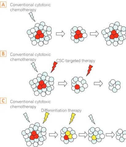
Fig. 2. Concept of cancer stem cell (CSC)-targeted therapy. Conven-tional chemotherapy does not target cancer stem cells (red), which is resistant to chemo and radiation therapy. The tumor is reduced in size in the short term, but eventually relapses driven by cancer stem cells (A). When CSC-targeted therapy (B) or CSC-differentiating therapy (C) is combined with conventional therapy, tumor will progressively exhaust its growth potential.
B
C
A
또한 WNT 활성인자인 R-spondin1은 장줄기세포군의 구간을 확 장시키고,37 최근 연구결과에 의하면 줄기세포 표지자인 LGR5를 발 현하는 CBC 세포 옆에 위치하는 Paneth 세포가 줄기세포의 유지와 증식에 관여하는 WNT3a의 주요한 공급 세포로 밝혀졌다.38 이와 같이 WNT 신호에 관련된 많은 인자들은 암줄기세포 유지 및 증식에 관여하고 있으므로 암줄기세포 억제를 위한 주요 치료 표적의 하나이다.39 또한 WNT 신호의 근원지로서 주위 niche 및 미세환경이 많이 관여하므로 미세환경 조절 또한 중요한 표적이다. 2. Notch 신호와 세포증식 및 분화 줄기세포로부터 분화에 이르는 과정은 줄기세포의 분화 프로그램 과 주위 환경으로부터의 여러 신호에 의해 결정된다. Notch 신호의 활성화는 hairy/enhancer of split (Hes) 유전자의 발현을 증가시키 며, Hes-1 발현 활성화는 분비세포 분화와 관련된 Math-1과 Neu-rogenin-3와 같은 basic helix-loop-helix (bHLH) 전사인자들의 활 성을 억제한다.
Notch 신호전달의 중간매개체인 γ-secretase를 억제하면 Hes-1
억제에 의해 장점막내 분비세포 과다 증식을 유발하며,40 발생 배아
에서 Hes-1이 결손된 마우스는 위와 소장의 내분비 세포의 조기 발
달을 보이고 장내 술잔세포와 장세포 수가 증가한다.3,41
Notch 분자는 delta, jagged로 알려진 ligand와 결합하여 활성화되 는데, 활성화된 세포는 자신의 Notch ligand를 억제함으로써 주위세 포의 Notch 활성화를 억제하게 되며, 이와 같은 기전으로 주위세포 의 분화 결정에 기여한다.42 장관에서는 Notch 활성화는 흡수장세포 와 분비세포 사이의 분화를 결정하여, Notch 신호전달의 억제는 술 잔세포의 증식을 유발하고, Notch 활성화는 술잔세포 결손을 유발 한다.43,44 또한 WNT 신호와 함께 작용 시 세포의 증식을 촉진하고, WNT 신호 없이 단독으로 작용할 때 흡수 장세포로의 분화를 유도 하여 장선와 항상성 유지에 관여한다.43-45 대부분의 대장암종양에서 WNT 신호가 증가되어 있으므로 Notch 신호에 의한 암줄기세포 증 식 관련성이 많이 보고되어 있고, Notch 신호 조절에 의한 줄기세포의 분화가 잘 알려져 있으므로, 암줄기세포 억제 및 분화치료에 주요 표 적인자가 될 수 있다. 3. BMP 및 Hh 신호와 선와 형성 BMP2와 BMP4는 장점막의 간엽세포에서 발현되며, 선와-융모 경계부위에서 세포의 증식 억제와 분화 유도에 관여한다. BMP 수용 체 결손 또는 BMP 억제인자인 noggin의 과발현 마우스는 장선와의 과증식 소견을 보인다.46,47 BMP는 선와의 상부 뿐만 아니라 기저부에 서도 분비되지만 장줄기세포 부위의 간엽세포에서 분비되는 noggin 등의 BMP 억제인자는 BMP의 분화유도 효과를 억제하게 된다.
상피유래의 platelet-derived growth factor (PDGF)-A는 융모 형 성과정에서 융모 중심부를 형성하는 간엽조직의 증식을 조절하는 데 관여하며, 특히 PDGF-A 자극에 의해 간엽세포로부터 활성화
된 BMP 신호는 상피세포의 증식구간 내 제한적 증식에 관여한다.48
BMP 신호의 이상은 familial juvenile polyposis syndrome (JPS)과 관 련이 있으며, BMP-4 억제 단백질인 noggin 과발현 마우스의 경우 JPS와 유사한 용종이 발생하고,47 BMP 수용체인 Bmpr1a의 불활성 화 역시 동일한 용종 발생을 유발한다.46 이와 같은 JPS와 유사한 용 종의 발생은 BMP 신호 억제에 의해 융모에서 선와생성의 비정상적 활성화 유발이 관여한다고 설명하고 있다. BMP 신호 활성화는 분화 유도와 밀접한 관련이 있으므로 암줄기세포의 분화유도를 통한 항암 제 저항성 억제에 기여할 수 있는 치료 표적이다. Hh 신호전달 역시 배아 발생의 여러 단계에서 관여하며, 위장관 발 생에서 sonic hedgehog (SHH)과 Indian hedgehog (IHH) 발현은 상 피 줄기세포의 증식과 분화에 관여하여, 발생기간 동안 Hh 신호 억 제는 선와-융모 발생의 이상을 초래한다.49-52 장선와의 Hh 신호전달 에서, IHH는 분화된 상피세포에서 발현되어 주위 간엽세포에 영향을 주어 BMP 분비를 유도한다.52,53 SHH의 역할에 대해서는 아직 이견 이 많으며 그 기능에 대한 자료가 불충분하다. 이상의 여러 신호전달계의 상호작용에 의하여 장선와의 분화와 증 식이 조절되며, 선와 증식은 점막 손상의 복구에도 관여하여, 방사선 조사에 의한 장점막 손상 후 많은 선와 모양의 구조를 포함하는 결절 들이 재생되는데,54 이런 재생 선와들은 작은 분지들을 형성하여 새로 운 선와를 형성하는 선와 생성 주기를 활성화시킨다. 이러한 선와 생 성 주기의 활성화는 점막손상 복구 뿐만 아니라 초기 종양 생성과정 에서 클론 확장에도 관여하여, 암줄기세포의 발생과 증식에 중요한 역할을 한다. 최근 이상과 같은 장선화 항상성 유지의 기전에 대한 이해와 함께 장기간 동안 실험실 배양조건에서 장선와 구조를 유지, 증식할 수 있 는 방법이 개발되었다.55,56 WNT 관련인자, R-spondin-1, noggin, 그 외 성장인자 등의 조건에서 장줄기세포의 증식과 장선와 형성, 유지, 증식 등을 가능하게 함으로써 줄기세포의 증식, 분화 및 niche의 상 호관계에 대해 배양상태에서의 연구가 가능하게 되었다. 이와 같은 줄기세포 연구방법의 발전은 암줄기세포의 실험실 내 연구 방법에서 도 적용되어 유용한 방법으로 이용되고 있다.15,16
암줄기세포와 미세환경의 상호작용
줄기세포의 기능은 장상피세포들의 항상성을 유지하는 것으로 자 기 복제, 증식, 그리고 분화과정을 통해 장상피세포를 일정하게 유지 한다. 이 과정은 niche라는 주위 구조 내에서 이루어지게 되는데 주위 의 간엽세포들과 증식 또는 분화 중인 상피세포들에 의해 영향을 받 게 된다. 비록 기저막(basement membrane)에 의해 상피세포와 분리 되어 있지만 이곳의 간엽세포(세포외 기질, 장신경계, 혈관, 상피내 임 파구, 선와주위 섬유모세포)들은 줄기세포 niche를 유지하기 위해 필 요한 상피-간엽(epithelial-mesenchymal) 간의 상호작용을 증진시킨 다. 아직 알려지지 않은 많은 신호전달과 분비 인자가 이런 환경을 유 지하는 데 중요할 것으로 생각하며, 선와의 바깥쪽을 감싸는 선와주 위 간엽(pericryptal mesenchyme) 내의 근섬유모세포(myofibroblast) 는 HGF, transforming growth factor beta (TGFβ) 그리고 keratino-cyte growth factor (KGF) 등의 여러 물질을 분비한다.3,57 그 외 선와주위 간엽 세포에서 분비되어 그에 인접하여 있는 상피세포에 영향을 주는 여러 분비물질들로서 PDGF, SHH, BMP, forkhead-6 (Fkh), WNT 그리고 Notch 등이 알려져 있다. 장관 선와 내 항상성 유지를 위해 선와 주위 간엽조직은 매우 중요한 역할을 할 것으로 생각하며, 정상적인 선와 유지 뿐만 아니라 암과 같은 병적 상태에서도 암세포 와의 상호관계를 통해 주요 역할을 한다. 최근 침습성 암종의 진행에 미세환경의 미분화 골수성세포가 주 요 역할을 함이 보고되었고,58 여러 장염증 모델에서의 염증성 매개체 에 의한 암화과정 촉진이 알려져 있다.59 종양내 염증세포들에 의한 TNF-α, IL-6, TGF-β 등의 성장인자 및 사이토카인들은 종양의 형 성과 증식에 주요역할을 하며,60 상피세포의 NFκB 활성화를 통해 종양 생성을 증가시킬 뿐 아니라 줄기세포가 아닌 암세포를 암줄기세 포로 역분화시키는 데에도 기여한다.19,61 이와 더불어 종양 내 섬유아세포도 종양 미세환경의 주요 세포로 서 여러 종류의 성장인자와 사이토카인을 분비하여 종양의 증식에 관 여한다.62 특히 종양 내 근섬유아세포에서 분비되는 HGF는 WNT 활 성화를 통해 종양의 증식과 암줄기세포 상태 유지에 관여하고, 또한 c-Met를 통해 분화된 암세포를 암줄기세포로 역분화시키는 데 주요 역할을 한다.18 이상과 같이 종양 미세환경의 여러 세포군과 분비인자들은 종양의 증식 뿐만 아니라 암줄기세포의 유지 및 증식에도 관여하고 있고, 특 정 미세환경 조건에서는 분화 상태의 암세포를 암줄기세포의 특성을 지닌 세포로 변형시키기도 하므로 암줄기세포 표적치료에서 미세환 경 및 niche 인자의 조절은 빠질 수 없는 표적이다.18
결 론
대장암을 포함한 모든 암질환의 공통적인 문제로서 약제 저항성, 재발, 전이의 문제가 암 완치를 위해 해결해야 할 가장 중요한 문제 일 것이다. 이러한 약제 저항성, 재발, 전이의 문제에 공통적으로 관여 하는 세포들의 특성 중 하나가 암줄기세포 특성이므로 암 완치의 꿈 을 위한 여러 연구들이 암줄기세포를 표적으로 하여 진행되고 있다. 특히 최근 정상 장줄기세포에 대한 연구의 발전과 함께 정상 줄기세 포의 유지 및 증식에서와 유사한 신호전달을 이용하는 암줄기세포에 대한 이해도 많이 발전하고 있다. 그러나 아직 정상 장줄기세포에 대 한 영향을 최소화할 수 있는 암줄기세포 특이적인 표지자의 발굴이 쉽지 않고, 암줄기세포의 유지 증식에 관여하는 영향 인자로서 암줄 기세포의 다양성, 미세환경 인자 및 역분화 관련 인자 등 더욱 복잡한 상호관계가 밝혀지면서 암줄기세포에 대한 표적 치료 개발도 많은 어 려움이 예상된다. 그러나 최근 암줄기세포에 대한 분화치료 개념 및 새로운 관련 인자 규명 등의 중요한 연구결과들과 함께 많은 노력이 집중되고 있으므로 암줄기세포를 표적으로 한 치료제 개발에 한걸음 가까워질 것을 기대한다.REFERENCES
1. Bjerknes M, Cheng H. The stem-cell zone of the small intestinal
epithelium. I. Evidence from Paneth cells in the adult mouse. Am J Anat 1981;160:51-63.
2. Bjerknes M, Cheng H. Clonal analysis of mouse intestinal epi-thelial progenitors. Gastroenterology 1999;116:7-14.
3. Brittan M, Wright NA. Stem cell in gastrointestinal structure and neoplastic development. Gut 2004;53:899-910.
4. Barker N, van Es JH, Kuipers J, et al. Identification of stem cells in small intestine and colon by marker gene Lgr5. Nature 2007;449:1003-1007.
5. Breault DT, Min IM, Carlone DL, et al. Generation of mTert-GFP mice as a model to identify and study tissue progenitor cells. Proc Natl Acad Sci U S A 2008;105:10420-10425.
6. Montgomery RK, Carlone DL, Richmond CA, et al. Mouse telomerase reverse transcriptase (mTert) expression marks slowly cycling intestinal stem cells. Proc Natl Acad Sci U S A 2011;108:179-184.
7. Sangiorgi E, Capecchi MR. Bmi1 is expressed in vivo in intestinal stem cells. Nat Genet 2008;40:915-920.
8. Lopez-Garcia C, Klein AM, Simons BD, Winton DJ. Intestinal stem cell replacement follows a pattern of neutral drift. Science 2010;330:822-825.
9. Snippert HJ, van der Flier LG, Sato T, et al. Intestinal crypt ho-meostasis results from neutral competition between symmetri-cally dividing Lgr5 stem cells. Cell 2010;143:134-144.
10. van der Flier LG, van Gijn ME, Hatzis P, et al. Transcription fac-tor achaete scute-like 2 controls intestinal stem cell fate. Cell 2009;136:903-912.
11. Zhu L, Gibson P, Currle DS, et al. Prominin 1 marks intestinal stem cells that are susceptible to neoplastic transformation. Na-ture 2009;457:603-607.
12. Huang EH, Hynes MJ, Zhang T, et al. Aldehyde dehydrogenase 1 is a marker for normal and malignant human colonic stem cells (SC) and tracks SC overpopulation during colon tumorigenesis. Cancer Res 2009;69:3382-3389.
13. Vaiopoulos AG, Kostakis ID, Koutsilieris M, Papavassiliou AG. Colorectal cancer stem cells. Stem Cells 2012;30:363-371. 14. Powell AE, Wang Y, Li Y, et al. The pan-ErbB negative regulator
Lrig1 is an intestinal stem cell marker that functions as a tumor suppressor. Cell 2012;149:146-158.
15. O’Brien CA, Pollett A, Gallinger S, Dick JE. A human colon can-cer cell capable of initiating tumour growth in immunodeficient mice. Nature 2007;445:106-110.
16. Ricci-Vitiani L, Lombardi DG, Pilozzi E, et al. Identification and expansion of human colon-cancer-initiating cells. Nature 2007;445:111-115.
17. Barker N, Ridgway RA, van Es JH, et al. Crypt stem cells as the cells-of-origin of intestinal cancer. Nature 2009;457:608-611. 18. Vermeulen L, De Sousa E Melo F, van der Heijden M, et al. Wnt
activity defines colon cancer stem cells and is regulated by the microenvironment. Nat Cell Biol 2010;12:468-476.
19. Schwitalla S, Fingerle AA, Cammareri P, et al. Intestinal tumori-genesis initiated by dedifferentiation and acquisition of stem-cell-like properties. Cell 2013;152:25-38.
of human colorectal cancer stem cells. Proc Natl Acad Sci U S A 2007;104:10158-10163.
21. Vermeulen L, Todaro M, de Sousa Mello F, et al. Single-cell clon-ing of colon cancer stem cells reveals a multi-lineage differentia-tion capacity. Proc Natl Acad Sci U S A 2008;105:13427-13432. 22. Kemper K, Sprick MR, de Bree M, et al. The AC133 epitope, but
not the CD133 protein, is lost upon cancer stem cell differentia-tion. Cancer Res 2010;70:719-729.
23. Todaro M, Alea MP, Di Stefano AB, et al. Colon cancer stem cells dictate tumor growth and resist cell death by production of in-terleukin-4. Cell Stem Cell 2007;1:389-402.
24. de Sousa EM, Vermeulen L, Richel D, Medema JP. Targeting Wnt signaling in colon cancer stem cells. Clin Cancer Res 2011;17:647-653.
25. Todaro M, Francipane MG, Medema JP, Stassi G. Colon can-cer stem cells: promise of targeted therapy. Gastroenterology 2010;138:2151-2162.
26. Mani SA, Guo W, Liao MJ, et al. The epithelial-mesenchymal transition generates cells with properties of stem cells. Cell 2008;133:704-715.
27. Yang MH, Hsu DS, Wang HW, et al. Bmi1 is essential in Twist1-induced epithelial-mesenchymal transition. Nat Cell Biol 2010;12:982-992.
28. Hoey T, Yen WC, Axelrod F, et al. DLL4 blockade inhibits tumor growth and reduces tumor-initiating cell frequency. Cell Stem Cell 2009;5:168-177.
29. Lombardo Y, Scopelliti A, Cammareri P, et al. Bone morphoge-netic protein 4 induces differentiation of colorectal cancer stem cells and increases their response to chemotherapy in mice. Gastroenterology 2011;140:297-309.
30. Gupta PB, Onder TT, Jiang G, et al. Identification of selective in-hibitors of cancer stem cells by high-throughput screening. Cell 2009;138:645-659.
31. Kodach LL, Jacobs RJ, Voorneveld PW, et al. Statins augment the chemosensitivity of colorectal cancer cells inducing epigenetic reprogramming and reducing colorectal cancer cell ‘stemness’ via the bone morphogenetic protein pathway. Gut 2011;60:1544-1553.
32. Hirsch HA, Iliopoulos D, Struhl K. Metformin inhibits the inflam-matory response associated with cellular transformation and cancer stem cell growth. Proc Natl Acad Sci U S A 2013;110:972-977.
33. Pinto D, Gregorieff A, Begthel H, Clevers H. Canonical Wnt sig-nals are essential for homeostasis of the intestinal epithelium. Genes Dev 2003;17:1709-1713.
34. Korinek V, Barker N, Moerer P, et al. Depletion of epithelial stem-cell compartments in the small intestine of mice lacking Tcf-4. Nat Genet 1998;19:379-383.
35. Andreu P, Colnot S, Godard C, et al. Crypt-restricted prolifera-tion and commitment to the Paneth cell lineage following Apc loss in the mouse intestine. Development 2005;132:1443-1451. 36. Sansom OJ, Reed KR, Hayes AJ, et al. Loss of Apc in vivo
imme-diately perturbs Wnt signaling, differentiation, and migration. Genes Dev 2004;18:1385-1390.
37. Kim KA, Kakitani M, Zhao J, et al. Mitogenic influence of human R-spondin1 on the intestinal epithelium. Science 2005;309:1256-1259.
38. Sato T, van Es JH, Snippert HJ, et al. Paneth cells constitute the niche for Lgr5 stem cells in intestinal crypts. Nature 2011;469: 415-418.
39. Anastas JN, Moon RT. WNT signalling pathways as therapeutic targets in cancer. Nat Rev Cancer 2013;13:11-26.
40. Milano J, McKay J, Dagenais C, et al. Modulation of notch pro-cessing by gamma-secretase inhibitors causes intestinal goblet cell metaplasia and induction of genes known to specify gut secretory lineage differentiation. Toxicol Sci 2004;82:341-358. 41. Jensen J, Pedersen EE, Galante P, et al. Control of endodermal
endocrine development by Hes-1. Nat Genet 2000;24:36-44. 42. Crosnier C, Stamataki D, Lewis J. Organizing cell renewal in the
intestine: stem cells, signals and combinatorial control. Nat Rev Genet 2006;7:349-359.
43. Fre S, Huyghe M, Mourikis P, Robine S, Louvard D, Artavanis-Tsakonas S. Notch signals control the fate of immature progeni-tor cells in the intestine. Nature 2005;435:964-968.
44. van Es JH, van Gijn ME, Riccio O, et al. Notch/gamma-secretase inhibition turns proliferative cells in intestinal crypts and adeno-mas into goblet cells. Nature 2005;435:959-963.
45. van Es JH, de Geest N, van de Born M, Clevers H, Hassan BA. Intestinal stem cells lacking the Math1 tumour suppressor are refractory to Notch inhibitors. Nat Commun 2010;1:18.
46. He XC, Zhang J, Tong WG, et al. BMP signaling inhibits intestinal stem cell self-renewal through suppression of Wnt-beta-catenin signaling. Nat Genet 2004;36:1117-1121.
47. Haramis AP, Begthel H, van den Born M, et al. De novo crypt formation and juvenile polyposis on BMP inhibition in mouse intestine. Science 2004;303:1684-1686.
48. Karlsson L, Lindahl P, Heath JK, Betsholtz C. Abnormal gastroin-testinal development in PDGF-A and PDGFR-(alpha) deficient mice implicates a novel mesenchymal structure with putative instructive properties in villus morphogenesis. Development 2000;127:3457-3466.
49. Ramalho-Santos M, Melton DA, McMahon AP. Hedgehog sig-nals regulate multiple aspects of gastrointestinal development. Development 2000;127:2763-2772.
50. Wang LC, Nassir F, Liu ZY, et al. Disruption of hedgehog sig-naling reveals a novel role in intestinal morphogenesis and intestinal-specific lipid metabolism in mice. Gastroenterology 2002;122:469-482.
51. Madison BB, Braunstein K, Kuizon E, Portman K, Qiao XT, Gu-mucio DL. Epithelial hedgehog signals pattern the intestinal crypt-villus axis. Development 2005;132:279-289.
52. van den Brink GR, Bleuming SA, Hardwick JC, et al. Indian Hedgehog is an antagonist of Wnt signaling in colonic epithelial cell differentiation. Nat Genet 2004;36:277-282.
53. van Dop WA, Heijmans J, Büller NV, et al. Loss of Indian Hedge-hog activates multiple aspects of a wound healing response in the mouse intestine. Gastroenterology 2010;139:1665-1676. 54. Inoue M, Imada M, Fukushima Y, et al. Macroscopic intestinal
colonies of mice as a tool for studying differentiation of multipo-tential intestinal stem cells. Am J Pathol 1988;132:49-58. 55. Ootani A, Li X, Sangiorgi E, et al. Sustained in vitro intestinal
epi-thelial culture within a Wnt-dependent stem cell niche. Nat Med 2009;15:701-706.
56. Sato T, Vries RG, Snippert HJ, et al. Single Lgr5 stem cells build crypt-villus structures in vitro without a mesenchymal niche. Nature 2009;459:262-265.
57. Marsh MN, Trier JS. Morphology and cell proliferation of subepi-thelial fibroblasts in adult mouse jejunum. II. Radioautographic studies. Gastroenterology 1974;67:636-645.
58. Kitamura T, Kometani K, Hashida H,et al. SMAD4-deficient
in-testinal tumors recruit CCR1+ myeloid cells that promote inva-sion. Nat Genet 2007;39:467-475.
59. Taketo MM, Edelmann W. Mouse models of colon cancer. Gas-troenterology 2009;136:780-798.
60. Grivennikov SI, Greten FR, Karin M. Immunity, inflammation, and cancer. Cell 2010;140:883-899.
61. Greten FR, Eckmann L, Greten TF, et al. IKKbeta links inflamma-tion and tumorigenesis in a mouse model of colitis-associated cancer. Cell 2004;118:285-296.
62. Shao J, Sheng GG, Mifflin RC, Powell DW, Sheng H. Roles of myofibroblasts in prostaglandin E2-stimulated intestinal epithe-lial proliferation and angiogenesis. Cancer Res 2006;66:846-855.