Jin W oong C hung
1, G eun-Young K im
1,
Y eung-C hul M un
2, Ji-Y oung A hn
2,
C hu-M yong S eong
2and Jae-H ong K im
1,3 1School of Life Sciences and BiotechnologyKorea University, Seoul 136-701, Korea 2Department of Oncology/Hematology
College of Medicine, Ewha Woman's University Seoul 158-056, Korea
3Coressponding author: Tel, 82-2-3290-3452; Fax, 82-2-3290-3960; E-mail, jhongkim@korea.ac.kr Accepted 31 January 2005
Abbreviations: FL, Flt-3 ligand; HSC, hematopoietic stem cell; LTB4, leukotriene B4; RT-PCR, reverse transcription PCR; TPO, thrombopoietin; UCB, umbilical cord blood
Abstract
Leukotriene B4 (LTB4), derived from arachidonic
acid, is a potent chem otactic agent and activat-ing factor for hem atopoietic cells. In addition to host defense in vivo, several eicosanoids have been reported to be involved in stem cell dif-ferentiation or proliferation. In this study, w e investigated the effect of LTB4 on hum an cord
blood C D 34+ hem atopoietic stem cells (H S C s).
LTB4 w as show n to induce proliferation of H S C
and exert anti-apoptotic effect on the stem cells. B lockade of interaction betw een LTB4 and its
receptor enhanced self-renewal of the stem cells. E ffect of LTB4 on differentiation of C D 34+ H SC s
w ere confirm ed by clonogenic assays, and in-duction of the expression of B LT2 (the low - affinity LTB4 receptor), during the ex vivo
expan-sion w as confirm ed by reverse transcription- P C R . O ur results suggest that LTB4-B LT2
inter-action is involved in the cytokine-induced dif-ferentiation and ex vivo expansion of hem ato-poietic stem cells.
Keywords: BLT2; CD34; apoptosis; cell differentiation;
hematopoietic stem cells; leukotriene B4; receptors
Introduction
Hematopoietic stem cells (HSCs) have ability to self renew while functionally repopulating the cells of the blood and lymph for the life of an individual. These cells are thought to retain a high capacity for plasticity that would contribute to their ability to differentiate into not only hematopoietic cells but also non-hemato-poietic tissues such as brain, heart, skeletal muscle, liver and endothelial cells, following injury or stress (Heike et al., 2004). Such capacity of these cells gives rise to the hope that hematopoietic stem cells may regenerate various damaged tissues. In that regard, ex vivo expansion of hematopoietic stem cells is a promising technology for many potential ap-plications from marrow reconstitution to gene therapy. Despite of considerable progresses gained during the past ten years in understanding the biology of hematopoietic stem cells and its ex vivo expansion, the success was limited (Piacibello et al., 1997; Shih et al., 1999; Lewis et al., 2001). Self-renewal of stem cells depends on several critical factors, including the hematopoietic microenvironment, interactions with sup-porting stromal cells (Charbord, 2001), features of the extracellular matrix (Williams et al., 1991), hemato-poietic growth factors (Feugier et al., 2002; Terada et al., 2003) and cytokines (Flores-Guzman et al., 2002).
Leukotriene B4 (LTB4), derived from arachidonic
acid, is a potent chemotactic agent and activating factor for hematopoietic cells. Until recently, two cell surface receptors for LTB4 have been identified;
BLT1, a high affinity receptor, and BLT2, a low-affinity receptor. BLT2 is expressed ubiquitously, in contrast to BLT1 expressed predominantly in leukocytes. The differences in the distribution and pharmacological characteristics of BLT1 and BLT2 suggest distinct role(s) for these receptors in vivo (Toda et al., 2002). Based on the previous observations, LTB4 seems to
play important roles in inflammation in addition to host defense in vivo (S.W. Crooks et al., 1998). Recently, it has been reported that cysteinyl leukotriene re-ceptor is involved in differentiation of HSCs into eosinophils (Hashida et al., 2001). In addition, several eicosanoids have been reported to be involved in stem cell differentiation or proliferation (Desplat et al., 2000; Braccioni et al., 2002). In this study, we have investigated the effect of LTB4, one of the potent lipid
mediators, on cell proliferation, differentiation, and self-renewal capacity during the long-term culture of HSCs.
Leukotriene B
4
pathway regulates the fate of the hematopoietic
bach, Germany) (Kim, 2003). The efficiency of purifica-tion and immunophenotype were verified with flow cytometry and counterstained with a FITC-anti-human CD34 (HPCA-2; Becton Dickinson; Mountain View, California) as previously described (Yoo et al., 1999).
Ex vivo expansion of CD34+ hem atopoietic stem
cells
CD34+ cells were placed into 24-well plates (Falcon;
Becton Dickinson Biosciences) at a concentration of 1×10³ cells per well. Each well contained 0.5 ml
liquid medium plus the growth factors of interest. The culture medium consisted of Iscove's modified Dul-becco's medium (IMDM; Gibco BRL) supplemented with 10% fetal bovine serum. The cytokines included recombinant human G-CSF (100 U/ml), TPO (10 U/ml), and FL (50 ng/ml). The cultures were incubated at 37oC in a humidified atmosphere containing 5%
CO2. On day 14, cells were harvested from the
culture and the number of viable cells was determined by trypan blue exclusion. Cell counts were also performed using a hemocytometer. The cells from these aliquots were assayed for the number of he-matopoietic progenitor cells. Apoptosis assay was performed using Annexin-V-Flous staining kit (Roche) following the manufacturer's protocol.
Clonogenic assays
Clonogenic assays were performed as follows: 1× 10³ CD34+ UCB cells were cultured at two plates per
point in complete methylcellulose medium (HCC-4434; Methocult; Stem Cell Technologies, Vancouver, Ca-nada). It was supplemented with 50 ng/ml stem cell factor (SCF), 10 ng/ml interleukin-3, GM-CSF, and 1 U/ml recombinant human erythropoietin (EPO) at 37oC in a humidified atmosphere at 5% CO2. After 14 days
of incubation, colonies were enumerated with the aid of an inverted microscope and counted by mor-phologic criteria.
GAPDH was heated at 94 C for 20 s, annealed at 58oC for 30 s and extended at 72oC for 30 s for 25
repetitive cycles. GAPDH was used as an internal control. The final PCR products were separated on a 1.2% agarose gel with ethidium bromide and visualized under UV light.
Data analysis and statistics
The results are presented as the mean ± SD of the data obtained from three or more experiments per-formed in duplicates. Statistical comparisons between groups were made with the Student's t-test. Values of P < 0.05 were considered significant.
R esults
LTB4 induces proliferation of CD34+ HSCs
The effect of LTB4 on the in vitro proliferation of
CD34+ hematopoietic progenitor cells from freshly
purified UCB CD34+ cells was assessed using trypan
blue exclusion assay. Earlier report of optimal culture condition for hematopoietic stem cells with combined supplementation of G-CSF, TPO and FL (Yoo et al., 1999) was used as a control culture and compared with the additional supplementation of LTB4. In the
absence of exogenous cytokines, no viable cells were observed after 2 weeks of culture, regardless of whe-ther or not LTB4 was present (data not shown). When
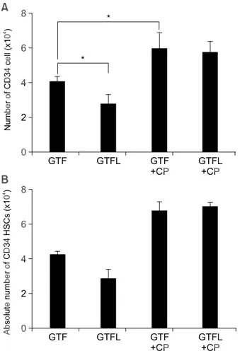
mixture of G-CSF, TPO, and FL was added to the medium, the number of total cells was increased approximately 350 folds during the culture as ex-pected (data not shown). Addition of LTB4 (300 nM)
significantly enhanced the growth of CD34+ HSCs
approximately by 1.5 fold when compared to the condition where no LTB4 was added (Figure 1A). The
effect of LTB4 was abolished by the addition of
CP105696 (10 µM), an antagonist of LTB4 receptors
(BLT) (Figure 1A), suggesting the specific effect of LTB4 on the proliferation of CD34+ hematopoietic stem
LTB4 exerts anti-apoptotic effect on CD34+
hem atopoietic stem cells
LTB4 was reported to be involved in cell survival by
its anti-apoptotic effect on various cells such as neutrophil (Hebert et al., 1996; Zhang et al., 2002). We performed Annexin-V staining to determine whe-ther LTB4 exerts anti-apoptotic effect also on CD34+
HSCs in a long term culture. As seen in Figure 2, addition of LTB4 decreased apoptosis when compared
to the control and CP105696 treatment inhibited the anti-apoptotic effect of LTB4 statistically significantly
(P < 0.05). These results suggest that the growth advantage of LTB4 on HSC is, at least partly, due
to its anti-apoptotic effect.
LTB4 induces differentiation of CD34+ HSCs
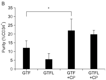
After 2 weeks of ex vivo expansion, purity of CD34+
HSCs was assesed by flow cytometry using CD34 antibody. Interestingly, the results were opposite to the growth experiments, where treatment of LTB4
showed lowest purity of HSCs in terms of retention of CD34 (Figure 1B). Accordingly, addition of BLT antagonist showed highest CD34-positive populations
(Figure 1B). The fact that the specific antagonist increased the purity of CD34+ HSCs even in the
absence of LTB4 might be due to the blockade of
interaction between BLT and the endogenously pro-duced LTB4 during differentiation of CD34+ HSCs
(Spanbroek et al., 2000). These results suggest that LTB4 induces differentiation of HSCs through its
re-ceptor during ex vivo expansion by autocrine or paracrinic manner, since the cells that lost CD34 might be differentiated or at least differentiating cells. Thus, we next performed clonogenic assays with methylcellulose cultures to confirm the involvement of LTB4 in HSC differentiation. In the methylcellulose
cultures, addition of LTB4 yielded approximately two-
fold increase in colony formations of CFU-GM and CFU-GEMM, when compared to the condition where no LTB4 was added (Figure 1C). Again, this effect
was inhibited by the addition of BLT antagonist, CP105696 (10 nM). In contrast, no significant dif-ferences in BFU-E-derived colony formation were observed in the presence or the absence of LTB4 or
CP105696 (Figure 1C). Taken together, these results suggest that LTB4 may induce differentiation of CD34+
HSCs by its receptor-mediated interaction.
Figure 1. Effects of LTB4 on differentiation of CD34+ hematopoietic stem cells. (A) Total number of mononuclear cells in liquid culture. Freshly isolated CD34+ cells were cultured in 10% IMDM with G-CSF (10 U/ml, G), TPO (T) and FL (F) in the absence or the presence of LTB4 (300 nM) and/or CP105696 (10 uM). On day 14, cells were harvested from each culture and the viable cells were counted using trypan blue. Values are the means ± SD of 3 separate experiments in duplicate cultures (*P < 0.05). (B) Purity of CD34+ HSCs after long term culture. The cells obtained after 2-week culture were stained with FITC-anti-CD34 (HPCA-2) antibody, and analyzed by flow cytometry. Data are presented as means of fluorescence (*P < 0.05). (C) Effects of LTB4 on in vitro clonal growth of hematopoietic progenitor cells from isolated human UCB CD34+ cells. Freshly isolated CD34+ cells (1×10³ cells/ml) were incubated in a liquid culture as described in the legend of (A). On day 14, cells harvested from each culture were plated into a methylcellulose culture supplemented with SCF, IL-3, GM-CSF, G-CSF and EPO. The culture was incubated for 14 days. Values are the means ± SD of three separate experiments in duplicated cultures.
Blockade of LTB4 pathway induces self-renewal of
CD34+ HSCs
From the above results, we assumed that it might be possible that self-renewal of CD34+ HSC can be
increased by blocking LTB4-BLT interaction. Thus, we
next determined the effect of BLT antagonist on self- renewal of CD34+ HSCs. After two weeks of ex vivo expansion, the cultures were collected and run through the MACS column as mentioned above. After purification of CD34+ cells, number of the cells was
assessed by trypan blue exclusion. As expected, treatment of CD105696 (10 µM) increased number of CD34+ HSCs by approximately two-fold, compared
to the control, whether LTB4 was added or not (Figure
3A). Even though there might be loss of a small portion of the cells during the purification, this result showed the similar tendency with absolute number of CD34+ cells (Figure 3B) calculated from total number of the cells (Figure 1A) and the purity of CD34+ HSCs
(Figure 1B), confirming blockade of BLT induces proliferation of CD34+ HSCs. Since these CD34+ cells
retained differentiating capacity as measured in clono-genic assays using methyl cellulose medium (data not shown), blockade of BLT may induce not only the proliferation but also the self-renewal of CD34+ HSCs.
Expression of BLT2, a receptor for LTB4, is
induced during differentiation of CD34+ HSCs
The induced expression of BLT1 during the cytokine- induced differentiation of CD34+ stem cells was re-ported by Pettersson et al. (2003). We next performed RT-PCR with BLT2-specific primers to investigate whether expression of BLT2, another receptor for LTB4 is also related to in vitro differentiation of CD34+
HSCs. As seen in Figure 4, BLT2 expression was increased during the long term culture when the
cytokines were added in the presence or absence of LTB4. However, the expression was decreased when
the LTB4 receptor antagonist was treated. Taken
together with previous report, these results show the expression of BLT maybe directly or at least indirectly cells. Human cord blood stem cells were isolated and cultured in the
same condition described in the legend of Figure 1 (A). After two-week culture, cells were harvested and subjected to Annexin V-staining and rate of apoptosis was calculated following the manu-facturer's protocol. Values are the means ± SD of 3 separate experiments in duplicate cultures (*P < 0.05).
Figure 3. Effects of BLT-antagonist on self-renewal of human UCB
CD34+ cells. A. Freshly isolated CD34+ cells (1×10³ cells/ml) were ex vivo expanded in a liquid culture as described in the legend of Figure 1 (A). On day 14, total cells in each well were harvested and subjected to MACS purification. Numbers of CD34+ cells in each well were determined under microscope, and the values were expressed as the means ± SD (*P < 0.05). B. Absolute number of CD34+ HSCs calculated from Figure 1A and 1B.
Figure 4. Expression of BLT2 in CD34+ HSCs during long term culture. Total RNAs were isolated from ex vivo expanded HSCs and subjected to RT-PCR as described in the Material and Methods.
related to the effect of LTB4 on HSCs' proliferation,
survival, differentiation and self-renewal.
D iscussion
Maintaining self-renewal and differentiation potential of hematopoietic stem cell (HSC) is a crucial component and a major challenge in stem cell research and potential clinical applications for HSC ex vivo ex-pansion including tumor cell purging, gene therapy, and stem cell transplantation. In the present study, effects of LTB4 and its receptor on ex vivo expansion,
differentiation and self-renewal of hematopoietic stem cells were determined.
LTB4 has been reported to enhance growth of a
variety of cells including human pancreatic cancer cells (Tong et al., 2002), and fibroblast (Woo et al., 2002). In addition, various cytokines, evaluated for their ability to support human HSC long-term ex vivo expansion, share many common characteristics with LTB4, such as proinflammatory effects, chemotactic
activities and growth-stimulating factors, and suggests that LTB4 might also be involved in the proliferation
phase of HSCs.
In our study, LTB4 itself was unable to induce
proliferation of HSC in the absence of other cytokines or even in the presence of any one or two com-binations of three cytokines used in this study (data not shown). This is consistent with previous report where LTB4 had no effect on proliferation of CD34+
HSCs in the absence of any cytokines (Desplat et al., 2000). However, when LTB4 was added to the culture
medium supplemented with all three cytokines includ-ing G-CSF, TPO and FL, the growth of HSCs was increased by approximately 50%, compared to the control. Thus, mechanism of LTB4 in HSC proliferation
is not solely due to self-mediated action but due to the concerted actions of other cytokines. For example, the cytokines might activate LTB4 signaling pathway
or induce the expression of the proteins involved in LTB4 signaling. Certain cykokines such as IL-13 up-
regulates leukotriene receptor in human lung fibroblast (Chibana et al., 2003). Indeed, the expression of BLT2, a LTB4 receptor, was induced during ex vivo
expansion of HSCs in the presence of the cytokines (Figure 4), suggesting the cytokines might up-regulate the expression of BLT2, directly or indirectly. Inter-estingly, the treatment of BLT2 antagonist induced down-regulation of the BLT2 itself expression impli-cating autocrine regulation of the receptor expression. Several eicosanoids, including prostaglandins and leukotrienes, have been reported to regulate cellular differentiation and apoptosis (Rudolph et al., 2001). Our results also showed anti-apoptotic effect of LTB4
on HSCs, suggesting the increase of the number of HSCs in the long term culture by LTB4 is, at least
in part, due to the retardation of apoptosis. Even though the total cell number was increased, popula-tion of CD34 positive cells in LTB4-treated cells was
lower than that of control. However, when the LTB4
receptor antagonist was treated, retention rate of
CD34 was even higher than that of control, sug-gesting LTB4 induces differentiation of HSCs during
ex vivo expansion. This was confirmed by the clono-genic assay, where LTB4 treatement enhanced
dif-ferentiation of HSC into various hematopoietic pro-genitors including CFU-GM and CFU-GEMM. The fact that absolute number of CD34-positive cells was higher when the HSCs were treated with BLT anta-gonist, even though the total cell number was re-latively lower, suggests that the regulation of LTB4
signaling pathway may contribute to the self-renewal as well as differentiation of HSCs. Taken together, LTB4-BLT interaction is closely related to the
cytoki-nes-induced hematopoeitic stem cell proliferation, dif-ferentiation, and self-renewal, and it might be pos-sible to determine the stem cell fate as needed, i.e. towards self-renewal or differentiation, during ex vivo expansion by controlling LTB4-BLT2 signaling
path-way.
Stem cells can be used to treat a variety of dise-ases and several recent studies in animal models demonstrate the potential of bioengineering strategies targeting adult and embryonic stem cells. In order to obtain the desired cells for transplantation, stem cell bioengineering approaches involve the manipulation of environmental signals influencing cell survival, proli-feration, self-renewal and differentiation. In that re-gard, even though the exact mechanisms are still to be elucidated, our findings will facilitate the studies and the clinical applications in the field of the stem cell-based regenerative therapies, as treatment acces-sibility will depend on the development of powerful technologies to produce sufficient cell numbers. Acknow ledgem ent
This work was supported by grants from Korea Re-search Foundation (KRF-2003-003-C00110).
R eferences
Braccioni F, Dorman SC, O'byrne PM, Inman MD, Denburg JA, Parameswaran K, Baatjes AJ, Foley R, Gauvreau GM. The effect of cysteinyl leukotrienes on growth of eosinophil progenitors from peripheral blood and bone marrow of atopic subjects. J Allergy Clin Immunol 2002;110:96-101
Charbord P. The hematopoietic stem cell and the stromal microenvironment. Therapie 2001;56:383-4
Chibana K, Ishii Y, Asakura T, Fukuda T. Up-regulation of cysteinyl leukotriene 1 receptor by IL-13 enables human lung fibroblasts to respond to leukotriene C4 and produce eotaxin. J Immunol 2003;170:4290-5
Crooks SW, Stockley RA. Leukotriene B4. Int J Biochem Cell Biol 1998;30:173-8
Desplat V, Ivanovic Z, Dupuis F, Faucher JL, Denizot Y, Praloran V. Effects of lipoxygenase matabilites of arachidonic acid on the growth of human blood CD34+ progenitors. Blood Cells Mol Dis 2000;26:427-36
Modulation by lipoxygenase-derived eicosanoids. J Immunol 1996;157:3105-15
Heike T, Nakahata T. Stem Cell plasticity in the hemato-poietic system. Int J Hematol 2004;79:7-14
Kim HL. Comparison of oligonucleotide-microassay and serial analysis of gene expression (SAGE) in transcript profiling analysis of megakaryocytes derived from CD34+ cells. Exp Mol Med 2003;35-460-6
Lewis ID, Almeida-Porada G, Du J, Lemischka IR, Moore KA, Zanjani ED, Verfaillie CM. Umbilical cord blood cells capable of engrafting in primary, secondary, and tertiary xenogeneic hosts are preserved after ex vivo culture in a noncontact system. Blood 2001;97:3441-9
Pettersson A, Richter J, Owman C. Flow cytometric mapping of the leukotriene B4 receptor, BLT1, in human bone marrow and periphiral blood using specific monoclonal antibodies. Int Immunopharmacol 2003;3:1467-75
Piacibello W, Sanavio F, Garetto L, Severino A, Bergandi D, Ferrario J, Ragioli F, Berger M, Aglietta M. Extensive am-plification and self-renewal of human primitive hematopoietic stem cells from cord blood. Blood 1997;89:2644-53 Rudolph IL, Kelley DS, Klasing KC, Erickson KL. Regulation of cellular differentiation and apoptosis by fatty acids and their metabolites. Nutr Res 2001;21:381-93
Tong WG, Ding XZ, Hennig R, Witt RC, Standop J, Pour PM, Adrian TE. Leukotriene B4 receptor antagonist LY293111 inhibits proliferation and induces apoptosis in human pan-creatic cancer cells. Clin Cancer Res 2002;8:3232-42 Williams DA, Rios M, Stephens C, Patel VP. Fibronectin and VLA-4 in haematopoietic stem cell-microenvironment interac-tions. Nature 1991;352:438-41
Woo CH, Yoo HJ, Cho SH, Eom YW, Chun JS, Yoo YJ, Kim JH. Leukotriene B4 stimulates Rac-ERK cascade to generate reactive oxygen species that mediates chemotaxis. J Biol Chem 2002;277:8572-8
Yokomizo T, Izumi T, Shimizu T Leukotriene B4: metabolism and signal transduction. Arch Biochem Biophys 2001;385: 231-41
Yoo ES, Ryu KH, Park HY, Seong CM, Chung WS, Kim SC, Choi YM, Hahn MJ, Woo SY, Seoh JY. Myeloid differentiation of human cord blood CD34+ cells during ex vivo expansion using thrombopoietin, flt3-lignad and/or gra-nulocyte-colony stimulation factor. Br J Haematol 1999;105: 1034-40
Zhang X, Moilanen E, Adcock IM, Lindsay MA, Kankaanranta H. Divergent effect of mometasone on human eosinophil and neutrophil apoptosis. Life Sci 2002;71:1523-34