Open Access
Overexpression of SOX2 Is Associated with Better Overall Survival in
Squamous Cell Lung Cancer Patients Treated with
Adjuvant Radiotherapy
Original Article
Purpose
The purpose of this study is to investigate the prognostic significance of SOX2 gene ampli-fication and expression in patients with American Joint Committee on Cancer stage III lung squamous cell carcinoma (SCC) who underwent surgery followed by adjuvant radiotherapy. Materials and Methods
Pathological specimens were obtained from 33 patients with stage III lung SCC treated with surgery followed by adjuvant radiotherapy between 1996 and 2008. SOX2 gene amplifica-tion and protein expression were analyzed using fluorescent in situ hybridizaamplifica-tion and immunohistochemistry, respectively. Patients were divided into two groups according to their SOX2 gene amplification and protein expression status. Kaplan-Meier estimates and a Cox proportional hazards model were used to identify the prognostic factors affecting patient survival.
Results
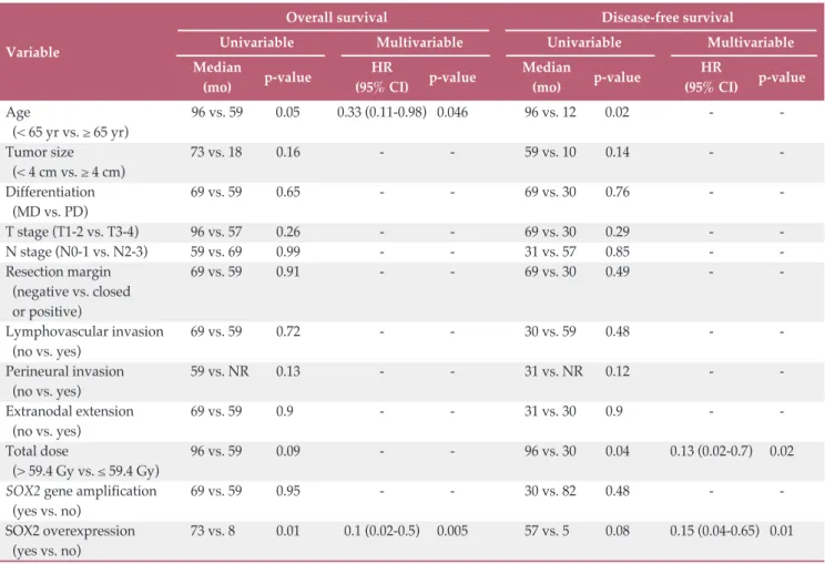
The median follow-up period for surviving patients was 58 months (range, 5 to 102 months). SOX2 gene amplification was observed in 22 patients and protein overexpression in 26 patients. SOX2 overexpression showed significant association with SOX2 gene amplification (p=0.002). In multivariate analysis, SOX2 overexpression was a significant prognostic factor for overall survival (OS) (hazard ratios [HR], 0.1; 95% confidence interval [CI], 0.002 to 0.5; p=0.005) and disease-free survival (DFS) (HR, 0.15; 95% CI, 0.04 to 0.65; p=0.01). Age (HR, 0.33; 95% CI, 0.11 to 0.98; p=0.046) and total radiation dose (HR, 0.13; 95% CI, 0.02 to 0.7; p=0.02) were the independent prognostic factors for OS and DFS. Patients with SOX2 amplification did not show a longer OS (p=0.95) and DFS (p=0.48).
Conclusion
Our data suggested that SOX2 overexpression could be used as a positive prognostic factor in patients with stage III lung SCC receiving adjuvant radiotherapy.
Key words
Overexpression, SOX-2, Carcinoma, Squamous cell, Lung neoplasms, Radiotherapy
Introduction
Lung cancer has one of the highest cancer mortality rates in several countries worldwide, including Korea [1,2]. Although cancer treatments have improved in recent decades, lung cancer remains non-responsive to curative
treatments. However, novel emerging molecular techniques have made it possible to investigate its molecular-level pathophysiology, identify specific causative oncogenes or tumor suppressor genes, and make progress toward new paradigms for cancer treatment. Compared with other can-cers, lung cancer has an increased number of well-defined genetic changes and/or abnormalities and more molecularly + + + + + + + + + + + + + + + + + + + + + + + + + + + + + + + + + + + + + + + + + + + + + + + + + + + + + + + + + + + + + + + + + + + + + + + + + + + + + + + + + + + + + + + + + + + + + + + + + + + + + + + + + + + + + + + + + + + + + + + + + + + + + + + + + + + + + + + + + + + + + + + + + + + + + + + + + + + + + + + + + + + + + + + + + + + + + + + + + + + + + + + + + + + + + + + + + + + + + + + + + + + + + + + + + + + + + + + + + + + + + + + + + + + + + + + + + + + + + + + + + + + + + + + + + + + + + + + + + + + + + + + + + + + + + + + + + + + + + + + + + + + + + + + + + + + + + + + + + + + +
Correspondence: Yong Bae Kim, MD, PhD Department of Radiation Oncology, Yonsei Cancer Center,
Yonsei University College of Medicine, 50-1 Yonsei-ro, Seodaemun-gu, Seoul 03722, Korea Tel: 82-2-2228-8095 Fax: 82-2-2227-7823 E-mail: [email protected] Received March 26, 2015 Accepted July 15, 2015
Published Online August 12, 2015
Hong In Yoon,
MD1,2Kyu Hyun Park,
PhD3Eun-Jung Lee,
PhD2Ki Chang Keum,
MD, PhD2Chang Geol Lee,
MD, PhD2Chul Hoon Kim,
MD, PhD1Yong Bae Kim,
MD, PhD2,31Department of Pharmacology,
Brain Korea 21 PLUS Project for Medical Science, Yonsei University College of Medicine, Seoul,
2Department of Radiation Oncology,
Yonsei Cancer Center,
Yonsei University College of Medicine, Seoul,
3Yonsei Song-Dang Institute for
Cancer Research, Yonsei University College of Medicine, Seoul, Korea
targeted agents have been applied in clinical practice for its treatment. However, most of the molecularly targeted agents used in clinical practice are for adenocarcinoma, and few are available for squamous cell carcinoma.
SOX2, a transcription factor encoded by the gene located at 3q26.33, plays a role in maintenance of embryonic stem cells. In addition, it is a “Yamanaka factor” which induces pluripotent stem cells from somatic cells, along with c-Myc, Oct4, and KLF4. It is also involved in morphogenesis and homeostasis of the esophageal, tracheobronchial, and bron-chiolar epithelium [3].
SOX2 is expressed in bronchial epithelial cells, though not in alveolar cells or adenocarcinoma precursor lesions [4-6]. SOX2 is found exclusively in squamous cell carcinoma, where it is amplified and overexpressed at the gene and pro-tein levels, respectively [3,6]. Previous studies have analyzed the prognosis of heterogeneously treated squamous cell car-cinoma patient populations. However, only a small number of studies have investigated the association between SOX2 and the clinical outcome of patients treated with radiother-apy [7,8]. The aim of this study was to assess the prognostic significance of SOX2 amplification and expression in squa-mous cell lung cancer patients treated using adjuvant radio-therapy.
Materials and Methods
1. Patient selectionThis retrospective study was approved by the Institutional Review Board of our institution (IRB No. 4-2012-0709). A total of 158 non-small cell lung cancer (NSCLC) patients who underwent pulmonary resection followed by adjuvant radio-therapy between 1996 and 2008 were identified. Squamous cell carcinoma was confirmed surgically in 71 patients, and clinical specimens were available from 36 patients. Of these, three patients were excluded from the analysis because of a lack of tumor tissue in their paraffin-embedded tissue blocks. Therefore, tumor samples from 33 patients were available for analysis. The patients’ medical records were reviewed retro-spectively for evaluation of clinicopathological characteris-tics and survival outcomes. After radical resection, all patients were determined to have pathologic TNM stage III tumors according to the sixth edition cancer staging guide-lines provided by the American Joint Committee on Cancer [9].
2. Fluorescent in situ hybridization
Fluorescent in situ hybridization (FISH) was performed for analysis of SOX2 gene amplifications on 4-!m-thick forma-lin-fixed paraffin embedded (FFPE) tissue sections. Briefly, the sections were deparaffinized in xylene (twice for 10 min-utes each), followed by immersion in 100% ethanol (twice for 5 minutes each) before hybridization. Pretreatment was per-formed according to the Vysis protocol for FFPE tissue spec-imens. A SOX2-specific DNA probe (green) and a centrom-ere 3-specific probe (red) wcentrom-ere used (ZytoLight SPEC SOX-2/CEN 3 Dual Color Probe, ZytoVision, Bremerhaven, Ger-many). The sections were then denatured and hybridized with ThermoBrite (Abbott Molecular, Des Plaines, IL) at 80°C for 5 minutes, followed by an overnight at 37°C. Post-hybr-idization washes were performed according to the Vysis pro-tocol for FFPE tissue specimens (Abbott Molecular), and DAPI counterstaining was then performed. Semi-quantita-tive analysis of the SOX2 amplification status was performed by comparing the number of green signals (SOX2 target regions) to the number of red signals in each sample (refer-ence regions). A non-amplified nucleus showed one green target signal for every corresponding red reference signal, with a green/red ratio of 1:1 (Fig. 1A). Cases containing 2-9 more green target signals than red signals in at least 30% of their tumor cells were defined as having low-level SOX2 amplification (Fig. 1B). Cases with an additional ! 10 green signals in a cluster-like formation were defined as having high-level SOX2 amplification (Fig. 1C). All tissue slides were analyzed under a 100" oil immersion objective lens using a fluorescence microscope equipped with the appro-priate filters. At least 100 nuclei per case were assessed.
3. Immunohistochemistry
Four-micrometer-thick tissue sections were deparaffinized, rehydrated, and washed twice in buffer. The slides were incubated in hydrogen peroxide for 10 minutes to reduce nonspecific background staining due to endogenous perox-idases, and then washed four times in buffer. Primary anti-bodies against human SOX2 (1:200, R&D Systems, Minnea-polis, MN) were then applied, and slides were incubated according to the manufacturer’s recommended protocols. The slides were washed four times in buffer, incubated with primary antibody enhancer for 20 minutes at room temper-ature, and then washed four times in buffer. Next, horserad-ish peroxidase polymer was applied to the slides, which were then incubated for 30 minutes at room temperature before washing four times in buffer, followed by incubation with hematoxylin and chromogen, washed four times in deion-ized water, and counterstained. The staining results were evaluated and each case was scored from 0 to 2, according to
the intensity of nuclear staining in the tumor cells. The stain-ing intensity of the normal bronchial epithelium served as the internal control and was given an arbitrary score of 1. Each tumor was compared to the internal control and given a score of 1 (moderate expression) (Fig. 1D) when the inten-sity was the same as that of the internal control, 2 (strong expression) (Fig. 1E) when stronger, and 0 (weak expression) (Fig. 1F) when weaker.
4. Statistical analyses
Disease-free survival (DFS) was estimated from the time of diagnosis to the time of initial tumor relapse (local recur-rence or distant) or death from any cause. Overall survival (OS) time was measured from the time of diagnosis to death or last follow-up date. For univariate analysis, Kaplan-Meier survival analyses were used to estimate OS and DFS, and dif-ferences in the survival rates were compared using log-rank tests. A Cox proportional hazards model was used for
mul-tivariate analysis to evaluate prognostic factors influencing OS and DFS. Multivariate analysis was performed using backwards elimination to stay in the model. Hazard ratios (HR) are given with 95% confidence intervals (95% CI). Cor-relations between categorical variables were examined using !2or Fisher exact tests. Continuous variables were compared to categorical variables using Mann-Whitney U tests. Statis-tical significance was defined as a p-value of < 0.05 for all analyses. SPSS ver. 20.0 (IBM Co., Armonk, NY) was utilized for all statistical analyses.
Results
1. Patient characteristics
The median patient age was 66 years (range, 48 to 73 A D B E C F Fig. 1. SOX2 amplification was assessed using fluorescence in situ hybridization (FISH, "1,000) and protein expression was
determined using immunohistochemistry ("200) in lung squamous cell carcinoma patients. SOX2-specific DNA probe in green combined with a centromere 3-specific probe in red was applied for FISH. (A) Nucleus without SOX2 amplification. (B) Nucleus with low-level SOX2 amplification (arrows). (C) Nucleus with high-level SOX2 amplification (arrows). (D) Mod-erate nuclear SOX2 expression (arrow). (E) Strong nuclear SOX2 expression (arrow). (F) Weak nuclear SOX2 expression.
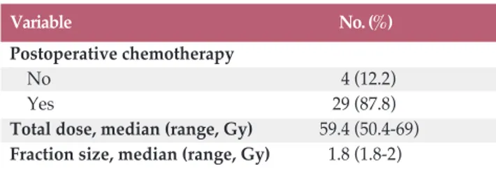
years), and 32 patients were male (97%). Most patients had an Eastern Cooperative Oncology Group (ECOG) perform-ance status of 0 or 1 (93.9%) and a positive smoking history (75.8%). Fourteen patients (42.4%) were classified as T3 or T4, and 27 patients (81.8%) as N2 or N3. A positive margin of resection was found in 11 patients (33.3%), lymphovascu-lar invasion and perineural invasion in seven patients (21.2%) and three patients (9.1%), and extranodal extension in four patients (12.1%). The median total dose and fraction size of radiotherapy were 59.4 Gy (range, 50.4 to 69 Gy) and 1.8 Gy (range, 1.8 to 2 Gy), respectively. The patient charac-teristics are listed in Table 1.
2. Correlation between SOX2 gene amplification and pro-tein expression and clinicopathological characteristics
SOX2 amplification was assessed using FISH and protein
expression was determined using immunohistochemistry in lung squamous cell carcinoma patients (Fig. 1). High- and low-level amplification were observed in four (12.2%) and 18 patients (54.5%), respectively (Table 1). SOX2 gene amplifi-cation group was defined as low or high level amplifiamplifi-cation. Strong and moderate expressions were observed in eight (24.3%) and 18 patients (54.5%), respectively (Table 1). SOX2 overexpression was defined as a score of 1 or 2, and no over-expression was defined as a score of 0.
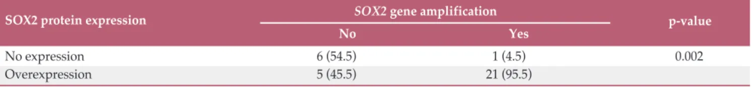
Analysis of the correlation between SOX2 gene amplifica-tion and protein expression showed significant associaamplifica-tion of SOX2 overexpression with SOX2 gene amplification (p=0.002) (Table 2). Next, we analyzed the association betw-een several clinicopathological characteristics and SOX2 gene amplification and protein expression. However, no clinico-pathological factor showed significant association with SOX2 gene amplification or protein expression (Table 3).
3. Prognostic factors for OS and DFS
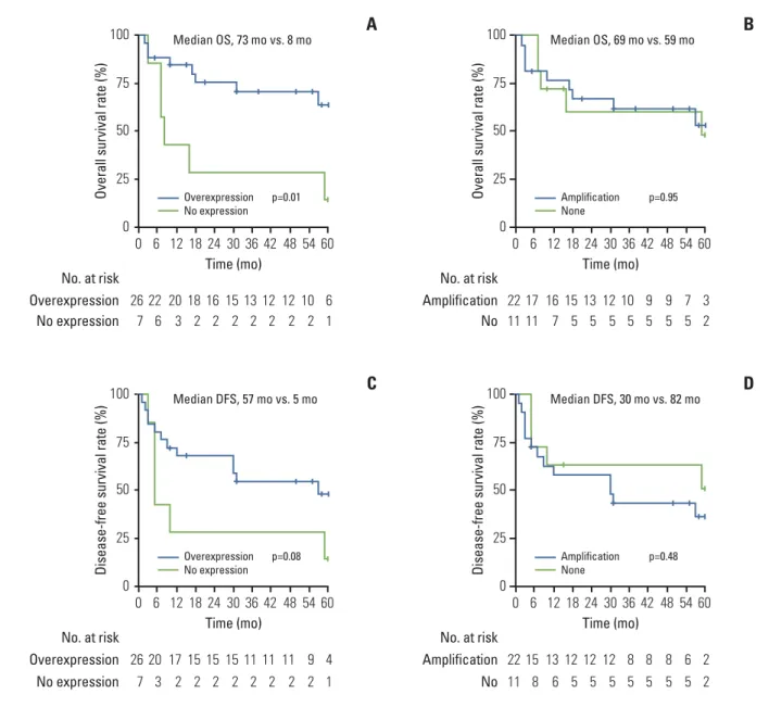
The median follow-up period for the surviving patients was 58 months (range, 5 to 102 months). The 5-year OS and DFS for all patients was 50.7% and 39.9%, respectively. In prognostic analysis using Kaplan-Meier curves, SOX2 over-expression was the only significant prognostic factor for OS (yes vs. no; median, 73 months vs. 8 months; p=0.01) (Table 4). Kaplan-Meier curves also showed that patients with SOX2 overexpression had significantly better OS, as shown in Fig. 2A. Age and total dose showed better OS with a statisti-cally significant trend. However, SOX2 amplification did not show association with OS (Table 4, Fig. 2B). In multivariate analysis, SOX2 overexpression (HR, 0.1; 95% CI, 0.02 to 0.5; p=0.005) and age (HR, 0.33; 95% CI, 0.11 to 0.98; p=0.046) were the independent prognostic factors for OS (Table 4). In univariate analysis, age (< 65 vs. ! 65; median, 96 months vs.
12 months; p=0.02) and total radiation dose (> 59.4 Gy vs. # 59.4 Gy; median, 96 months vs. 30 months; p=0.04) were the significant prognostic factors for DFS (Table 4). SOX2 overexpression showed a significant trend toward better DFS (p=0.08) (Table 4, Fig. 2C). SOX2 amplification also did not show association with DFS (Table 4, Fig. 2D). In multivariate analysis, SOX2 overexpression (HR, 0.15; 95% CI, 0.04 to 0.65; p=0.01) and total radiation dose (HR, 0.13; 95% CI, 0.02 to 0.7; p=0.02) were the independent prognostic factors for DFS (Table 4).
Discussion
SOX2 plays roles in a variety of pathological processes from normal development to cancer. Overexpression of SOX2 is not associated with epithelial cell differentiation in the lungs, but its expression shows a notable increase in basal cell precursors (p63+ Krt14$ cells) [10]. Therefore, SOX2 reg-ulates the balance between self-renewal and differentiation by controlling the fate of basal cells and also triggers lung epithelial carcinogenesis [11].
SOX2 play a role in various signal transduction pathways and exerts various biological effects via extensive networks involving upstream and downstream molecules and protein interactions [12]. Many proteins upstream of SOX2 have been identified. EGFR-Src-Akt signaling is relevant to the self-renewal of cancer stem cells and has an effect on side popu-lation cells in lung cancer [13]. Several downstream effectors of SOX2 have been identified including those involved in cancer stem cell-related signal transduction pathways. For example, C-MYC, WNT1, WNT2, and NOTCH1 play roles in non-small cell lung cancer cells [14]. SOX2 was linked with apoptosis in non-small cell lung cancer via the MAPK4-sur-vivin pathway [15]. SOX2 forms a network with cell cycle regulators including cyclin D1, cyclin E, and p27, as well as other biologically important pathways such as "-catenin and transforming growth factor " [16-18].
Little is known about the relationship between radioresis-tance and SOX2. An analysis of the SOX2 interactome using immunoprecipitation coupled with mass spectrometry analysis by Fang et al. [19] found interactions between SOX2 and proteins involved in DNA repair, such as XRCC1, 5, and 6, which were associated with radioresistance in vitro and in human tissues [20-22]. Future studies are needed to investi-gate the direct relationship between radioresistance and these genes. In addition, cancer stem cell radioresistance can be explained by an increased recovery from DNA damage and a decrease in production of reactive oxygen species [23,24]. Therefore, SOX2 might influence radioresistance
through regulation of cancer stem cell activity. If SOX2 increases radioresistance, SOX2 overexpression would result in worse prognosis in irradiated patients, in contrast to our findings. However, all patients enrolled in our study under-went surgery followed by postoperative radiotherapy, and most patients received adjuvant systemic chemotherapy. Thus, we concluded that our findings were not appropriate for investigating the relation between SOX2 and radioresis-tance. Instead, we suggest that further studies designed for decisive examination of this point, such as a prospective study with lung squamous cell carcinoma patients who receive definitive radiotherapy, are required.
Several previous studies investigated whether SOX2 amp-lification was associated with prognosis in non-small cell lung cancer. Although some reported that SOX2 amplifica-tion showed close correlaamplifica-tion with poor prognosis, others reported that patients with amplification had a good survival outcome. These conflicting results arise from the limited availability of patient tissues and methods for investigation of SOX2 amplification. However, our findings showed that
SOX2 amplification is not associated with survival.
A meta-analysis utilizing the eight published series found significantly greater expression of SOX2 in squamous carci-noma than in adenocarcicarci-noma, which was associated with improved OS. Similarly, multivariate analysis also showed that SOX2 overexpression is the significant prognostic factor for OS and DFS. Therefore, we suggest that overexpression of SOX2 might be a positive prognostic factor in patients with lung squamous cell carcinoma. However, as several studies showed that the use of small interfering RNA to knockdown SOX2 decreased the growth and radioresistance of cancer cells in contrast to clinical studies [18,25], it remains unclear whether this could result in a feasible therapeutic approach. Therefore, further studies involving well-designed transla-tional research from benchside to bedside are needed.
The current study included a homogenous NSCLC patient population treated with adjuvant radiotherapy. To the best of our knowledge, this is the first study to investigate whe-ther SOX2 overexpression has prognostic significance in
Table 1. Patient characteristics
Variable No. (%)
Age, median (range, yr) 66 (48-73) Tumor size, median (range, cm) 4 (2-7) Sex Male 32 (97) Female 1 (3) ECOG 0 25 (75.7) 1 6 (18.2) 2 2 (6.1) Smoking history No 8 (24.2) Yes 25 (75.8) Radical resection Lobectomy 23 (69.7) Pneumonectomy 10 (30.3) pT stage T1 3 (9.1) T2 16 (48.5) T3 7 (21.2) T4 7 (21.2) pN stage N0 1 (3.0) N1 5 (15.2) N2 24 (72.7) N3 3 (9.1) pStage IIIa 23 (69.7) IIIb 10 (30.3) Grade MD 15 (45.5) PD 18 (54.5) Resection margin Negative 21 (63.6) Closed 1 (3.0) Positive 11 (33.3) Lymphovascular invasion No 26 (78.8) Yes 7 (21.2) Perineural invasion No 30 (90.9) Yes 3 (9.1) Extranodal extension No 29 (87.9) Yes 4 (12.1)
SOX2 gene amplification
None 11 (33.3)
Low 18 (54.5)
High 4 (12.2)
SOX2 protein expression
Weak 7 (21.2) Moderate 18 (54.5) Strong 8 (24.3) Table 1. Continued Variable No. (%) Postoperative chemotherapy No 4 (12.2) Yes 29 (87.8)
Total dose, median (range, Gy) 59.4 (50.4-69) Fraction size, median (range, Gy) 1.8 (1.8-2)
ECOG, Eastern Cooperative Oncology Group; MD, mod-erately differentiated; PD, poorly differentiated.
Table 2. Relationship between SOX2 gene amplification and SOX2 protein expression
Table 3. Clinicopathologic characteristics according to SOX2 amplification and expression
Variable SOX2 gene amplification SOX2 protein expression
None (n=11) Amplification (n=22) p-value No (n=7) Overexpression (n=26) p-value Sex Male 10 (90.9) 22 (100) 0.33 6 (85.7) 26 (100) 0.2 Female 1 (9.1) 0 (0.0) 1 (14.3) 0 (0.0) Age (yr) < 65 7 (63.6) 8 (36.4) 0.14 4 (57.1) 11 (42.3) 0.67 ! 65 4 (36.4) 14 (63.6) 3 (42.9) 15 (57.7) Smoking history No 4 (36.4) 4 (18.2) 0.39 3 (42.9) 5 (19.2) 0.32 Yes 7 (63.6) 18 (81.8) 4 (57.1) 21 (80.8) Tumor size (cm) < 4 4 (36.4) 9 (40.9) 1 1 (14.3) 12 (46.2) 0.2 ! 4 7 (63.6) 13 (59.1) 6 (85.7) 14 (53.8) Differentiation MD 5 (45.5) 10 (45.5) 1 4 (57.1) 11 (42.3) 0.67 PD 6 (54.5) 12 (54.5) 3 (42.9) 15 (57.7) TNM stage IIIa 9 (81.8) 14 (63.6) 0.43 6 (85.7) 17 (65.4) 0.4 IIIb 2 (18.2) 8 (36.4) 1 (14.3) 9 (34.6) T stage T1-2 6 (54.5) 13 (59.1) 1 3 (42.9) 16 (61.5) 0.42 T3-4 5 (45.5) 9 (40.9) 4 (57.1) 10 (38.5) N stage N0-1 3 (27.3) 3 (13.6) 0.38 2 (28.6) 4 (15.4) 0.58 N2-3 8 (72.7) 19 (86.4) 5 (71.4) 22 (84.6) Resection margin Negative 7 (63.6) 14 (63.6) 1 5 (71.4) 16 (61.5) 1 Closed or positive 4 (36.4) 8 (36.4) 2 (28.6) 10 (38.5) Lymphovascular invasion No 8 (72.7) 18 (81.8) 0.66 5 (71.4) 21 (80.8) 0.62 Yes 3 (27.3) 4 (18.2) 2 (28.6) 5 (19.2) Perineural invasion No 11 (100) 19 (86.4) 0.53 7 (100) 23 (88.5) 1 Yes 0 (0.0) 3 (13.6) 0(0.0) 3 (11.5) Extranodal extension No 10 (90.9) 19 (86.4) 1 6 (85.7) 23 (88.5) 1 Yes 1 (9.1) 3 (13.6) 1 (14.3) 3 (11.5)
Values are presented as number (%). MD, moderately differentiated; PD, poorly differentiated.
SOX2 protein expression SOX2 gene amplification p-value
No Yes
No expression 6 (54.5) 1 (4.5) 0.002
Overexpression 5 (45.5) 21 (95.5)
patients who underwent radiotherapy in NSCLC, albeit only a small number of patients were included. Nevertheless, despite the high possibility, we cannot assume that postop-erative radiotherapy changed the prognosis of patients and that SOX2 is involved in this effect. We were also unable to confirm a relationship between SOX2 and radioresistance. Thus, further studies are certainly needed in order to validate
the effect of postoperative radiotherapy on prognosis and the relationship between radiotherapy and SOX2. Still, based on our findings, it is meaningful that the prognostic value of SOX2 overexpression was verified by utilizing the homoge-nous database of all patients treated with the whole defini-tive treatment scheme including radical surgery followed by postoperative radiotherapy. Ov er al l s ur vi va l r at e (% ) 100 75 50 25 0 100 75 50 25 0 100 75 50 25 0 100 75 50 25 0 Overexpression No. at risk 60 Time (mo) 24 30 36 42 48 54 18 12 6 0 6 16 15 13 12 12 10 18 20 22 26 1 2 2 2 2 2 2 2 3 6 7 No expression p=0.01 Median OS, 73 mo vs. 8 mo
A
Overexpression No expression Ov er al l s ur vi va l r at e (% ) Amplification No. at risk 60 Time (mo) 24 30 36 42 48 54 18 12 6 0 3 13 12 10 9 9 7 15 16 17 22 2 5 5 5 5 5 5 5 7 11 11 No p=0.95 Median OS, 69 mo vs. 59 moB
Amplification None Di se as e-fre e su rv iv al ra te (% ) Overexpression No. at risk 60 Time (mo) 24 30 36 42 48 54 18 12 6 0 4 15 15 11 11 11 9 15 17 20 26 1 2 2 2 2 2 2 2 2 3 7 No expression p=0.08 Median DFS, 57 mo vs. 5 moC
Overexpression No expression Dise as e-fre e su rv iv al ra te (% ) Amplification No. at risk 60 Time (mo) 24 30 36 42 48 54 18 12 6 0 2 12 12 8 8 8 6 12 13 15 22 2 5 5 5 5 5 5 5 6 8 11 No p=0.48 Median DFS, 30 mo vs. 82 moD
Amplification NoneFig. 2. Kaplan-Meier curves for overall survival and disease-free survival rates. (A) Patients with SOX2 overexpression
showed a significantly longer overall survival rate compared to those without (median, 73 months vs. 8 months, respectively; p=0.01). (B) Patients with SOX2 amplification did not show a better overall survival rate than those without amplification (median, 69 months vs. 59 months; p=0.95). (C) Patients with SOX2 overexpression showed a significant trend toward longer disease-free survival compared to those without (median, 57 months vs. 5 months; p=0.08). (D) Patients with SOX2 ampli-fication did not show a longer disease-free survival than those without ampliampli-fication (median, 30 months vs. 82 months; p=0.48).
This study has several shortcomings. Primarily, it was ret-rospective in design and included only a small number of patients who underwent postoperative radiotherapy. We thought that SOX2 overexpression showed a significant trend toward better DFS on the Kaplan-Meier curve in Fig. 2C due to the small number of patients in spite of signif-icant relationship between SOX2 overexpression and pro-gression-free survival in multivariate analysis. A well-designed large-scale prospective study is necessary to con-firm our results. In addition, despite a very strong positive relationship between SOX2 protein overexpression and gene amplification, gene amplification of SOX2 did not show sig-nificant association with OS or DFS, in contrast to SOX2 over-expression. Similarly, in a recent large retrospective study, only SOX2 overexpression showed significant association with better overall survival, although significant positive cor-relation was observed between SOX2 protein overexpression and gene amplification [8]. Furthermore, from an indepth review of our data, five patients with SOX2 overexpression
without gene amplification showed an excellent outcome of median OS (60 months). Based on these findings, we suggest that a high SOX2 protein level may affect the prognosis of lung squamous cell carcinoma patients more acutely than
SOX2 gene amplification. Consequently, we recommend an
evaluation of the mechanism of SOX2 overexpression with no gene amplification. Thus, further studies investigating other mechanisms of SOX2 protein overexpression, such as chromosomal translocation or mutation without gene ampli-fication, would also be necessary.
Conclusion
In conclusion, SOX2 overexpression could be a positive prognostic factor in lung squamous cell carcinoma patients treated with postoperative adjuvant radiotherapy. Further
Table 4. Stepwise uni- and multi-variate analysis using Cox regression model for overall survival and disease-free survival
Overall survival Disease-free survival
Variable Univariable Multivariable Univariable Multivariable
Median
p-value HR p-value Median p-value HR p-value
(mo) (95% CI) (mo) (95% CI)
Age 96 vs. 59 0.05 0.33 (0.11-0.98) 0.046 96 vs. 12 0.02 - -(< 65 yr vs. ! 65 yr) Tumor size 73 vs. 18 0.16 - - 59 vs. 10 0.14 - -(< 4 cm vs. ! 4 cm) Differentiation 69 vs. 59 0.65 - - 69 vs. 30 0.76 - -(MD vs. PD) T stage (T1-2 vs. T3-4) 96 vs. 57 0.26 - - 69 vs. 30 0.29 - -N stage (-N0-1 vs. -N2-3) 59 vs. 69 0.99 - - 31 vs. 57 0.85 - -Resection margin 69 vs. 59 0.91 - - 69 vs. 30 0.49 - -(negative vs. closed or positive) Lymphovascular invasion 69 vs. 59 0.72 - - 30 vs. 59 0.48 - -(no vs. yes) Perineural invasion 59 vs. NR 0.13 - - 31 vs. NR 0.12 - -(no vs. yes) Extranodal extension 69 vs. 59 0.9 - - 31 vs. 30 0.9 - -(no vs. yes) Total dose 96 vs. 59 0.09 - - 96 vs. 30 0.04 0.13 (0.02-0.7) 0.02 (> 59.4 Gy vs. # 59.4 Gy)
SOX2 gene amplification 69 vs. 59 0.95 - - 30 vs. 82 0.48 -
-(yes vs. no)
SOX2 overexpression 73 vs. 8 0.01 0.1 (0.02-0.5) 0.005 57 vs. 5 0.08 0.15 (0.04-0.65) 0.01 (yes vs. no)
1. Jung KW, Park S, Won YJ, Kong HJ, Lee JY, Seo HG, et al. Pre-diction of cancer incidence and mortality in Korea, 2012. Can-cer Res Treat. 2012;44:25-31.
2. Jemal A, Bray F, Center MM, Ferlay J, Ward E, Forman D. Global cancer statistics. CA Cancer J Clin. 2011;61:69-90. 3. Bass AJ, Watanabe H, Mermel CH, Yu S, Perner S, Verhaak
RG, et al. SOX2 is an amplified lineage-survival oncogene in lung and esophageal squamous cell carcinomas. Nat Genet. 2009;41:1238-42.
4. Sutherland KD, Berns A. Cell of origin of lung cancer. Mol Oncol. 2010;4:397-403.
5. Long KB, Hornick JL. SOX2 is highly expressed in squamous cell carcinomas of the gastrointestinal tract. Hum Pathol. 2009; 40:1768-73.
6. Yuan P, Kadara H, Behrens C, Tang X, Woods D, Solis LM, et al. Sex determining region Y-Box 2 (SOX2) is a potential cell-lineage gene highly expressed in the pathogenesis of squa-mous cell carcinomas of the lung. PLoS One. 2010;5:e9112. 7. Velcheti V, Schalper K, Yao X, Cheng H, Kocoglu M,
Dhodap-kar K, et al. High SOX2 levels predict better outcome in non-small cell lung carcinomas. PLoS One. 2013;8:e61427. 8. Wilbertz T, Wagner P, Petersen K, Stiedl AC, Scheble VJ, Maier
S, et al. SOX2 gene amplification and protein overexpression are associated with better outcome in squamous cell lung can-cer. Mod Pathol. 2011;24:944-53.
9. Greene FL, Page DL, Fleming ID, Fritz AG, Balch CM, Haller DG, et al. AJCC cancer staging manual. 6th ed. New York: Springer; 2002.
10. Gontan C, de Munck A, Vermeij M, Grosveld F, Tibboel D, Rottier R. Sox2 is important for two crucial processes in lung development: branching morphogenesis and epithelial cell differentiation. Dev Biol. 2008;317:296-309.
11. Hussenet T, du Manoir S. SOX2 in squamous cell carcinoma: amplifying a pleiotropic oncogene along carcinogenesis. Cell Cycle. 2010;9:1480-6.
12. Liu K, Lin B, Zhao M, Yang X, Chen M, Gao A, et al. The mul-tiple roles for Sox2 in stem cell maintenance and tumorigene-sis. Cell Signal. 2013;25:1264-71.
13. Singh S, Trevino J, Bora-Singhal N, Coppola D, Haura E, Altiok S, et al. EGFR/Src/Akt signaling modulates Sox2 expression and self-renewal of stem-like side-population cells in non-small cell lung cancer. Mol Cancer. 2012;11:73. 14. Chen S, Xu Y, Chen Y, Li X, Mou W, Wang L, et al. SOX2 gene
regulates the transcriptional network of oncogenes and affects tumorigenesis of human lung cancer cells. PLoS One. 2012;7: e36326.
15. Chen S, Li X, Lu D, Xu Y, Mou W, Wang L, et al. SOX2 regu-lates apoptosis through MAP4K4-survivin signaling pathway in human lung cancer cells. Carcinogenesis. 2014;35:613-23. 16. Wu F, Zhang J, Wang P, Ye X, Jung K, Bone KM, et al.
Identi-fication of two novel phenotypically distinct breast cancer cell subsets based on Sox2 transcription activity. Cell Signal. 2012; 24:1989-98.
17. Lin F, Lin P, Zhao D, Chen Y, Xiao L, Qin W, et al. Sox2 targets cyclinE, p27 and survivin to regulate androgen-independent human prostate cancer cell proliferation and apoptosis. Cell Prolif. 2012;45:207-16.
18. Chen Y, Shi L, Zhang L, Li R, Liang J, Yu W, et al. The molec-ular mechanism governing the oncogenic potential of SOX2 in breast cancer. J Biol Chem. 2008;283:17969-78.
19. Fang X, Yoon JG, Li L, Tsai YS, Zheng S, Hood L, et al. Land-scape of the SOX2 protein-protein interactome. Proteomics. 2011;11:921-34.
20. Niu Y, Zhang X, Zheng Y, Zhang R. XRCC1 deficiency incr-eased the DNA damage induced by gamma-ray in HepG2 cell: involvement of DSB repair and cell cycle arrest. Environ Tox-icol Pharmacol. 2013;36:311-9.
21. Chang HW, Kim SY, Yi SL, Son SH, Song DY, Moon SY, et al. Expression of Ku80 correlates with sensitivities to radiation in cancer cell lines of the head and neck. Oral Oncol. 2006;42: 979-86.
22. Ayene IS, Ford LP, Koch CJ. Ku protein targeting by Ku70 small interfering RNA enhances human cancer cell response to topoisomerase II inhibitor and gamma radiation. Mol Can-cer Ther. 2005;4:529-36.
23. Bao S, Wu Q, McLendon RE, Hao Y, Shi Q, Hjelmeland AB, et
References
studies are needed to investigate whether SOX2 is associated with radioresistance and how this could be exploited thera-peutically.
Conflicts of Interest
Conflict of interest relevant to this article was not reported.
Acknowledgments
This study was supported by a faculty research grant from Yonsei University College of Medicine for 2012(6-2012-0187). We thank Jin-Kyu Park and Korea CFC pathology laboratory to support our study.
al. Glioma stem cells promote radioresistance by preferential activation of the DNA damage response. Nature. 2006;444: 756-60.
24. Diehn M, Cho RW, Lobo NA, Kalisky T, Dorie MJ, Kulp AN, et al. Association of reactive oxygen species levels and
radiore-sistance in cancer stem cells. Nature. 2009;458:780-3. 25. Wang X, Ji X, Chen J, Yan D, Zhang Z, Wang Q, et al. SOX2
enhances the migration and invasion of ovarian cancer cells via Src kinase. PLoS One. 2014;9:e99594.