Efficient differentiation of human pluripotent
stem cells into neural lineage by modulation of
BMP and Activin/Nodal signaling pathways
Dae-Sung Kim
Department of Medical Science
Efficient differentiation of human pluripotent
stem cells into neural lineage by modulation of
BMP and Activin/Nodal signaling pathways
Directed by Professor Dong-Wook Kim
The Doctoral Dissertation
submitted to the Department of Medical Science
the Graduate School of Yonsei University
in partial fulfillment of the requirements for the degree of
Doctor of Philosophy
Dae-Sung Kim
This certifies that the Doctoral Dissertation
of Dae-Sung Kim is approved
Thesis Supervisor : Dong-Wook Kim
Thesis Committee Member : Kook In Park
Thesis Committee Member : Jin Woo Lee
Thesis Committee Member : Dong-Youn Hwang
Thesis Committee Member : Han-Sung Jung
The Graduate School
Yonsei University
Acknowledgements
I am really grateful to my thesis advisor, Prof. Dong-Wook Kim for
his continuous support, encouragement and precious guidance through my
graduate study. And I appreciate Dr. Dong-Yeon Hwang, Dr. Han Soo Kim
who provided helpful advice in preparing manuscript and Prof. Kook In Park,
Prof. Jin Woo Lee and Prof. Han-Sung Jung for serving on my committee. I
would like to thank to colleagues in my laboratory, Dr. Hye-Yeong Ha, Dr.
Hoon Chul Kang, Ji Young Kim, Yong Joon Huh and Young-Eun Lee for
many technical assistance and showing the companionship just like a family. I
also want to express thanks to my old high school teacher, Jae Ho Yu who
inspired me to open my eyes to biology.
And finally, I would like to express all my gratitude to my parents and
sisters who give me infinite support and sincere care through my life.
Table of contents
Abstract---1
I. Introduction---5
II. Materials and Methods ---10
1. Human ES and iPS cell culture ---10
2. Differentiation of Human ES and iPS cells ---10
3. Western Blotting for detection of phosphorylation of Smad 1/5/8 ---11
4. Immunostainig and Quantitative analysis ---12
5. Quantitative RT-PCR and data analysis ---13
III. Results
1. Inhibition of BMP signaling by a small molecule, Dorsomorphin, enhances early neuroectodermal marker expression, but decrease other lineage marker expression in human embryonic stem cells---16
2. Suppression of Activin/Nodal signaling pathway inhibits mesodermal, endodermal and undifferentiated marker expressions, but not ectodermal marker expression ---24
3. Simultaneous inhibition of BMP and Activin/Nodal signals exclusively induce
neural differentiation from human ES cells---
---27
4. Simultaneous inhibition of BMP and Activin/Nodal signals facilitates neural differentiation from human pluripotent stem cells regardless of their own differentiation nature
---31
5. Neural precursors generated by treatment of BMP and Avtivin/Nodal signal inhibitors could differentiate into mature dopamine neurons with high yield
---41
IV. Discussion---47
V. Conclusion ---54
References ---55
Abstract (In KOREAN) ---60
LIST OF FIGURES
Figure 1. Dorsomorphin inhibits BMP-mediated activation of
Smad 1/5/8 by inhibiting BMP type I receptor function in human
ES cells.---19
Figure 2. Quantitative analyses of expression of markers
associated with the early embryonic development in EBs.---22
Figure 3. Effects of Activin/Nodal signal blockade by SB431542
on the expression of three germ layer markers in human ES cells.
---26
Figure 4. Simultaneous suppression of BMP and Activin/Nodal
signal pathway is sufficient to direct neural differentiation of
human ES cells.---29
Figure 5. Differentiation propensities among human pluripotent
stem cells.---33
Figure 6. Comparison of neural differentiation propensity among
cell lines by a well-characterized neural differentiation protocol
---36
Figure 7. Simultaneous blockade of BMP and Activin/Nodal
signal efficiently induced neural differentiation from various
human pluripotent stem cell lines with distinct differentiation
propensities.---39
Figure 8. Neural precursors generated by dual signal modulation
could differentiate efficiently into dopaminergic neurons.---44
Figure 9. Efficient induction of TH-positive neurons from human
pluripotent stem cells.---45
Efficient differentiation of human pluripotent stem cells into neural
lineage by modulation of BMP and Activin/Nodal signaling pathways
Dae-Sung Kim
Department of Medical Science
The Graduate School, Yonsei University
(Directed by Professor Dong-Wook Kim)
For therapeutic applications, efficient differentiation of human pluripotent stem cells [i.e. human embryonic stem cells (hESCs) and induced pluripotent stem (iPS) cells] into the specific cell type or cell lineage of interest is a prerequisite. Recent report demonstrated that each hESC line is skewed towards a certain differentiated lineage. We could also detect marked differences in differentiation propensity among 6 hESC lines generated by 4 institutions as well as 3 human iPS cell lines derived from 3 different types of somatic cells.
Since this innate propensity can often negatively affect differentiation into another cell lineage, all the hESCs as well as iPS cells need to be examined for their
differentiation propensity and then appropriate cell lines can be selected for each therapeutic application.This might be troublesome because thorough examinations of differentiation propensity of ever-increasing hESCs and iPS cell lines are laborious, time-consuming, and costly. Therefore, it would be of great benefit if there were a way to induce differentiation of all human pluripotent stem cells into a specific cell lineage of interest, irrespective of their original differentiation propensity.As a proof-of-principle experiment, we set out to establish a universal protocol that drives all the human pluripotent stem cell lines with various differentiation propensities toward the neural lineage. Our strategy to generate such a broadly applicable protocol for neural differentiation was by manipulating signaling pathways critically implicated in embryonic neural induction.
Since inhibition of the BMP signaling has been known to play a role in neural induction, BMP signal was suppressed by a specific inhibitor, dorsomorphin (DM) during spontaneous differentiation as embryoid bodies (EBs) in H9 hESC line. Treatment of 5µM DM during EB differentiation significantly increased neuroectodermal differentiation of hESCs, however it was not sufficient to reduce differentiation into meso/endodermal lineage and remnants of undifferentiated cells. Since Activin/Nodal signal is implicated in meso/endodermal differentiation and maintenance of pluripotency, the specific inhibitor of Activin/Nodal pathway, 10µM of SB431542 (SB) was added into the EB culture to reduce non-neural derivatives in
differentiating cells. Although the expression of meso- (Brachyury and Cerberus), endodermal (AFP and GATA4) and undifferentiation markers (Oct3/4 and Nanog) was dramatically reduced by treatment of SB, the neuroectodermal marker (Sox1, Pax6 and nestin) expression was not enhanced. We found that H9 hESCs could exclusively and efficiently differentiate into neuroectodermal lineage, if only BMP and Activin/Nodal signals were simultaneously blocked by combination of DM and SB during EB differentiation. The simultaneous modulation of BMP and Activin/Nodal signaling pathways with the small molecules could also efficiently overcome markedly different differentiation inclination from all 6 hES and 3 iPS cell lines tested. In addition, neural cells generated by the modulation of BMP and Activin/Nodal signaling also could efficiently generate dopaminergic neurons by a simple and commonly used dopaminergic differentiation protocol. In summary, human pluripotent stem cells with a variety of differentiation propensity can be efficiently differentiated into neural lineage by modulation of BMP and Activin/Nodal pathways. This study is very important from a clinical point of view since it would provide not only an efficient method to produce neural cells for treating neurological diseases but also a broadly applicable strategy to generate any type of desirable cells irrespective of innate differentiation potential of human ES and iPS cells.
____________________________________________________________________ Key Words : human embryonic stem cells (hESCs), induced pluripotent stem (iPS) cells, differentiation propensity, BMP signaling, Activin/Nodal signaling, small molecule, neural differentiation, dopaminergic neuron
Efficient differentiation of human pluripotent stem cells into neural
lineage by modulation of BMP and Activin/Nodal signaling pathway
Dae-Sung Kim
Department of Medical Science
The Graduate School, Yonsei University
(Directed by Professor Dong-Wook Kim)
I. Introduction
Human embryonic stem (ES) cells are pluripotent cells derived from the inner cell mass of preimplantation embryos1. Due to their origin and nature, ES cells provide a good model system for exploring human early development as well as the generation of cellular sources used for replacement therapies2. However, several limitations such as ethical problems, teratoma formation and immune rejection remained to be solved for therapeutic application.
Recently, new type of human pluripotent stem cell, induced pluripotent stem (iPS) cells, has been derived and was being charactereized3-5. These cells were created
by reprogramming somatic cells via viral introduction of defined genetic factors, and were known to share many characteristics of ES cells. Since they could be generated from patients, the advent of iPS cells was considered as a breakthrough for immune rejection faced in ES cells. Furthermore, as iPS cells do not require human eggs or destruction of preformed embryos, it is relatively free from ethical problems.
One of the practical goals of studying the differentiation of pluripotent stem cells is generating cells that can replace the function of disabled cells or tissues in incurable disease such as parkinson’s disease and spinal cord injury6. In case of the application of central nervous system (CNS) disorders, cells should be differentiated into neural lineage cells prior to transplantation. Up to date, a lot of differentiation techniques to specify human ES cells into neural lineages in vitro have been developed, including EB-based methods7 and coculture methods with stromal cells8. Although current advances are encouraging, it needs to be ameliorated in several aspects. For example, a few procedures take long time (over one month or more) to generate neural precursors8,9 or utilize non-human origin components concerning xeno-contamination8. Certain protocols are not applicable to all ES or pluripotent stem cell lines but limited to a particular cell line with low efficiency in generating neural precursors10. Furthermore, recent report demonstrated that each human ES cell line has its own differentiation inclination toward specific cell lineage, moreover directed differentiation methods could not efficiently overcome such propensity of
each cell line11. Since this innate propensity is often negatively affect differentiation into another cell lineage, all the hESCs as well as iPSCs need to be examined for their differentiation propensity and then appropriate cell lines are to be selected for each therapeutic application.This might be troublesome because thorough examinations of differentiation propensity of ever-increasing hESCs and iPSC lines are laborious, time-consuming, and costly.Therefore, it would be of great benefit if there is a way to induce differentiation of all pluripotent stem cells into a specific cell lineage of interest, irrespective of their original differentiation propensity. Establishment of standardized universal differentiation methods would also provide a great advance for practical application to every cell lines regardless of their origin (donor, ethnic background, derivation methods or source of pluripotent stem cells as in iPS cells) as well as differentiation propensity. In addition, a homogeneous population of neural precursors in a large scale would be a more desirable approach for both basic and applied scientific research12.
Here, we set out to develop a new and simple technique for neural differentiation of two types of human pluripotent stem cells – ES cells and iPS cells. To realize our purpose, two important issues were estimated for efficient differentiation. First, since differentiation of the pluripotent stem cells might be controlled through specific signaling pathways in similar manners of early embryogenesis, we assumed that neural differentiation would be facilitated through
the modulation of cellular signaling pathways involved in neural induction mechanism. Second, small molecules were used to control the differentiation of stem cells13. Small molecules are membrane permeable and freely diffused in differentiating cells irrespective of intracellular dimensions (e.g the 3 dimensional spherical structures or 2 dimensional-monolayer culture). Hence, it turn-on or -off the cellular signals so tightly that it could regulate the timing and process of differentiation.
We represent here that two important signaling pathway, BMP and Activin/Nodal signaling pathways are critical for the neuroectodermal differentiation in human ES and iPS cells, and simultaneous suppression of two signaling exclusively induce neural differentiation with less concerning about meso/endodermal contamination along with the remnants of undifferentiated cells. In addition, we also detected significant differences in differentiation propensity among 6 hESC lines generated by 4 institutions as well as 3 human iPSC lines derived from 3 different types of somatic cells. Moreover, our differentiation system was also efficient for directing pluripotent stem cells showing the inclination to meso- and/or endodermal differentiation toward neural lineage cells. Differentiated neural precursors could be efficiently induced neurons showing dopaminergic phenotypes by the treatment of signaling molecules and neurotrophic factors. Collectively, our results show here that simultaneous suppression of BMP and Activin/Nodal signals could exclusively
differentiate human pluripotent stem cells into neuroectodermal lineage. In addition modulation of cell signaling by small molecule is a very useful tool for the generation of homogeneous population of particular cell type and an alternative technique to overcome differentiation propensity among various pluripotent stem cell line, which provide a valuable mean to step toward the clinical application of human pluripotent stem cells.
II. Materials and Methods
1. Human ES and iPS cell culture
Human ES cell lines, H9 (p31-45, WiCell Inc., Madison, Wisconsin, USA), Miz-hES4 (p67-75) and Miz-hES6 (p34-45) (MizMedi Hospital, Seoul, Korea), CHA-hES3 (p88-93, Pocheon CHA Hospital, Seoul, Korea), SNU-hES3 (p30-36) and SNU-hES16 (p71-76) (Seoul National University Hospital, Seoul. Korea), were routinely cultured in Knockout-Serum Replacement (KSR) medium containing DMEM-F12 supplemented with 20% KSR (Invitrogen, Carlsbad, CA, USA), 1 x non-essential amino acid (Invitrogen), 0.1mM beta-mercaptoethanol (Sigma, St. Louis, MO, USA), and 4ng/ml of basic fibroblast growth factor (bFGF) (Invitrogen) on the layer of mitotically-arrested mouse embryonic fibroblasts (MEF), except SNU-hES3 and 16 cultured on STO (ATCC, Manassas, VA, USA) as feeder cells. Human ES cell colonies were transferred onto the fresh feeder cells in every 5~7 day by mechanical passaging. Human iPS cells, dH1f2-2-iPS, MSC-Y2-3-iPS, and BJ1-12-iPS5, which provided by Dr. George Q. Daley in Harvard Medical School, were cultured in the same condition as human ES cells.
2. Differentiation of Human ES and iPS cells
isolated from feeder cells by treatment of 2mg/ml of type IV collagenase (Invitrogen) for 30 minute, then transferred to Petri dish in ES culture medium without bFGF. Forming EBs, human ES cell clumps were cultured with or without various concentration of dorsomorphin (Sigma) and SB431542 (Calbiochem, San Diego, CA, USA). Culture media was changed in the next day of EB formation, then in every 2 days. After 10 day of differentiation, to expand the number of neural precursors, EB was cultured in N2 media consisting of DMEM-F12, 1 x N2 supplement and 20ng/ml of bFGF for 8~10 days. During expansion process, media was changed every other day. To further differentiate into dopaminergic neurons, expanded neural precursors were triturated by mild pipetting, then seeded on plates coated with hESC-qualified Matrigel (BD Scientific, Bedford, MA, USA) at a density of 1 ~ 2 x 10⁶ cells/Cm, and cultured in N2 media supplemented with 200 ~ 500 ng/ml of Sonic Hedgehog (Shh) (R&D Systems, Minneapolis, MN, USA) and 100 ng/ml of FGF8 (R&D Systems) for 8 days. For dopaminergic maturation, regionalized precursors were cultured in Neurobasal media (Invitrogen) or DMEM-F12 supplemented with 1x N2 and containing 20 ng/ml of GDNF (R&D Systems), 20 ng/ml of BDNF (R&D Systems) and 200mM of ascorbic acid (Sigma).
3. Western Blotting for detection of phosphorylation of Smad 1/5/8
concentration of dorsomorphin and 1μg/ml of noggin (R&D Systems) for 30 minutes, then treated with 50ng/ml of BMP4 (R&D Systems) for 30 minutes. Cells were lysed with RIPA buffer (Sigma) containing phosphatase inhibitor cocktail (Sigma) and protease inhibitor cocktail (Roche Applied Science, Mannheim, Germany). 30μg of total protein were electrophoresed on a 10% sodium dodecylsulfate gel and transferred onto the nitrocellulose membrane (Bio-Rad, Hercules, CA, USA) for western blot. The antibodies against phospho-Smad1/5/8 (Cell Signaling Technology, Danvers, MA, USA), Smad1/5/8 (Santa-Cruz Biotechnology, Santa-Cruz, CA, USA),
β-tubulin (Sigma) were subjected to bind each antigens at 4 ℃ for the overnight.
After secondary antibodies bound, the blots were visualized using ECL substrate solution (Pierce, Rockford, IL, USA).
4. Immunofluorescent stainig and Quantitative analysis
Cells were fixed in 4% paraformaldehyde in phosphate-buffered saline (PBS). EBs were fixed in 4% paraformaldehyde, transferred to 20% sucrose, frozen in O.C.T. compound (Tissue Tek, Torrance, CA, USA) and sectioned at 10 μm of thickness. Fixed cells and sections were permeablized 0.01% Triton X-100 in PBS, only in case of intracellular antigens, followed by blocking for 1 hour at room temperature with 5% normal donkey serum. Primary antibody against the following proteins were bound at 4℃ for the over-night: Oct4 (1:100, Santa Cruz
Biotechnology); SSEA4 (1:500, Santa Cruz Biotechnology); Sox1 (1:200, Millipore, Billerica, MA, USA); Pax6 (1:200, DSHB, Iowa, IA, USA), nestin (1:1000, Millipore); Brachyury (1:100, R&D Systems); a-fetoprotein (AFP) (1:200, Santa Cruz Biotechnology); Tuj1 (1:1000, Covance, Berkeley, CA, USA); tyrosine hydroxylase (TH) (1:500, Millipore or 1:300, Pel-freez, Rogers, AR, USA); En1 (1:50, DSHB). Appropriate fluorescence-tagged secondary antibodies (Molecular Probes, Eugene, OR, USA) were used for visualization. Mounting medium containing 4’, 6-diamidino-2-phenylindole (DAPI; Vector, Burlingame, CA, USA) was used for nuclei counter staining. Cell images were captured with Olympus IX71 microscope and DP71 digital camera and analysed by Image-Pro Plus ver5.1 (Media Cybernetics, Silver Spring, MD, USA). Quantitative evaluation was performed by counting immunolabeled cells or colonies from at least three independent experiments. Values were expressed as means ± S.E.M. Student’s t-test and ANOVA test using the SPSS software Version 12.0 was used to determine statistical significance.
5. Quantitative RT-PCR and data analysis
Total RNA was extracted using a Easy-Spin® total RNA purification kit (iNtRON Biotechnology, Seoul, Korea) according to the manufacturer’s instructions, Then 1µg RNA was reverse transcribed to cDNA with Power cDNA synthesis kit (iNtRON Biotechnology). Quantitative RT-PCR was performed using iQTM
SYBR®Green Supermix (Bio-Rad) on the CFX96 Real-Time System (Bio-Rad) under the following conditions; 1 minute at 95℃; 40 cycles of 20 seconds at 95℃, 20 seconds at 60℃, 20 second at 72℃; and 1 minute extension at 72℃. GAPDH or β-actin transcript was used as an endogeneous reference to calculate Ct values and relative expression level (value of 2-∆∆Ct) of target genes according to Bio-Rad’s instruction. All treated samples are represented as the expression level of the gene relative to their corresponding untreated control (control value equals to one). Differentiation propensity to differentiate into three germ layers during spontaneous or directed differentiation among human ES and iPS cell lines were compared as described11. Briefly, ∆Ct was calculated by the substraction of Ct of each gene and that of reference gene for the same sample. To compute ∆∆Ct value, the mean and standard error (S.E.M) of ∆Ct values calculated from biologically triplicated samples for each cell line was normalized to the lowest ∆Ct value among 9 sets of EBs from 9 cell lines tested. Therefore, the difference value represents the fold increase of gene expression above the lowest level. The primer sequences are as follows: Sox1-F 5’-gag att cat ctc agg att 5’-gag att cta-3’; Sox1-R 5’-ggc cta ctg taa tct ttt ctc cac t-3’; Pax6-F 5’-gcg gaa gct gca aag aaa ta-3’; Pax6-R 5’-ttt ggc tgc tag tct ttc tcg-3’; Nestin-F 5’-tgc ggg cta ctg aaa agt tc-3’; Nestin-R 5’-agg ctg agg gac atc ttg ag-3’; Brachury-F 5’-agg tac cca acc ctg agg a-ag-3’; Brachury-R 5’-gca ggt gag ttg tca gaa tag gt-3’; Cerberus-F 5’-aca gtg ccc ttc agc cag act-3’; Cerberus-R 5’-aca act act ttt
tca cag cct tcg t-3’; AFP-F5’- tgc aaa cga tga agc aag ag-3’; AFP-R 5’-aac agg cct gag aaa tct gc-3’; GATA4-F 5’-gtc atc tca cta cgg gca ca-3’; GATA4-R 5’-ctt cag ggc cga gag gac-3’;Sox17-F 5’-ggc gca gca gaa tcc aga-3’; Sox17-R 5’-cca cga ctt gcc cag cat-3'; Nanog-F 5’-cca aca tcc tga acc tca gc-3’; Nanog-R 5’-gct att ctt cgg cca gtt gt-3’; Oct4-F 5’-tgg gct cga gaa gga tgt g-gt-3’; Oct4-R 5’-gca tag tcg ctg ctt gat cg-gt-3’; Id1-F 5’-ggt gcg ctg tct gtc tga g-3; Id1-R 5’- ctg atc tcg ccg ttg agg-3’; Id3-Id1-F 5’-ctg gac gac atg aac cac tg-3’; Id3-R 5’-gta gtc gat gac gcg ctg ta-3’; GCM1-F 5’-ctc tga agc tca tcc ctt gcc-3’; GCM1-R 5’-tgg acg cct tcc tgg aaa gac-3’; GATA2-F 5’-aga acc gac cac tca tca agc c-3’; GATA2-R 5’-tgc tct tct tgg act tgt tgg ac-3’; β-ACT-F 5’-gct ctt ttc cag cct tcc
III. Results
1. Inhibition of BMP signaling by a small molecule, Dorsomorphin, enhances early neuroectodermal marker expression, but decrease other lineage marker expression in human embryonic stem cells
In present study, two well-characterized human ES cell lines (H9 and Miz6-hESCs) were investigated for exploring the optimal differentiation conditions. Since H9 and Miz6-hESCs exhibited a comparable degree of differentiation, data collected from H9 were shown throughout the study unless specified in the text. To initiate the differentiation of ES cells into the neural lineage, cell colonies were detached from feeder layers and cultured as the spherical aggregates, embryoid bodies (EBs). Since spontaneous differentiation of EB yields only a small fraction of neuroectodermal cells, specific processes for either the promotion of neural induction or enrichment of neurally restricted precursors are required14. To facilitate neural differentiation, we attempted to modulate the signaling pathway involved in embryonic neural induction.
TGF-β superfamily signals have been implicated in initial neural fate selection. From classical experiment using Xenopus embryos in 1920’s, inhibition of the bone morphogenetic protein (BMP) signaling pathway has been thought to play a crucial role in neural induction15. In line with this concept, previous reports showed
that direct treatment of noggin, one of the BMP signal antagonists, into mouse and human ES cell culture could facilitate neural fate specification16-18. Although treatment of this molecule in ES cell differentiation was apparently effective in neural differentiation, however leakage of BMP signaling and enhanced endogeneous BMP expression in differentiating cells were still observed18. In addition, treatment at the high concentration (~ 700ng/ml) was required for neural specification in suspended culture of EBs17. It may be due to the nature of noggin, high molecular-recombinant protein. Since noggin accomplish the blockade of BMP signaling only by disturbing extracellular BMP molecules to bind their receptors, it is limited to modulate intracellular events (unable to modulate endogenous and/or paracrine BMP signaling between cells of EBs). Furthermore, EBs are compact 3-dimensional masses of differentiating cells and even laid epithelial components in their outer portion19. Thus, it is difficult for exogenously added proteins to diffuse internally and modulate cellular signaling. For this reason, membrane diffusible small molecules, instead of protein type of modulators, could be an alternative for modulating the cellular signaling thereby determining the cellular fate.
Dorsomorphin, the first small-molecular inhibitor of BMP signaling (Fig. 1a), was recently identified through screening system using zebra fish embryos20. It was known to inhibit selectively the function of BMP type I receptors ALK2, ALK3 and ALK6 thereby to block BMP-mediated SMAD1/5/8 phosphorylation20. To begin with
application for differentiation, we assessed the effectiveness of this molecule and the optimal dosage in human ES cells. To test the efficacy of dorsomorphin, human ES cell colonies detached from feeder cells were pretreated with dorsomorphin for 30 minutes or vehicle only (control), followed by treatment of 50 ng/ml BMP4 for 30 minutes, which was sufficient to phosphorylate Smad1/5/8, downstream effecter molecules of BMP signals in human ES cells. Western blot analysis demonstrated that pretreatment of dorsomorphin effectively blocked BMP4-induced Smad 1/5/8– phosphorylation dose-dependently, compared with untreated control without any effect on the expression of Smad proteins (Fig. 1b). Next, we investigated whether continuous treatment of dorsomorphin could inhibit BMP signaling that was induced spontaneously during EB differentiation and could thereby give rise to neural induction from ES cells. After 4 days of spontaneous differentiation of EBs in the presence or in the absence of dorsomorphin, endogenous expression of Id1 and Id3 transcripts, the indicators of BMP signaling activity21 were examined by quantitative real-time RT-PCR (qRT-PCR). Continuous treatment of dorsomorphin diminished endogenous Id1 and Id3 transcripts dose-dependently in differentiating EBs. Evenly at 5µM, dorsomorphin was more significantly effective on the inhibition of expression of Id1 transcript than 1µg/ml noggin-treated group as positive control (Fig. 1c). To assess the effect of dorsomorphin on neural induction in addition to blockade of BMP signals, expression of early neural markers were examined from the same samples.
0 00 0 0.2 0.20.2 0.2 0.4 0.40.4 0.4 0.6 0.60.6 0.6 0.8 0.80.8 0.8 1 11 1 1.2 1.21.2 1.2 Id1 Id3 R el a ti v e E x p re ss io n 0 0 0 0 1 1 1 1 2 2 2 2 3 3 3 3 4 4 4 4 5 5 5 5 6 6 6 6 7 7 7 7 pax6 nestin R e la ti v e E x p r e ss io n vehicle DM 0.1µM DM 1µM DM 5µM NOG 1µg/ml
c
c
c
c
d
d
d
d
** ** ** ** ** ** ** ** ** ** ** ** ** **** ** ** ** * * * * ** ** ** ** * ** * * * * * * * * *Figure 1. Dorsomorphin inhibits BMP-mediated activation of Smad 1/5/8 by inhibiting BMP type I receptor function in human ES cells. (a) Structure of dorsomorphin (b) Phosphorylation of Smad 1/5/8 detected by immunoblot after pretreatment with dorsomorphin for 30min followed by treatment of 50ng/ml BMP4 for additional 30 min in human ES cells. Equivalent protein loading was confirmed by detection of total Smads and β-actin. (c) After 4 day of spontaneous differentiation in the presence or absence of dorsomorphin, transcripts of Id1 and Id3, indicator molecules of BMP signal, were quantified by qRT-PCR. Transcription of Ids by spontaneous activation of BMP signal during differentiation was inhibited by dorsomorphin in a dose-dependent manner. One μg/ml of noggin protein was used as a positive control. (d) Enhanced expression of early neural markers in the same samples as shown in (c) exhibited a correlation with the reduction of Id transcripts. Bar diagrams shown in (c) and (d) represent fold induction of each maker by treatment normalized against those of vehicle treated group. DM, dorsomorphin; NOG, noggin. (* p<0.05, ** p<0.01 to vehicle group, ANOVA test)
The result showed that the expression of Pax6 and nestin was increased by dorsomorphin in a dose-dependent manner (Fig. 1d). Together, these results demonstrate that continuous treatment of dorsomorphin effectively inhibited the signal transduction induced by auto- and/or paracrine- BMPs, thereby augmented the neural differentiation in differentiating EBs. Next, to further investigate the effects of dorsomorphin on three germ layer differentiation of human ES cells, EBs were cultured with or without the various concentration of dorsomorphin for 10 day and the expression of three germ layer as well as undifferentiation markers was investigated by qRT-PCR and immunohistochemical analysis. The continuous treatment of 5μM dorsomorphin significantly enhanced the expression of neuroectodermal markers (Sox1 and nestin), while suppressed that of mesodermal markers (Brachyury, Cerberus and smooth muscle actin (SMA)) endodermal markers (alpha-peptoprotein (GATA4) and undifferentiation markers (Oct3/4 and Nanog) in 10 day EBs (Fig. 2a-d). Mesodermal marker expression in immunohistochemical staining was weak either in dorsomorphin-treated or control group suggesting that the expression level of these markers are extremely low in differentiating EBs at day 10. On the contrary, the relatively high expression of basal level of endodermal and undifferentiation markers was diminished to up to 50% by dorsomorphin treatment that made cells still highly labeled with anti-AFP and Oct3/4 antibody (Fig. 2e-j). These results suggest that although strong and continuous suppression of BMP signal by treatment of
Oct4 mergedmergedmergedmerged
DAPI DAPI DAPI
DAPI mergedmergedmergedmerged
DM 5µM DM 5µM
e
e
e
e
ffff
g
g
g
g
h
h
h
h
iiii
jjjj
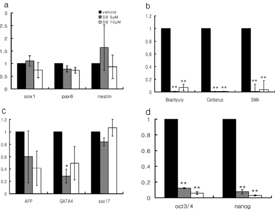
AFP AFPAFP AFP SSEA4 SSEA4SSEA4 SSEA4Figure. 2 Quantitative analysis of expression of markers associated with the early embryonic development in differentiating EBs. The effects of BMP signal blockade on the expression of three germ layer markers were examined in EBs cultured for 10 days in the presence or absence of various concentrations of dorsomorphin. (a) Continuous treatment of dorsomorphin efficiently enhanced the expression of neuroectodermal markers (Sox1, nestin and Pax6), and (b) suppressed that of medodermal marker (brachyury, Cerberus and) and (d) undifferentiation markers (Oct4 and Nanog) in 10 day EBs. (c) The expression of endodermal markers (AFP, GATA4 and Sox17) was slightly decreased. Immunohistochemical analysis clearly showed that there are remaining undifferentiated cells (Oct3/4 and SSEA4-double positive cells) (e-g) and endodermal derivatives (AFP-positive cells, red arrow heads) (h-j) in sections of EBs treated with 5µM dorsomorphin for 10 days. Histograms with error bars represent the mean of three experiments and s.e.m.; EBM, EB media control; DM, dorsomorphin; NOG, noggin (* p<0.05; **p<0.001 compared with control).
dorsomorphin may not only induce neuroectodermal differentiation, but reduce mesodermal differentiation, it is still concerns of the existence of undifferentiated cells as well as derivatives of endodermal lineage within the differentiating cells.
2. Suppression of Activin/Nodal signaling pathway inhibits mesodermal, endodermal and undifferentiated marker expressions, but not neuroectodermal marker expression
One of the prerequisites for efficient differentiation protocol is to accomplish the pure population of target cells (differentiated cells or precursors) devoid of undesired cells. However, our previous results showed that despite augmented neural differentiation from human ES cells by BMP signaling modulation, differentiated cells were contaminated by remnants of endodermal derivatives and undifferentiated cells. To overcome this complication, another signal should be also adjusted to enhance the purity of neural population in differentiated cells. Activin/Nodal pathway, another member of TGF-β superfamily, is known to plays a critical role for the differentiation of the endoderm and mesoderm in the developing embryos22, and in absence of this signal, precocious neural differentiation was promoted in mouse embryo23. Furthermore, Activin/Nodal signaling is required for the maintenance of human ES cells24. More recently, human ES cells were predominantly differentiated
into neuroectodermal lineage by inhibition of Activin/Nodal signal pathway25. Therefore, we postulated that inhibition of Activin/Nodal signaling could solve the complications caused by single inhibition of BMP signal.
To verify this hypothesis, we treated SB431542, a selective inhibitor of Activin/Nodal signaling, during 10 days of EB differentiation at 5~10μM concentration which were used to inhibit Activin/Nodal signaling in human ES cells26. Consistent with data presented in earlier reports25, expression of mesodermal (Brachyury, Cerberus and SMA) and endodermal markers (AFP and GATA4) along with undifferentiation markers (Oct3/4 and Nanog) was dramatically reduced (Fig. 3b-d). However, this molecule did only slightly but not significantly enhance the neuroectodermal marker (Sox1, Pax6 and nestin) expression at 5~10 μM of concentration (Fig. 3a). It is assumed that this discrepancy was due to difference of microenvironment in the differentiation condition. While ES cells were differentiated in chemically defined media showing BMP quiescent condition in previous report, our differentiation system may still possess BMP-like activity in KSR-containing media21. In a recent report, inhibition of Activin/Nodal by 10 μM SB431542 only negligibly increased neuroectodermal markers in EB culture, which is consistent with our data27. These results showed that attenuation of Activin/Nodal signal is appropriate for the reducing meso-, endodermal differentiation together with the traces of undifferentiated cells, but not sufficient for neural differentiation of human ES cells
Figure 3. Effects of Activin/Nodal signal blockade by SB431542 on the expression of three germ layer markers in differentiating EBs. (a) Inhibition of Activin/Nodal signal pathway by the treatment of a selective inhibitor, SB431542, did not enhance the neuroectodermal differentiation, even though there was a slight increase of nestin transcriptsin 10 day cultured EBs. (b and d) Continuous treatment of SB431542 for 10 day EB culture showed dramatic effects on the inhibition of mesodermal differentiation as well as the maintenance of undifferentiation state during differentiation of human ES cells. (c) Expressions of endodermal markers (GATA4) were slightly decreased (* p<0.05; **p<0.001 compared with vehicle group (DMSO only)).
3. Simultaneous inhibition of BMP and Activin/Nodal signals exclusively induce neural differentiation from human ES cells
Based on our results, we postulated that the simultaneous inhibition of two signals by treating both dorsomorphin and SB431542 could exclusively enhance neuroectodermal differentiation of human ES cells. To verify our assumption, we treated 5μM of dorsomorphin alone or in combination with a series of SB431542 concentration (5 and 10 μM) during EB differentiation. Due to toxicity of chemicals, cell death was observed after 7~8 days of differentiation in group treated with two chemicals. EBs differentiated for 10 days in the presence of two chemical inhibitors showed a compact spherical shape and relatively small size compared to that of control. At 10 day of differentiation, while cystic EBs were frequently observed in control group and to some extent in dorsomorphin-only treated, it was hardly seen in group treated with two chemicals. Gene expression studies by qRT-PCR in 10-day EBs indicated that levels of neuroectodermal markers were increased by treatment of dorsomorphin but did not shown significantly additive effects in response to the graded effect of SB431542 (Fig. 4a). However, the diminution of meso-, endodermal, and undifferentiation marker expression were deeper in the combinatorial treatment of two chemicals than in dorsomorphin treatment only. (Fig.4b-d). Since recent report showed the possibility that inhibition of Activin/Nodal signal committed human ES
cells into trophoblate phenotypes27, we additionally checked the expression of trophoblast specific markers (GATA2, GCM1). As expected, expression of trophoblast markers also inhibited, because BMP signal which is critical for trophoblast differentiation was suppressed along with Activin/Nodal signal in our condition (Fig. 4e). Immunohistochemical analysis with section of 10-day EBs showed that though a majority of EB cells were nestin and Pax6 double-positive compared to control group exhibiting small fraction of neural phenotypes (Fig. 4f), which supported our gene expression data. Careful analysis of the expression pattern of three germ layer markers by treatment of two chemicals revealed that there are additive effects in respect to the combinatorial inhibition of two signaling pathways, but not synergistic effects. For example, increment of sox1 expression seemed to be mainly due to the attenuation of BMP signal by dorsomorphin. On the other hand, S431542, Activin/Nodal-inhibitor, had slightly additive effect on nestin (or Pax6) when combined with dorsomorphin. Expression of Brachyury, AFP and Oct3/4 exhibited also similar pattern with nestin (or Pax6) where the expression was additively affected by two chemicals. These results represent the possibility that simultaneous modulation of BMP and Activin/Nodal signaling may be sufficient to the exclusive conversion into neuroectoderm fate during ES cell differentiation, and that two signaling pathways seems to affect independently and cooperatively in cell fate determination during the early neural induction.
(continued) 0 0.2 0.4 0.6 0.8 1 1.2 Brachyury Cerberus 0 00 0 1 11 1 2 22 2 3 33 3 4 44 4 5 55 5 6 66 6 7 77 7
Sox1 Pax6 Nestin
Vehicle DM5 DM5+SB5 DM5+SB10 0 0.2 0.4 0.6 0.8 1 1.2 AFP GATA4 0 0.2 0.4 0.6 0.8 1 1.2 Oct3/4 Nanog
a
b
c
d
e
0 0.2 0.4 0.6 0.8 1 1.2 GATA2 GCM1f
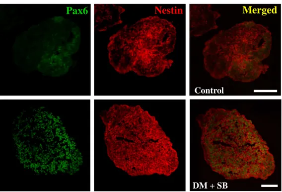
Figure 4. Simultaneous suppression of BMP and Activin/Nodal signal pathway is sufficient to direct neural differentiation of human ES cells. To investigate whether simultaneous inhibition of BMP and Activin/Nodal signals could facilitate neural differentiation of human ES cells, EBs were subjected to vehicle (DMSO), dorsomorphin (5µM) and SB431542 (5 and 10 µM) for 10 days. (a) Gene expression studies demonstrated that increment of neuroectodermal markers and (b-d) diminution of meso-, endodermal and undifferentiation markers were more consolidated by simultaneous treatment of dorsomorphin and SB431542. (e) Expression of trophoblast markers (GATA2 and GCM1) were also diminished through directed neural differentiation of ES cells by the modulation of two signals. (c) Immunocytochemical analysis confirmed that expression of Pax6 and Nestin were enhanced in EBs differentiating in the presence of dorsomorphin and SB431542 compared with control EBs. Bars in graph (a)-(e) represent the fold increase of each maker by chemical treatment against that of vehicle (DMSO) treatment as means ± s.e.m by three independent repeats; DM, dorsomorphin; SB, SB431542, scale bar: 100 µm
Control
DM + SB
Merged
Nestin
Pax6
4. Simultaneous inhibition of BMP and Activin/Nodal signals facilitates neural differentiation from human pluripotent stem cells regardless of their own differentiation nature
It was recently reported that there are marked differences in differentiation propensities among human ES cell lines, particularly into the endodermal and the mesodermal lineages. Moreover, such tendency was still preserved when cells were subjected by protocols directing into particular cell fates11. Although this report provided important information to human ES cell research, there is little knowledge regarding to the difference of differentiation potential among human iPS cells or between human ES cells and iPS cells. Hence, we compared the differentiation potential among 6 human ES cell lines and 3 iPS cell lines. Furthermore, we explored particularly whether our neural differentiation protocol could overcome these differentiation inclinations.
To compare the differentiation propensities among 9 human pluripotent stem cell lines, colonies detached from feeder cells were cultured for 10 days as EBs in conventional EB media without any additives and followed by analyses of three germ layer and undifferentiation markers by qRT-PCR. Consistent with previous report11, there are marked differences in differentiation propensities among human ES cells as well as iPS cells tested (Fig. 5a-d). For instance, Miz-hES4 and BJ1-12-iPS cells
showed marked inclination to differentiate toward meso- and endodermal lineages during 10 day EB differentiation (Fig. 5b-c). In contrast, H9 and Miz-hES6 each showed the highest expression levels of neuroectodermal markers, Sox1 and Pax6, with moderate meso-, and endodermal inclination (Fig. 5a-c). Surprisingly, SNU-hES3, 16 and CHA-hES3 cells exhibited less germ layer marker expression than others with aberrantly more undifferentiation marker expression at day 10 of differentiation, suggesting that they might differentiate more slowly than other cell lines probably due to different culture condition (eg. feeder layer) or higher passage number (Fig.5d). To investigate whether different propensities among human pluripotent stem cells are still maintained by directed differentiation protocol toward neural fate, we applied H9, Miz-hES4, BJ1-12-iPS and MSC-Y2-3-iPS cells to well-characterized neural differentiation protocol7. EBs derived from each cell lines cultured in EB media for 4 days, followed by adherent culture in N2 media supplemented with 20ng/ml of bFGF. After 5~6 days from EB attachment, a lot of colonies showing rosette structures appeared as described in previous report7. While H9 cells efficiently produced columnar rosette cells (Fig. 6a and e), other cell lines generated a lot of flattened / fibroblast-like cells and small fraction of rosette cells surrounded by mixed population of other lineage cells, which could make subsequent procedure for isolating neural precursor difficult (Fig. 6a, b). Immunocytochemical and quantitative analysis also demonstrated that more Pax6 and nestin double-positive
a. (neuroectodermal markers)
b. (mesodermal markers)
c. (endodermal markers)
Figure. 5 Differentiation propensities among humanpluripotent stem cells. To explore whether there are differentiation propensities among human ES and iPS cells, 6 human ES cell lines (H9, Miz-hES4 and 6, SNU-hES3 and 16, CHA-hES3) and 3 iPS cell line (BJ1-12-iPS, MSC-Y2-3-iPS, dH1f2-2-iPS) were tested. After 10 day spontaneous differentiation as EBs, mRNA expression levels of neuroectodermal (Sox1 and Pax6) (a), mesodermal (Brachyury and Cerberus) (b), endodermal (AFP and GATA4) (c) and undifferentiation markers (Oct3/4 and Nanog) (d) were assessed by qRT-PCR. Data were represented as means ± s.e.m of relative expression level of each gene normalized against the lowest one (designated as value of 1) among tested cell lines. Statistical significance was estimated using one-way ANOVA (analysis of variance) test with multiple comparisons among cell lines. To reduce a type I error rate, it was applied Bonferroni correction as Post Hoc. Miz6, Miz-hES6; Miz4, Miz-hES4; SNU3, SNU-hES3; SNU16, SNU-hES16; CHA3, CHA-hES3; BJ1-2, BJ1-12-iPS; MSC-Y2-3, MSC-Y2-3-iPS; dH1f2-2, dH1f2-2-iPS
a
H9 H9 H9 H9 Miz4Miz4Miz4Miz4 BJ1 BJ1BJ1 BJ1---12-121212 Y2Y2Y2Y2----333 3 0 20 40 60 80 100 H9 Miz4 BJ1-12 Y2-3
b
** ** ** ** ******** ** ** ** ** nestin/Pax6 H9 Miz-hES4 MSC-Y2-3-iPSCc
BJ1-12-iPSCFigure 6. Comparison of neural differentiation propensity among cell lines by a well-characterized neural differentiation protocol. Three human pluripotent stem cell lines (H9, Miz-hES4, BJ1-12-iPS and MSC-Y2-3-iPS) showing distinct differentiation propensity were specified into neural lineage as previously reported7. EBs generated from each cell lines were cultured in EB media for 4 days and subjected to neural differentiation by adherent culture in N2 media for additional 6 days. (a) Morphologically, neural rosette structures (arrow head) were efficiently generated in H9, while these were hardly detected in Miz-hES4, BJ1-12-iPS and MSC-Y2-3-iPS cells (a and b). Non-neural cells exhibiting flatten or fibroblast-like shapes were prevalent in those cell lines instead. (b) Quantitative analysis of colonies containing rosette structure demonstrated that Miz-hES4, BJ1-12-iPS and MSC-Y2-3-iPS cells exhibiting poor propensity to neural lineage could not efficiently specify into neural fate (c). Immunofluorescence staining also demonstrated that H9 cells could differentiate into neural lineage (high yield of nestin and Pax6 double positive cells) (e), whereas Miz-hES4, BJ1-12-iPS and MSC-Y2-3-BJ1-12-iPS cells still reserved endo- and mesodermal differentiation propensities with poor neural differentiation by one of well-characterized neural differentiation protocols (** p<0.01 compared with H9, ANOVA test). Scale bar : 50µ m.
neural precursors were shown in H9, whereas these were hardly detected in Miz-hES4 and BJ1-12-iPS cells. In contrast, more cells labeled with anti-Brachyury or AFP antibodies were detected in Miz-hES4, BJ1-12-iPS and MSC-Y2-3-iPS cells, respectively (Fig. 6c). These data indicated that the neural differentiation propensity among these cell lines were still preserved even when cells were directly differentiated.
Next, we investigated whether forced differentiation by simultaneous modulation of BMP and Activin/Nodal signals could efficiently specify pluripotent stem cells into neural fate with less variation among cells lines. EBs from 9 pluripotent stem cell lines were cultured in EB media supplemented with or without 5µM dorsomorphin plus 10µM SB431542 for 10 days and subjected to gene expression analyses of three germ layer and undifferentiation markers through qRT-PCR and immunohistochemisty analysis. Surprisingly, combinatorial treatment of two chemicals dramatically increased the expression of neuroectodermal markers in all 9 cell lines. Sox1 expressions were increased by 4~100 fold among cell lines. Moreover, Pax6 expression showed the remarkable increase in majority of cell lines around 100 folds (Fig. 7a). Concommitently with upregulation of neuroectodermal genes, expressions of mesodermal, endodermal and undifferentiation markers were markedly decreased (Fig. 7a). Interestingly, the pattern, increment of neuroectodermal marker and concurrent diminution of other lineage marker plus undifferentiation
0.0001 0.001 0.01 0.1 1 10 100 1000 10000 R e la ti v e e x p r e ss io n o n a l o g s c a le (D M + S B -t r e a te d g r o u p /v e h ic le -t r e a te d g r o u p ) Sox1 Pax6 ne stin Brachyury Ce rbe rus AFP GATA4 Oct3/4 Nanog
H9 Miz6 Miz4 H9 Miz6 Miz4 H9 Miz6 Miz4 SNU3 H9 Miz6 Miz4 SNU3 SNU3 SNU16 SNU3 SNU16 SNU16 SNU16 CHA3 CHA3 BJ1CHA3 CHA3 BJ1BJ1BJ1---12 -12 Y212 12 Y2Y2Y2---3 -3 dH1f23 3 dH1f2dH1f2dH1f2----222 2
a
Figure 7. Simultaneous blockade of BMP and Activin/Nodal signal efficiently induced neural differentiation from various human pluripotent stem cell lines with distinct differentiation propensities. (a) In differentiating EBs derived from various human pluripotent stem cell lines, addition of dorsomorphin along with SB431542 dramatically increased the expression of neuroectodermal markers, whereas meso-, endodermal and undifferentiation marker expressions were markedly decreased. In particular, the fold induction of neuroectodermal markers was more distinctively observed in cell lines showing differentiation propensities toward non-neural lineages (eg, Miz-hES4 and BJ1-12-iPS). Bars diagrams represent means ± s.e.m of the fold increase of each maker by chemical treatment against that of vehicle (DMSO) treatment on log scale. Data were obtained from at lease three independent repeats. (b) EB sections from four cell lines (H9, Miz-hES4; BJ1-12-iPS and MSC-Y2-3-iPS) were subjected to immunocytochemical analysis with anti-nestin antibody. While Miz-hES4, BJ1-12-iPS and MSC-Y2-3-iPS cells poorly labeled with anti-nestin antibody compared to H9 in spontaneous differentiation (upper lane), simultaneous treatment of dorsomorphin and SB431542 markedly increased the number of Nestin positive cells (lower lane). DM, dorsomorphin; SB, SB431542, Scale bar: 100µm.
marker expression, was more distinctively observed in cell lines showing unfavorable propensities to neural fate. For instance, chemical treatment strongly forces neuroectodermal differentiation of Miz-hES4 cells that innately differentiated into meso- and endodermal lineages in spontaneous differentiation condition than that of other cell lines exhibiting neuroecotdermal propensity. This was also the case for BJ1-12-iPS cell line that displayed a similar pattern of propensity. Although these cells expressed significantly lower level of Sox1 and Pax6 compared to H9 after 10 days spontaneous differentiation, expression levels of these markers were comparable to that of H9 by the treatment of dorsomorphin and SB431542. We also confirm these results through immunofluroscence staining with EB sections from these cell lines (Fig. 7b). Collectively, these results suggested that differentiation inclination among cell lines could be overcome through the simultaneous modulation of BMP and Activin/Nodal signaling pathways with the small molecules, thereby most of human pluripotent stem cells could be selectively fated into neural lineage cells.
5. Neural precursors generated by treatment of BMP and Avtivin/Nodal signal inhibitors could differentiate into dopamine neurons with high yield
To further explore the differentiation potentials of neural precursors generated by simultaneously blocking BMP and Activin/Nodal signals, we developed the protocol for directed differentiation of neural precursors into dopaminergic
neurons (Fig. 8a). For the expansion of neural precursors, 10 day EBs in the absence (control-NPs) or presence of signal inhibitors (DMSB-NPs) were cultured in N2 media supplemented with 20ng/ml of bFGF. Proliferating DMSB-NPs showed the similar shape of spherical neural mass (SMN) known to be consisted with pure neural precursor population (Fig. 8b)12. After expansion, EBs were triturated by mild pipetting and seeded onto Matrigel-coated culture dish in N2 media. As soon as neural precursor clumps attached, a lot of rosette structures were observed in the center of colonies and neuronal processes were begun to put out inthe margins (Fig. 8C). During attachment differentiation, 200 ~ 500ng/ml Shh and 100ng/ml FGF8 were added into N2 media to regionalize neural precursors into the ventral mesencephalic progenitors. After 8 days regionalization, morphogenic factors (Shh and FGF8) were substituted with 20ng/ml of GDNF, 20ng/ml of BDNF, and 200mM of ascorbic acid for dopaminergic maturation.
Immunocytochemical analyses showed marked increase in the number of Tuj1-positive cells from the DMSB-NPs of MSC-Y2-3-iPS cells (50.7±2.2 % of total cells) compared to the vehicle-treated cells (2.6± 0.5 % of total cells) (Fig. 9a, d and j). A significant portion (49.5±6.8 %) of the Tuj1-positive cells were DA neurons (Fig. 9d-f and j). H9 and BJ1-12-iPS cells also generated a comparable amount of DA neurons among the total Tuj1-positive cells: H9, 53±7.4%; BJ1-12-iPS, 43±1.3%) (Fig. 9k). A lot of TH positive cells expressed En1, a midbrain specific transcription
factor (Fig. 9g), and there are no TH positive cells co-expressing DβH, a marker for noradrenergic neurons (Fig. 9h). Some TH-positive cells (~10%) were merged with cells showing GABAergic phenotype (Fig. 9i), which means that small fraction of TH-positive cells may be olfactory neurons. Semi-quantative RT-PCR analysis confirmed the step-wise neuronal differentiation from undifferentiated ES (iPS) cells to midbrain dopaminergic neurons (Fig. 9l). These data indicated that neural cells generated by the modulation of BMP and Activin/Nodal signaling also retain the capability to be differentiated into a specific neuronal type such as DA neuron.
Figure. 8 Neural precursors generated by dual signal modulation could differentiate efficiently into dopaminergic neurons. (a) Schematic representation of dopaminergic differentiation protocol from neural precursor generated by modulation of BMP and Activin.Nodal signals. (b) During expansion step, neural precursors induced by two chemicals proliferated as forms of large spherical structure, just like neurosphere or spherical neural mass (SNM) as previously described12. Red arrow heads indicate neural rosette structures. (c) As soon as attached on substratum, clumps of expanded neural precursors began to differentiate to typical neuronal cells.
Fig. 9. Efficient induction of TH-positive neurons from human pluripotent stem cells. Representative images of differentiated neurons from MSC-Y2-3-iPS (a-f) and counting data (j). Compared to control group in which neuronal cells were derived from spontaneously differentiated EBs (a-c), highly efficient Tuj1-positive and TH and Tuj1-double positive were induced from neural precursors generated by the dual signal modulation (d-f and j). Phenotypic characterization of TH-positive neurons (g-i). (g) Many of TH-positive neurons stained with anti-En1 antibody, which represent midbrain characters, (h) there were not DβH positive cells among TH double positive cells, which means that these cells were not adrenergic / noradrenergic neurons. (i) Small fraction of TH-positive cells were merged with cells showing GABAergic phenotype (indicated by yellow arrow heads), implying that some of these TH-positive cells may be olfactory. (k) Efficient differentiation of TH-positive neurons was induced from two cell lines, H9 and BJ1-12-iPSCs. (i) RT-PCR analyses of cells at neuronal differentiation process. ES, ES cells; nEB, EBs induced into neural fates by treatment of chemicals; hNP, expanded neural precursors in N2 media; pDA, dopaminergic precursors induced by Shh and FGF8; DA, dopaminergic neurons differentiatied without mitogens for 10 days. Scale bar : 50µm.
IV. Discussions
The generation of large number of pure neural precursors from pluripotent stem cells has important issues not only in therapeutic purposes but also in developmental biology. Current trials for neural differentiation methods have contributed partially to fulfill these purposes, however have inappropriate limitations to solve for application of human pluripotent stem cells to clinical uses. Therefore, a more simple and efficient method to produce abundant pure neural precursors from a lot of cell lines is cardinal for the realization of cell replacement therapy through human pluripotent stem cells.
Our primary strategy for efficient neural differentiation from human pluripotent stem cells was modulating signaling pathways critical for embryonic neural induction. In contrast to current approaches which tried to optimize culture conditions or to modify methods used to differentiate mouse ES cells, our differentiation system via modulation of cellular signaling can be commonly applied to various cell lines cultured in different conditions or by various methods with less variation. There has been significant progress in our understanding of neural induction mechanism in the past few decades, and a lot of developmental studies suggested that various cellular and molecular mechanisms are involved in vertebrate neural induction including BMP, Wnt, Activin/Nodal and FGF signals15. Of these,
BMP signaling has been mainly considered to play a pivotal role for neural induction, and several reports have provided evidences that treatment of BMP signal antagonist, noggin, could facilitate neural differentiation of human ES cells16-18. These reports favorably support the traditional neural induction model – the default theory of vertebrate neuralization15, 28. Through removal of BMP signals inhibiting neural induction by treatment of noggin, they induced ES cells to differentiate into neural lineage. Though these reports provided promising results, they overlooked the possibilities that other lineage cells along with undifferentiated cells might still be mixed with differentiated neural precursors. In present study, we presented evidences that endodermal lineage cells and undifferentiated cells existed with neural precursors. Residual undifferentiated cells in neural precursors would be a serious demerit of differentiation protocols, since these cells can form teratoma when transplanted. These results suggest that modulation of another signal along with BMP signal is required for obtaining pure population of neural precursors from ES cells.
Activin/Nodal signaling is known to be essential for maintaining the pluripotency in both human and mouse, though effective time window is not synchronous between two species29. In addition, they also were known to be responsible for the endodermal and mesodermal differentiation, as well as play a negative role for neural induction30. Here, we presented that blockade of Activin/Nodal signal efficiently diminished meso-, endodermal differentiation as well
as reducing undifferentiated markers. However, inhibition of Activin/Nodal signaling alone was not sufficient for the induction of efficient neural differentiation in our differentiation system. On the contrary, previous studies demonstrated that the inhibition of Activin/Nodal signaling during EB differentiation augmented neural fate specification25. The most obvious difference between two experiments is the components of media used during differentiation. Previous study had used lab-made chemically defined media (CDM) exhibiting BMP signal-quiescence, whereas we used the conventional EB media (see the Materials & Methods). EB media contained 20% of knockout serum replacement, which is known to contain BMP-like differentiation-inducing activity21. Compared with the former where BMP signaling has negligible effects, our differentiation system would still allow the BMP-like activities of media component to inhibit the neural differentiation to some extent, so the inhibition of Activin/Nodal alone would not be sufficient to induce neural differentiation in human ES cells. Therefore, we next postulated if BMP and Activin/Nodal signaling simultaneously were blocked, neural differentiation could be efficiently augmented. Interestingly, simultaneous inhibition of two signals during EB differentiation did not enhance the expression of neuroectodermal markers as much as single inhibition of BMP signal, suggesting that neural inducing effects seemed to be contributed mainly by blockade of BMP signal. However, effects by inhibition of Activin/Nodal signal, suppressing meso-, endodermal and undifferentiation marker
expression, were also added up by inhibiting BMP signal. Therefore, we suggested here that two signaling pathways seems to affect independently and cooperatively in cell fate determination during the early neural induction and exclusive neural differentiation of human pluripotent stem cells also would require inhibition of both BMP and Activin/Nodal signals.
As shown in previous report11, we confirmed here the existence of differentiation propensity across human pluripotent stem cell lines. In addition, differences of neuroectodermal marker expression among cell lines also showed statistical significance. Current neural differentiation protocols seem not to be efficient in diverse cell lines or somehow depend on their own differentiation propensities of cell lines. Here, we showed, through well-characterized neural differentiation protocol, that it is not easy to convert efficiently cells showing non-neural propensity into non-neural fate. In this method, cells were initially subjected to differentiate for 4~6 days in spontaneous differentiation condition, then neural induction and selection steps were followed7. In our experiences, however, ES cell lines exhibiting high propensity into the endodermal lineage gave rise to a lot of cystic EBs at 4~6 days of differentiation. Technically, through adherent culture of cystic EB, it is not only difficult to differentiate into neural lineage cells, but also hard to isolate neural precursors from mixed cell population. It was recently reported that differentiation of ES cells into primitive endodermal lineage cells contributing to
cystic structures in EBs was influenced with BMP signaling16. Since BMP signal was inhibited throughout the initial differentiation process in our method, it was not favorable for ES cells to fate into the primitive endodermal. Simultaneously, the suppression of Activin/Nodal signals did not allow ES cells to differentiate into the mesodermal and endodermal lineage as well as to remain in undifferentiation state. Thus, ES and iPS cells would only take neuroectodermal lineage when both BMP and Activin/Nodal signals are inhibited. In that respect, it was reasonable that most of cell lines tested elicit the efficient neural differentiation devoid of other lineage cells beyond their differentiation propensity. In other words, these results suggest that selective removal of unwanted lineage cells are not required for the enrichment of neural lineage cells.
We observed that cell lines which were innately inclined to meso- and/or endodermal differentiation could be boosted up to specify into neural fate by modulation of BMP and Activin/Nodal signal. One of possible explanation for our results is that endogeneous BMP or Activin/Nodal signal may be so strong in those cell lines that they could have been inclined to differentiate into meso/endodermal fate. Therefore, strong and sustained inhibition of these signals might be able to convert them to neuroectodermal fate. Second possibility is that those cell lines might have high sensitivity to BMP and Activin/Nodal signal, which probably made them highly susceptible to activation/inhibition of those signals.