Introduction
An isthmus, defined as narrow extension from either 1 or 2 main root canals,1 is a routinely encountered anatomic
complexity in human permanent teeth, especially in mandi bular first molars,2 with the highest prevalence at the apical
level.3,4 According to Srivastava et al.(2018)5, the preva
lence of isthmuses in the mesial roots is 78.4%. Isthmuses that are not detected in radiographs are difficult to reach
by instrumentation, irrigation, and/or medicaments,6 and
therefore function as reservoirs of microorganisms, vital tissue, or necrotic tissue.7 The apical level is critical for
endodontic treatment and its insufficient cleaning may be responsible for treatment failure.
With the recent advent of microcomputed tomography (microCT), it has become possible to obtain more detailed qualitative and quantitative descriptions of root anatomy, as microCT has important advantages over other techniques, such as the possibility of reproducing the internal dental anatomy in multiple planes without destroying the samples; however, it is unsuitable for clinical use.7,8 Because micro
CT provides highresolution, detailed, and accurate infor mation on root canal morphology, it can be used as a refer ence standard in anatomical in vitro studies.911
Comparison of limited- and large-volume cone-beam computed tomography using a small
voxel size for detecting isthmuses in mandibular molars
Elen de Souza Tolentino
1, Pablo Andrés AmorosoSilva
2, Murilo Priori Alcalde
3,
Fernanda Chiguti Yamashita
1,*, Lilian Cristina Vessoni Iwaki
1, Izabel Regina Fischer RubiraBullen
4,
Marco Antônio Húngaro Duarte
31Department of Dentistry, State University of Maringá, Maringá, PR, Brazil
2Department of Restorative Dentistry, State University of Londrina, Londrina, PR, Brazil
3Department of Operative Dentistry, Endodontics and Dental Materials, Bauru School of Dentistry, University of Sao Paulo, Bauru, Brazil 4Department of Surgery, Stomatology, Pathology and Radiology, Bauru School of Dentistry, University of Sao Paulo, Bauru, SP, Brazil ABSTRACT
Purpose: This study was performed to compare the ability of limited and largevolume conebeam computed
tomography(CBCT) to display isthmuses in the apical root canals of mandibular molars.
Materials and Methods: Forty human mandibular first molars with isthmuses in the apical 3 mm of mesial roots
were scanned by microcomputed tomography(microCT), and their thickness, area, and length were recorded.
The samples were examined using 2 CBCT systems, using the smallest voxels and field of view available for each
device. The MannWhitney, Friedman, and Dunn multiple comparison tests were performed(α=0.05).
Results: The 3D Accuitomo 170 and iCat devices detected 77.5% and 75.0% of isthmuses, respectively(P>0.05).
For length measurements, there were significant differences between micro-CT and both 3D Accuitomo 170 and
iCat(P<0.05).
Conclusion: Both CBCT systems performed similarly and did not detect isthmuses in the apical third in some cases.
CBCT still does not equal the performance of microCT in isthmus detection, but it is nonetheless a valuable tool in
endodontic practice.(Imaging Sci Dent 2021; 51: 27-34)
KEY WORDS: ConeBeam Computed Tomography; Endodontics; Molar; XRay Microtomography
Copyright ⓒ 2021 by Korean Academy of Oral and Maxillofacial Radiology
This is an Open Access article distributed under the terms of the Creative Commons Attribution NonCommercial License(http://creativecommons.org/licenses/bync/3.0) which permits unrestricted noncommercial use, distribution, and reproduction in any medium, provided the original work is properly cited.
Imaging Science in Dentistry·pISSN 22337822 eISSN 22337830
This study was supported by the Conselho Nacional de Desenvolvimento Científico
e Tecnológico(CNPq) Foundation(Brazil) for scholarship support(grant number
151990/20166).
Received June 10, 2020; Revised September 3, 2020; Accepted September 23, 2020 *Correspondence to : Prof. Fernanda Chiguti Yamashita
Department of Dentistry, State University of Maringá, Avenida Mandacaru, 1550, Bloco S08, Maringá PR, CEP 87080000, Brazil
Although radiographs used for clinical procedures show important details such as the number of root canals, they do not provide enough details on the internal anatomy.12
Conebeam computed tomography(CBCT) has gained broad acceptance in dentistry in recent years as it generates 3dimensional data at a lower radiation dose and cost and a higher spatial resolution than helical CT.13,14 However,
CBCT involves a considerably higher radiation dose than conventional radiographs.15
Several CBCT systems are currently on the market. Those systems vary in terms of image quality and performance. The voxel size, slice thickness, spatial resolution, and the size of the scan volume(field of view; FOV) are impor tant parameters that influence the quality of the images. Since voxels are isotropic in CBCT, images can be con structed in any plane with high fidelity.16 A smaller voxel
size provides a higher resolution, and, in CBCT, voxel sizes range from 0.075mm3 to 0.4mm3.16
FOV selection is equally important, as the FOV is directly related to voxel size and influences spatial and contrast res olution. Besides providing higher radiation doses, a larger FOV offers lower resolution than a smaller FOV, which directly influences the visibility of anatomical structures on CBCT. FOV selection is indeed a limiting factor for visual izing the canal space, and the smallest FOV available is usually indicated for an endodontic exam.17 In this setting,
some studies have compared different CBCT systems with different FOV and voxel sizes in the assessment of root canals, mainly for detecting root fractures.16,17 However,
few studies have used CBCT images to detect and describe isthmuses in root canal systems.1,2,4,9 Furthermore, the
extant studies assessed only a single CBCT system, FOV, and voxel size, and did not incorporate an analysis of micro CT images.
The use of CBCT could contribute to the detection and location of isthmuses in the root canal. Thus, the aim of this study was to assess the detection of isthmuses in the apical third of mesial root canals of human mandibular molars, by comparing limited and largevolume CBCT systems while using their highestresolution settings.
Materials and Methods
Sample and reference group
After receiving approval from the ethics committee(CEP #1.929.037), a microCT system(SkyScan 1174v2; Bruker micro CT, Kontich, Belgium) was used to scan mandibular first molars extracted from a Brazilian population. The teeth that presented an isthmus in the apical third of the mesial
root on microCT were selected.
The teeth were extracted for periodontal disease, exten sive caries, or coronal fractures, and after extraction, they were immediately stored in 10% neutralbuffered formalin solution(Kolplast CI ltda, Itupeva, São Paulo, Brazil). Based on the reconstructed microCT images, teeth with in
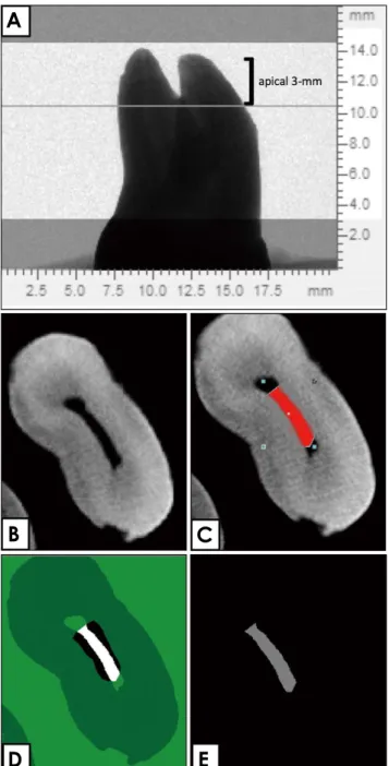
Fig. 1. Visualization and qualitative evaluation of 3dimensional
models(microCT). A. Location of the isthmus while viewing the apical 3mm of the mesial root. B. Raw image preview showing the type II isthmus at the point of its major thickness. C. Region of interest preview. D. Binary selection preview. E. Morphometry pre view allowing measurements of the thickness, area, and length of the isthmus(CTVol v.2.2.1 and data viewing software).
A
B
D
E
complete root development, a Cshaped root canal, calci fied root canals, internal or external resorption, root fractures, and/or endodontic treatment were excluded. The sex and age of the patients were unknown. The microCT para meters used were 50kV, 80μA, 360° of rotation, and an isotropic resolution of 19.6μm. The system included a chargecoupled device camera(1304×1024 pixels). The images of each specimen were reconstructed with dedicated software(NRecon v.1.6.3, BrukermicroCT, Kontich, Bel gium) that provided axial crosssections of the inner struc tures of the roots in the BMP format.
A total of 40 teeth with isthmuses in the apical third of mesial roots were selected after microCT scanning. Sample size was performed using repeatedmeasures analysis of variance with a significance level of 5% and a power of 95%. Threedimensional models were reconstructed using automatic segmentation and the surfacemodeling software CTAn v.1.12(BrukermicroCT, Kontich, Belgium). CTVol v.2.2.1 and data viewing software(BrukermicroCT, Kon tich, Belgium) were used for visualization and qualitative evaluation of the specimens. For each tooth, the isthmus was located by viewing axial images of the apical 3mm of the mesial root. At the point of the major thickness of the isthmus in the apical 3mm, the thickness, area, and length of the isthmus were recorded using these specific soft ware tools(Fig. 1). Since the rate of isthmus detection on microCT was 100%, these images served as a reference group.
CBCT scanning
The samples were examined using 2 CBCT systems(3D Accuitomo® 170(J. Morita Corp., Kyoto, Japan, and New
Generation iCat®, Imaging Sciences International, Hat
field, PA, USA), according to the protocols recommended by the manufacturer. The specifications of each system, energy parameters, and scan settings are shown in Table 1. For the 2 systems, the smallest voxel size and FOV were used(4cm×4cm/0.08mm3 and 8cm×8cm/0.125mm3,
respectively), and the images were obtained in different planes(sagittal, coronal, and axial) with a 1mm slice thick
ness and 0.5mm slice interval for both devices.
The teeth were covered with a thin layer of utility wax (Tenatex Red; Kemdent, Swindon, UK) to simulate the periodontal space. They were then fixed in prefabricated sockets in dry human mandibles, which were coated with 3 layers of wax buccally and lingually to provide softtissue simulation. Each tooth was scanned separately.
Two independent external observers(radiologists, both with 8 years of experience with CBCT) were calibrated based on the criteria and variants established prior to their evaluation, using 25% of the sample(both were calibrated by evaluating 10 randomly selected CBCT scans, which were measured twice within a 15day interval). The images were examined using the scanners’ proprietary software (Xoran 3.1.62 version, Xoran Technologies, Ann Arbor, MI, USA, and One Volume Viewer, J. Morita MFG. Cor poration, Kyoto, Japan) in a Intel® CoreTM 2 Duo 1.86
Ghz6300(Intel Corporation, Santa Clara, CA, USA) PC workstation with a NVIDIA GeForce 6200 turbo cache video card(NVIDIA Corporation, Santa Clara, CA, USA) running Windows XP Professional SP2(Microsoft Corpo ration, Redmond, WA, USA) and with an EIZOFlexScan S2000 monitor at a resolution of 1600×1200 pixels(EIZO NANAO Corporation, Hakusan, Japan). All analyses were performed in a semidark and silent room and examiners were instructed to take breaks between analyses to avoid eye fatigue. Software tools such as filter, zoom, and con trast could be used, allowing image optimization at the discretion of each examiner. All analyses were performed independently and the examiners did not communicate.
Each tooth was analyzed by axial reconstructions of 0.1 mm/0.1mm, moving from the coronal to apical region. The apical thirds were then evaluated. The blinded examiners analyzed the 1 to 3mm apical level in the axial recon structions and recorded whether they detected isthmuses in each tooth in both software programs. Isthmuses were recorded as being present when the scans showed a narrow ribbonshaped communication between the mesiobuccal and mesiolingual canals on an axial image. When differ ences were found, a consensus was reached after the image
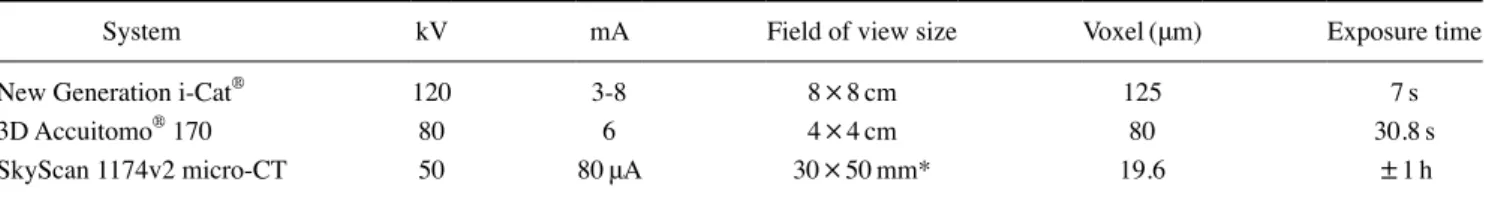
Table 1. Specifications and energy parameters of the cone-beam computed tomography systems and micro-computed tomography
System kV mA Field of view size Voxel(μm) Exposure time
New Generation iCat® 120 38 8×8cm 125 7s
3D Accuitomo® 170 80 6 4×4cm 80 30.8s
SkyScan 1174v2 microCT 50 80μA 30×50mm* 19.6 ±1h
was discussed with a third observer, an endodontist with 10 years of experience. When isthmuses were detected, their length(extension) was measured using the ruler tool in each CBCT proprietary software(mm). These values were converted to percentages and compared with the microCT records.
Statistical analysis
The results of the diagnostic CBCT methods were repor ted as percentages for the detection of isthmuses, consi dering microCT as the reference method. The CBCT sys tems were compared using the MannWhitney test, and the isthmus length measurements made using the microCT and CBCT devices were compared using the Friedman and Dunn multiple comparison tests, at α=0.05. The kappa test was used to assess interexaminer variability in detecting isthmuses18 and the intraclass correlation coefficient(ICC)19
was used to test the interexaminer reliability for length measurements and repeated exams. The data were analyzed using Prism 5.0 software(GraphPad Software Inc., La Jolla, CA, USA).
Results
In the microCT images, the major thickness of the isth muses at the apical 1 to 3mm level varied from 0.09 to 0.37mm(mean, 0.15mm). The volume, area, and length varied from 0.80 to 2.23mm3 (mean, 1.21mm3); 0.07 to 0.60
mm2 (mean, 0.29mm2); and 0.91 to 2.68mm(mean, 1.89
mm), respectively. The isthmuses that were not detected on the CBCT scans had volume, area, and length varying from 0.68 to 1.87mm3 (mean 0.98mm3); 0.07 to 0.47mm2
(mean 0.21mm2); and 1.13 to 2.43mm(mean 1.71mm),
respectively. The apical major thickness of these isthmuses
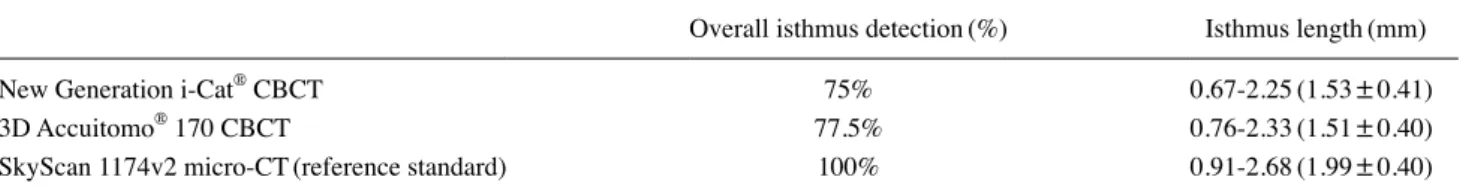
Table 2. Overall detection of isthmuses converted into percentages and length values(minimum, maximum, mean±standard deviation) on
microcomputed tomography(CT) and conebeam computed tomography(CBCT)
Overall isthmus detection(%) Isthmus length(mm)
New Generation iCat® CBCT 75% 0.672.25(1.53±0.41)
3D Accuitomo® 170 CBCT 77.5% 0.762.33(1.51±0.40)
SkyScan 1174v2 microCT(reference standard) 100% 0.912.68(1.99±0.40)
Fig. 2. Isthmuses identified in the mesial
roots of mandibular first molars on micro- computed tomography and on limited and largevolume conebeam computed tomographic scans.
ranged from 0.07 to 0.17mm(mean, 0.10mm).
Considering microCT as the reference standard, images acquired with the 3D Accuitomo 170 detected 77.5%(31 of 40) of isthmuses in the sample. For the iCat system, the detection rate was 75.0%(30 of 40, Table 2). The differ ence between the systems was not significant(P>0.05).
Figure 2 shows isthmuses identified in both CBCT images. Nondetected isthmuses are illustrated in Figure 3.
Significant differences were found between microCT both and 3D Accuitomo 170 and iCat(P<0.05), indi
cating that both CBCT scanners did not detect some isth muses, impeding their length measurements. When 3D Accuitomo 170 and iCat were compared, no statistically significant difference was found(P>0.05). Figure 4 shows
the median and interquartile values of the isthmus length measurements made using microCT, 3D Accuitomo 170, and iCat.
The kappa values for interobserver agreement in the detection of isthmuses ranged from 0.89 to 1.00(perfect). Interexaminer agreement was excellent for length mea surements, with replicability at a significance level of 5% (ICC>0.90, Table 3).
Discussion
Anatomical complexities such as isthmuses are not un common2,3,20,21 and may represent a challenge in endodontic
therapy. As in other studies,9,10 this study used microCT
as a reference standard, since it enables nondestructive 3dimensional microscopy of the internal dental anatomy.22
Nevertheless, it is not available for clinical use, which is where CBCT may come into play.23
Fig. 3. Isthmuses identified in the mesial
roots of the mandibular first molars on microcomputed tomography, but not detected on limited and largevolume conebeam computed tomographic scans.
Fig. 4. Median(gray line) and interquartile values of the isthmus
length measurements analyzed using microCT, 3D Accuitomo 170, and Next Generation iCat(P<0.05). CT: computed tomography.
Familiarity with the specific features(such as FOV and voxel sizes) of different CBCT systems is important for both clinicians and radiologists, who often decide the acquisition protocol to be used in each specific case. A larger FOV pro vides less resolution and contrast,24 whereas limitedvolume
units tend to offer the highest image resolution,25 making
them preferable for endodontic applications.17 For this rea
son, the smallest FOV and voxel size available on each device were used to provide the sharpest image in an attempt to achieve the most comparable results possible. The objec tive was to compare the highestresolution settings avail able on each device, not to compare the equivalent settings on both devices.
Since no previous study has compared different CBCT units for the detection of isthmuses, the purpose of compar ing 2 different systems is to simulate a situation similar to that encountered in clinical practice, where an endodontist does not always have the option of choosing a particular system to request an exam. The iCat system is among the more commonly used units in the world26 and the 3D
Accuitomo 170 is characterized as one of the bestquality CBCT machines currently on the market,24 since it presents
the smallest FOV available and a superfine voxel size of 80 μm. Nonetheless, both units performed similarly(P>0.05).
When comparing the accuracy of Accuitomo, iCat, and other 3 CBCT systems in detecting root fractures, Hassan et al.27 found that iCat was the most accurate. In the auth
ors’ opinion, the detector design might explain the super ior ity of this system. The iCat is a flatpanel detector(FPD) based system, while Accuitomo is an image intensifier tube/ charged coupled device combination, which has been repor ted to be inferior to FPD in terms of contrast and spatial res olution.24
The present methodology was based on previous in vivo2
and ex vivo1,9,10 investigations. For each tooth, the isthmus
was located by viewing the apical 3mm axial images of the mesial root,9,10 using a mapreading strategy in axial
reconstructions from the coronal to apical region.1,2,9,10 All
isthmuses were classified according to the criteria of Hsu and Kim28 as type II(definite connection is present between
the 2 main canals).
Some studies found that small isthmuses could not be detected on CBCT.2931 In the present study, the narrower
isthmuses could not be detected. The overall mean apical thickness of the isthmuses was 0.10mm, while that of the isthmuses detected on CBCT was 0.15mm. It is known that partial volume averaging can limit the ability of CBCT to detect thinner objects.25 When comparing microCT and
CBCT, OrdinolaZapata et al.(2016)29 found that CBCT
was not accurate for detecting the correct anatomy when variable anatomical configurations were present. Marca et al.(2013)30 compared CBCT and microCT for evaluating
variations in 3rooted maxillary premolars and concluded that CBCT produced poorer image details. In addition, a recent systematic review31 revealed that CBCT can be con
sidered for root canal evaluations; however, some morpho logical aspects and voxel size can influence the ability of CBCT to detect certain features.
Previous in vivo assays2,4 using CBCT showed the pre
sence of isthmuses in up to 87.9%2 of mandibular molars.
However, those studies investigated a single type of CBCT system and did not compare different acquisition settings. In the present ex vivo study, considering only the apical 1 to 3mm level, CBCT scans showed isthmuses in more than 75% of the sample. Patient movement and position, root canal fillings, and pins and posts, which are important sources of artifacts in vivo were not an issue, and their absence most likely improved observer performance. Nonetheless, soft tissue simulation was used, and the CBCT scans were gener ated by applying clinical scanning protocols.
CBCT has been shown to improve decisionmaking in complex clinical cases in endodontics.32 However, opera
tional errors due to misinterpretation have negative impacts on endodontic planning. There is a consensus that imaging artifacts play an important role in endodontic diagnoses. Recently, special attention has been given to software pro grams and their ability to reduce these artifacts.32 According
to Bueno et al.32, future technology advancements for CBCT
equipment may include artifact reduction, cleaner images with sharper and highermagnification images, highquality
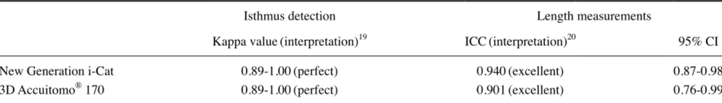
Table 3. Interobserver agreement for isthmus detection and length measurements on conebeam computed tomographic images
Isthmus detection Length measurements
Kappa value(interpretation)19 ICC(interpretation)20 95% CI
New Generation iCat 0.891.00(perfect) 0.940(excellent) 0.870.98
3D Accuitomo® 170 0.891.00(perfect) 0.901(excellent) 0.760.99
diagnostic tools, and mechanical motion stabilizers.32
Although the use of a small FOV reduces the radiation dose, the fact that CBCT involves a higher radiation dose than conventional radiographs restricts the application of CBCT to selected cases, and a thorough clinical examination is mandatory before choosing to perform CBCT. Post erior teeth with complex anatomy, symptoms, and/or persistent periapical lesions are strong candidates for a CBCT exam ination for the presence of isthmuses. Both limited and largevolume CBCT units have a similar ability to exhibit isthmuses, allowing both clinicians and radiologists to care fully study the root anatomy and its variations even when not all CBCT options are available. This technology still does not outperform microCT, since neither CBCT scanner detected isthmuses in the apical third in some cases. None theless, CBCT represents a valuable tool in endodontic practice.
Conflicts of Interest: None
References
1. Pécora JD, Estrela C, Bueno MR, Porto OC, Alencar AH, Sousa Neto MD, et al. Detection of root canal isthmuses in molars by mapreading dynamic using CBCT images. Braz Dent J 2013; 24: 56974.
2. Estrela C, Rabelo LE, de Souza JB, Alencar AH, Estrela CR, Sousa Neto MD, et al. Frequency of root canal isthmi in human permanent teeth determined by conebeam computed tomogra phy. J Endod 2015; 41: 15359.
3. Mannocci F, Peru M, Sherriff M, Cook R, Pitt Ford TR. The isthmuses of the mesial root of mandibular molars: a micro computed tomographic study. Int Endod J 2005; 38: 55863. 4. Tahmasbi M, Jalali P, Nair MK, Barghan S, Nair UP. Prevalence
of middle mesial canals and isthmi in the mesial root of mandi bular molars: an in vivo conebeam computed tomographic study. J Endod 2017; 43: 10803.
5. Srivastava S, Alrogaibah NA, Aljarbou G. Conebeam computed tomographic analysis of middle mesial canals and isthmus in mesial roots of mandibular first molars - prevalence and related factors. J Conserv Dent 2018; 21: 52630.
6. de Pablo OV, Estevez R, Péix Sánchez M, Heilborn C, Cohenca N. Root anatomy and canal configuration of the permanent mandibular first molar: a systematic review. J Endod 2010; 36: 191931.
7. Carr GB, Schwartz RS, Schaudinn C, Gorur A, Costerton JW. Ultrastructural examination of failed molar retreatment with secondary apical periodontitis: an examination of endodontic biofilms in an endodontic retreatment failure. J Endod 2009; 35: 13039.
8. AmorosoSilva PA, OrdinolaZapata R, Duarte MA, Gutmann JL, del CarpioPerochena A, Bramante CM, et al. Microcom puted tomographic analysis of mandibular second molars with Cshaped root canals. J Endod 2015; 41: 8905.
9. Tolentino ES, AmorosoSilva PA, Alcalde MP, Honório HM, Iwaki LC, RubiraBullen IR, et al. Accuracy of highresolution smallvolume conebeam computed tomography in detecting complex anatomy of the apical isthmi: ex vivo analysis. J Endod 2018; 44: 18626.
10. Tolentino ES, AmorosoSilva PA, Alcalde MP, Honório HM, Iwaki LC, RubiraBullen IR, et al. Limitation of diagnostic value of conebeam CT in detecting apical root isthmuses. J Appl Oral Sci 2020; 28: e20190168.
11. Domark JD, Hatton JF, Benison RP, Hildebolt CF. An ex vivo comparison of digital radiography and conebeam and micro computed tomography in the detection of the number of canals in the mesiobuccal roots of maxillary molars. J Endod 2013; 39: 9015.
12. VillasBôas MH, Bernardineli N, Cavenago BC, Marciano M, Del CarpioPerochena A, de Moraes IG, et al. Microcomputed tomography study of the internal anatomy of mesial root canals of mandibular molars. J Endod 2011; 37: 16826.
13. Jain S, Choudhary K, Nagi R, Shukla S, Kaur N, Grover D. New evolution of conebeam computed tomography in dentistry: combining digital technologies. Imaging Sci Dent 2019; 49: 17990.
14. Miracle AC, Mukherji SK. Conebeam CT of the head and neck, part 1: physical principles. AJNR Am J Neuroradiol 2009; 30: 108895.
15. Wrzesień M, Olszewski J. Absorbed doses for patients under going panoramic radiography, cephalometric radiography and CBCT. Int J Occup Med Environ Health 2017; 30: 70513. 16. Kamburoglu K, Onder B, Murat S, Avsever H, Yuksel S, Paksoy
CS. Radiographic detection of artificially created horizontal root fracture using different cone beam CT units with small fields of view. Dentomaxillofac Radiol 2013; 42: 20120261. 17. Hassan BA, Payam J, Juyanda B, van der Stelt P, Wesselink PR.
Influence of scan setting selections on root canal visibility with cone beam CT. Dentomaxillofac Radiol 2012; 41: 6458. 18. Mchugh ML. Interrater reliability: the kappa statistic. Biochem
Med(Zagreb) 2012; 22: 27682.
19. Koo TK, Li MY. A guideline of selecting and reporting intra class correlation coefficients for reliability research. J Chiropr Med 2016; 15: 15563.
20. Harris SP, Bowles WR, Fok A, McClanahan SB. An anatomic investigation of the mandibular first molar using micro-computed tomography. J Endod 2013; 39: 13748.
21. Vertucci FJ. Root canal morphology and its relationship to endo dontic procedures. Endod Topics 2005; 10: 329.
22. Endal U, Shen Y, Knut A, Gao Y, Haapasalo M. A highresolu tion computed tomographic study of changes in root canal isth mus area by instrumentation and root filling. J Endod 2011; 37: 2237.
23. Van Dessel J, Huang Y, Depypere M, RubiraBullen I, Maes F, Jacobs R. A comparative evaluation of cone beam CT and microCT on trabecular bone structures in the human mandible. Dentomaxillofac Radiol 2013; 42: 20130145.
24. Katsumata A, Hirukawa A, Okumura S, Naitoh M, Fujishita M, Ariji E, et al. Relationship between density variability and imaging volume size in conebeam computerized tomographic scanning of the maxillofacial region: an in vitro study. Oral Surg Oral Med Oral Pathol Oral Radiol Endod 2009; 107: 4205.
25. Scarfe WC, Farman AG. What is conebeam CT and how does it work? Dent Clin North Am 2008; 52: 70730.
26. Kau CH, Richmond S, Palomo JM, Hans MG. Threedimen sional cone beam computerized tomography in orthodontics. J Orthod 2005; 32: 28293.
27. Hassan B, Metska ME, Ozok AR, van der Stelt P, Wesselink PR. Comparison of five cone beam computed tomography sys tems for the detection of vertical root fractures. J Endod 2010; 36: 1269.
28. Hsu YY, Kim S. The resected root surface. The issue of canal isthmuses. Dent Clin North Am 1997; 41: 52940.
29. OrdinolaZapata R, Bramante CM, Versiani MA, Moldauer BI, Topham G, Gutmann JL, et al. Comparative accuracy of the clearing technique, CBCT and microCT methods in studying
the mesial root canal configuration of mandibular first molars. Int Endod J 2017; 50: 906.
30. Marca C, Dummer PM, Bryant S, VierPelisser FV, Só MV, Fontanella V, et al. Threerooted premolar analyzed by high resolu tion and cone beam CT. Clin Oral Investig 2013; 17: 153540.
31. Borges CC, Estrela C, Decurcio DA, Pécora JD, SousaNeto MD, RossiFedele G. Conebeam and microcomputed tomo graphy for the assessment of root canal morphology: a systematic review. Braz Oral Res 2020; 34: e056.
32. Bueno MR, Estrela C, Azevedo BC, Diogenes A. Development of a new conebeam computed tomography software for endo dontic diagnosis. Braz Dent J 2018; 29: 51729.