석 사 학 위 논 문
Niemann Pick Disease , type C 생쥐 해마로
투사하는 중추신경로의 변화에 관한 연구
제 주 대 학 교 대 학 원
의 학 과
김 중 모
ABSTRACT
Retrograde transneuronal labelling of the pseudorabies virus was used to investigate the differences of the CNS hippocampal pathways among NPC +/+, +/-, and -/- mice. For hippocampal pathways, a total of 3-5 ul Bartha strain of pseudorabies virus were injected into the hippocampus using stereotaxic instrument in 40 adult NPC +/+, +/-, and -/- mice. After 48 hours post pseudorabies virus injection, the brains were removed and the tissue sections were processed for immunohistochemistry using polyclonal antibody against the pseudorabies virus. Using streological counting methods, the PRV positive cells were counted in the positive nuclei of the whole brain of the NPC +/+, +/-, and -/- mice. The relative number of PRV positive cells in labelled nuclei were same in the hippocampus and medial septal nuclei of the NPC +/+, +/-, -/-. But there were significant decrease of PRV positive cell number in entorhinal cortex, cerebral cortex including piriform
cortex, lateral septal nucleus, thalamus, lateral hypothalmic nucleus, nucleus of the diagonal band, superior mammilary nucleus, locus cereuleus, medial geniculate nucleus, anterior olfactory nucleus in NPC -/- compared to the +/+ and +/-, and some showed no positive labeling. In summary, the CNS hippocampal pathways of the NPC -/- were reduced and the number of the cells within the pathways decreased in a circuit specific manner.
Our data suggests that the discontinuity of CNS hippocampal pathways in NPC -/- mice may contribute to the symptoms and functional disabilities especially memory and learning.
Keywords : Niemann-Pick type C, hippocampus, CNS pathway, pseudorabies virus
CONTENTS
ABTRACT ⅰ CONTENTS ⅲ LIST OF FIGURES ⅳ LIST OF TABLES ⅴ INTRODUCTION 1MATERIALS AND METHOD 3
RESULT 7
DISCUSSION 9
CONCLUSION 12
REFERENCES 13
LIST OF FIGURES
Fig. 1. PRV-immunoreactive neurons in the brain 18
Fig. 2. Schematic drawings illustrating the positive nuclei after PRV injection into the hippocampus of the NPC +/+
mouse 20
Fig. 3. Schematic drawings illustrating the positive nuclei after PRV injection into the hippocampus of the NPC -/-
LIST OF TABLE
Table 1. The average PRV positive cell number per each section at 48 hrs after PRV injection into the hippocampus of
INTRODUCTION
Niemann-Pick type C disease (NP-C) is a fatal, autosomal recessive disorder that results in premature death from progressive neuro-degeneration. (Penchev et al., 1995) Recently, researchers have discovered two genes that can produce the NP-C phenotype when mutated. In 1997, NPC1 was identified and sequenced (Carstea et al., 1997; Loftus et al., 1997) and in 2000, a second gene (HE1/NPC2) was identified (Naureckiene et al., 2000). At the cellular level, mutations in the NPC1 and NPC2 genes result in the late lysosomal accumulation of lipids [Sokol et al., 1988; Blanchette et al., 1988] and ultimately lead to disorders of cholesterol imbalance. NPC patients suffer from such symptoms as ataxia, impaired vertical gaze, and dementia. The common neuropathological features of NPC include neuronal ballooning, axonal spheroid formation, and neuro-degeneration (Tanaka et al., 1988; Penchev et al, 1995, Higashi et al., 1991, Higashi et al., 1993). The extent and specificity of neurodegeneration is still largely unknown.
The hippocampus is widely believed to exert an important influence on memory. And several neuronal pathways including the septohippocampal system are thought to play a crucial role in the
performance of a variety of complex learning and memory tasks[Coyle et al., 1983; Lamour et al., 1984]. This specific hippocampal function can be performed by neuronal circuits which connected in the brain. In cognitive and memory processes, cholinergic involvement has been known as one of the most important transmitter system.
The BALB/c-npc1nih mouse model for Niemann-Pick type C
has facilitated closer examination of this phenomenon of abnormal cholesterol storage and neuronal lose in the central nervous system. As in the human phenotype, central neurological symptoms such as ataxia and hind limb paresis dominate the clinical picture.
In order to assess the neuro-degenerative effects of NP-C on hippocampal afferent pathways, we performed a pseudorabies virus neurotracing study on wild-type NPC1 mice (+/+) and NPC1 knockout mice (+/-)/(-/-).
Materials and Method
1. Animals The BALB/c mice adult wild-type control(NPC +/+),
hetereozygous(NPC +/-) and homozygous(NPC-/-) animals of both sexes were used in this study. We have established a NPC mice colony, BALB/c mice carrying the genetic mutation for NPC1, and are ready to be used for our experiments. NPC heterozygous mice are being bred to acquire NPC (+/+), NPC (+/-), and NPC (-/-) mice. The genotypes of the mice are determined from genomic DNA isolated from tail-snip DNA using a polymerase chain reaction (PCR)-based method and oligonucleotide primers described previously.
2. Pseudorabies virus The Bartha strain of the pseudorabies
virus(PRV) was used in this study. The titer of the virus stock was, determined on PK15 cell line, was 1X10⁸ plaque forming unit(pfu)/ml.
Aliquots of the PRV were stored at – 80 oC, and vials were thawed immediately prior to the injection. Excess virus were inactivated with Clorox and discarded.
3. Surgery Animals were anesthetized with ketamine HCl(0,75
mg/kg) and xylazine(1 mg/kg) prior to surgical procedures The injection of pseudorabies virus was made into the hippocampus with an aid of the stereotaxic instrument at the point from bregma [dentate gyrus , 3.7
mm(AP), 2.4 mm(Lateral)]and Hamilton syringe was lowered vertically until it reaches the injection areas. Total 3 ul of the pseudorabies virus was injected slowly at the speed of 1 ul per minute. Then syringe was removed slowly and surgical wounds were sutured with wound clips. Most of the rats were allowed to survive a total of 2 days postinjection.
4. Immunohistochemistry All mice were reanesthetized in same
manner and perfused transcardially with 100-200 ml of heparinized saline(18 oC) followed by 400 ml of 4% paraformaldehyde-lysine periodate. The brains were removed, and cut in transverse plane at 30 um on a freezing microtome. The free floating tissue sections were processed for double immunofluorescence procedure using polyclonal antibodies to pseudorabies virus or choline acetyl transferase. One every six sections was incubated overnight at 38 oC with the mixture of rabbit anti pseudorabies virus(1:200) and goat anti choline acetyl transferase(1:200, Chemicon Int’l Inc., diluted in 0.1 M sodium phosphate buffer containing 1% normal donkey serum and 0.3% Triton X-100). After 14 hours, the sections were reacted for 2 hours with a cocktail of FITC labelled donkey anti-rabbit IgG(1:50, Jackson Immunoresearch Lab.) and TRITC labelled donkey anti-goat IgG(1:50, Jackson Immunoresearch Lab.). Cells were counted as positive if a reasonable portion of the cell body was visible in the section.
5. Stereology CAST stereolgy system with Olympus BX-51
microscope and CAST software were used for stereological cell counting of the PRV positive neurons in all mice. The unbiased stereological estimation of the total number, cell size and axon populations were made the optical fractionator. All the nuclei containing positive enurons were outlined from front to back through the nucleus/region of positive labelled. The PRV positive labelled cells were counted in regions outlined. Sampling was done using the Olympus CAST(Computer Assisted Stereological Toolbox) system version 2.1.0(Olympus Denmark A/S, Albertslund, Denmark). A counting frame(2025 um2 )was placed
randomly on the first counting area and systematically moved through all counting areas until the entire delineated area was sampled. The sampling frequency was chosen so that about 140 PRV positive cells were counted in each specimen. Average thickness of the sections were 12-17 um. A neuron was defined as a cell with a clearly visible cell body within the 10 um Z-plane of the counting frame using a 100X oil immersion objective. An upper guard zone of 2 um was used, so the cells were excluded from both surfaces to avoid the problem of the lost caps, and only the profiles that came into focus within the counting volume were counted. The estimate of the total number of the PRV containing neurons were calculated according to the optical fractionator formula.
percentage cell number (mean + S.E.M.) of the NPC+/+, +/-, and -/- animals. The P<0.05 level was used determine statistical significance.
RESULT
1. Cresyl violet staining in the NPC +/+, +/- and -/- mouse hippocampus
As shown in Table 1, at 4 weeks and 8 weeks of age both NPC -/- and NPC +/+ mice showed no significant differences in stereological cell counts of cresyl stained cells in the CA1, CA2/3 and dentate gyrus regions of the hippocampus.
2. PRV positive cells in the NPC +/+, +/- and -/- mouse hippocampus
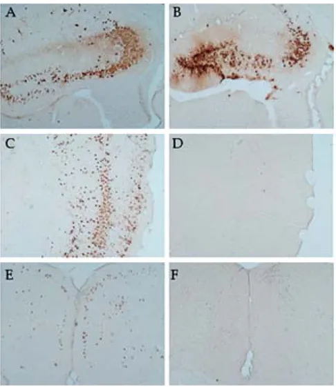
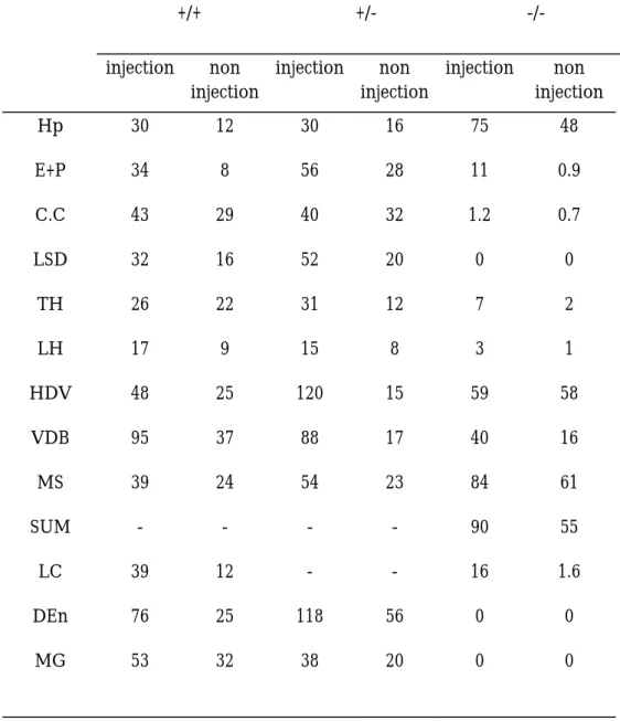
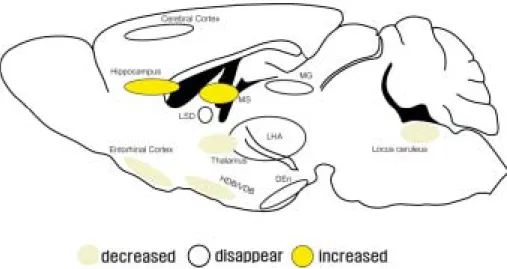
After injection of pseudorabies virus(PRV) into the hippocampus, the PRV immuno-positive cells were observed in several different areas of the brain. The labeling pattern of NPC -/- was different to NPC +/+ and +/-. The relative number of PRV positive cells in labelled nuclei were increased in the hippocampus(Hp), (HDV), and medial septal nucleus(MS) of the NPC-/- mouse.
But there were significant decrease of PRV positive cell number in entorhinal cortex(E), Cerebral cortex(CC) including
piriform cortex(P), lateral septal nucleus(LSD), thalamus(TH), lateral hypothalmic nucleus(LH), nucleus of the diagonal band(VDB), locus ceruleus(LC), medial geniculate nucleus(MG), dorsal entorhinal nucleus(DEn) in NPC -/- compared to the +/+ and +/-, and some showed no positive labeling.
DISCUSSION
The Pseudorabies virus (PRV) is a swine neurotropic alpha-herpesvirus that has been used for over twenty years to map neural circuits due to its ability to cross synaptic junctions. (Loewy et al., 1998) The Bartha strain, an attenuated strain used for retrograde tracing, can be injected into the peripheral nervous system or intracerebrally (Card J.P. et al., 1999; Aston-Jones G. et al., 2000). PRV has been used to map such pathways as: the retinal projections in mice (Provencio I. et al., 1998), the rat nucleus accumbens and thalamus (O'Donnell P. et al., 1997), and the rat prefrontal cortex to the ventral tegmental area (Carr D. B. et al., 2000). But care must be taken when using this virus to label neural circuits that are undergoing neuro-degeneration as is the case with NPC.
Research on PRV-Bartha’s replication, assembly and egress in neurons (Card et al., 1993) can shed light on its uses and limitations as a neurotracer in neurodegenerative cases. Viral replication occurs within the nucleus of the infected cell. From here, the naked capsid buds from the nuclear envelope and transverses the endoplasmic reticulum (ER). As the nuclear envelope fuses with the ER membrane the naked capsid is freed into the cytoplasm near
the Golgi complex. The Golgi complex then wraps around the naked capsid to form a bilaminar envelope. The enveloped capsids then enter the dendritic portion of the neurons where the outer membrane of the envelope can fuse with the post-synaptic terminal. The capsid, now surrounded by a single membrane, crosses the synaptic junction and fuses to the pre-synaptic terminal. The naked capsid then travels up the axon towards the nucleus to repeat the process of replication and egress. This process of infection must be kept in mind when using it as a neurotracer in NPC tissue
There are several possible explanations for the loss of PRV labeling in NPC1 (-/-) mice. (1) NPC1 (-/-) mice accumulate a large number of cholesterol-loaded vesicles within the cell body that can hinder viral movement throughout the neuron. This traffic jam effect could have an affect on viral access to the nucleus and/or on viral egress from the cell. (2) Intracellular trafficking of cholesterol is hindered in NPC (-/-) mice. If Golgi derived enveloping of viral capsids is cholesterol dependant then this process would be hindered in NPC1 (-/-) mice. (3) If neurodegeneration has reached a point where synapses are lost then the virus would be unable to enter the crippled neuron. (4) If non-labeled neurons have undergone nuclear lysis this would hinder viral entry, which appears to be receptor mediated, and would hinder viral replication.
In the present study, we examined the transsynaptic cholinergic circuitry projecting to the hippocampus using the pseudorabies virus as a neurotracer.Decreased nuclei are entorhinal cortex(E), Cerebral cortex(CC) including piriform cortex(P), lateral septal nucleus(LSD), thalamus(TH), lateral hypothalmic nucleus(LH), nucleus of the diagonal band(VDB), locus ceruleus(LC), medial geniculate nucleus(MG), dorsal entorhinal nucleus(DEn). This may be because of the difficulties in the viral transport due to the cholesterol accumulation in the npc -/- hippocampal cell body.
Interestingly, hippocampus(Hp), (HDV), and medial septal nucleus(MS) showed increased immunoreactivity in npc-/- mouse. This may help the explanation that virus had difficulty in the remote transport but icreased in the same cells which they infected first. So these nuclei could be candidates of the closely related nuclei to the hippocampus.
Totally, these changes tells that the CNS pathway to the
hippocampus has been broken occurs along the pathway, and cause memory disturbance
CONCLUSION
Our data suggests that the CNS pathways innervating the hippocampus were different among NPC +/+, +/-, -/- mice and the hippocampal innervation also showed degenerative changes in a transsynaptic manner in NPC -/- mice. The degeneration, disturbance of the transport in the axon or abnormal function of the synapse could be possible candidate for NPC mice symptoms.
REFERENCES
Aston-Jones, G., Card, J.P. 2000. Use of pseudorabies virus to delineate multisynaptic circuits in brain: opportunities and limitations. J Neurosci Methods.103 (1):51-61.
Blanchette-Mackie, E.J., Dwyer, N.K., Amende, L.M., et al. 1988. Type-C Niemann-Pick disease: low-density lipoprotein uptake is associated with premature cholesterol accumulation in the Golgi complex and excessive cholesterol storage in lysosomes. Proc. Natl. Acad. Sci. USA 85:8022–8026.
Card, J.P., Enquist, L.W., Moore, R.Y. 1999. Neuroinvasiveness of pseudorabies virus injected intracerebrally is dependent on viral concentration and terminal field density. J Comp Neurol. 407 (3): 438-452.
Carr, D.B., Sesack, S. R. 2000. Projections from the Rat Prefrontal Cortex to the Ventral Tegmental Area: Target Specificity in the Synaptic Associations with Mesoaccumbens and Mesocortical Neurons. J. Neurosci. 20 (10):3864 - 3873.
Coyle, J.T., Price, D.L., Delong, M.R. 1983. Alzheimer’s disease : a disorder of cortical cholinergic innervation, Science 219:1184-1190.
Carstea, E.D., Morris, J.A., Coleman, K.G., et al. 1997. Niemann–Pick C1 disease gene: homology to mediators of cholesterol homeostasis. Science 277:228–231.
Dougherty, K.D., Turchin, P.I., Walsh, T.J. 1998. Septocingulate and septohippocampal cholinergic pathways: involvement in working/episodic memory, Brain Res. 810(1-2):59-71.
German, D.C., Quintero, E.M., Liang, C.-L., et al. 2001. Selective neurodegeneration, without neurofibrillary tangles, in a mouse model of Niemann–Pick C disease. J. Comp. Neurol. 433:415–425
Higashi, Y., Pentchev, P.G., Murayama, S., et al. 1991. Pathology of Niemannn-Pick type C studies of murine mutants. In: F. Ikuta, Editor, Neuropathology in Brain Research, Elsevier Science, Amsterdam, pp. 85–102.
Higashi, Y., Murayama, S., Pentchev, P.G. et al. 1993. Cerebellar degeneration in the Niemann–Pick type C mouse. Acta Neuropathol.
Lamour, Y., Dutar, P., Jobert, A. 1984. Septo-hippocampal and other medial septum-diagonal band neurons: electrophysiological and pharmacological properties, Brain Res. 309(2):227-239.
Loewy, A.D. 1998. Viruses as transneuronal tracers for defining neural circuits. Neurosci Biobehav Rev. 22 (6): 679-684.
Loftus, S.K., Morris, J.A., Carstea, E.D., et al. 1997. Murine model of Niemann–Pick C disease: mutation in a cholesterol homeostasis gene. Science 277 :232–235.
Mesulam, M.M., Mefson, E.J., Wainer, B.H. et al. 1983. Central cholinergic pathways in the rat : an overview based on an alternate nomenclature(Ch1-Ch6), Neuroscience 10:1185-1201.
Naureckiene, S., Sleat, D.E., Lackland, H. 2000. Science 290:2298-2301
O'Donnell, P., Lavin, A., Enquist, L.W., et al. 1997. Interconnected parallel circuits between rat nucleus accumbens and thalamus revealed by retrograde transynaptic transport of pseudorabies virus. J Neurosci. 17 (6):2143-2167.
Penchev, P. G., Vanier M. T., Suzuki K., et al. 1995. Niemann-Pick disease type C: a cellular cholesterol lipidosis, in The Metabolic and Molecular Bases of Inherited Diseases (Scriver C. R., Stanbury J. B., Wyngaarden J. B., and Frederickson D. S., eds), pp. 2625-2639. McGraw-Hill, New York.
Provencio, I., Cooper, H.M., Foster, R.G. 1998. Retinal projections in mice with inherited retinal degeneration: implications for circadian photoentrainment. J Comp Neurol. 395 (4):417-439.
Sokol, J., Blanchette-Mackie, E.J., Kruth, H.S., et al. 1988. Type C Niemann-Pick disease: lysosomal accumulation and defective intracellular mobilization of low-density lipoprotein cholesterol. J. Biol. Chem. 263:3411–3417.
Tanaka, J., Nakamura, H., Miyawaki, S. 1988. Cerebellar involvement in murine sphingomyelinosis: a new model of Niemann-Pick disease. J. Neuropathol. Exp. Neurol. 47:291–300.
Vanier, M.T., Millat, G. 2003. Niemann-Pick disease type C. Clinical Genetics 64 (4):269-281.
Voykto, M.L. 1996. Cognitive functions of the basal forebrain cholinergic system in monkeys : memory or attention?, Behav. Brain Res. 75:13-25.
Fig. 1. PRV-immunoreactive neurons in the brain (× 40) a, b: hippocampus; c, d: cerebral cortex; e, f : cingulated cortex. a,c,e : npc +/+, b,d,f : npc-/-
Table 1. The average PRV positive cell number per each section at 48 hrs after PRV injection into the hippocampus of the mouse brain
+/+ +/- -/- injection non injection injection non injection injection non injection Hp 30 12 30 16 75 48 E+P 34 8 56 28 11 0.9 C.C 43 29 40 32 1.2 0.7 LSD 32 16 52 20 0 0 TH 26 22 31 12 7 2 LH 17 9 15 8 3 1 HDV 48 25 120 15 59 58 VDB 95 37 88 17 40 16 MS 39 24 54 23 84 61 SUM - - - - 90 55 LC 39 12 - - 16 1.6 DEn 76 25 118 56 0 0 MG 53 32 38 20 0 0
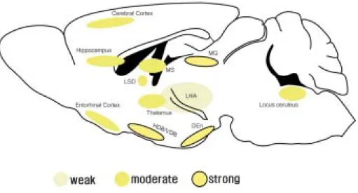
Fig. 2. Schematic drawings illustrating the positive nuclei after PRV injection into the hippocampus of the NPC +/+ mouse
Fig. 3. Schematic drawings illustrating the positive nuclei after PRV injection into the hippocampus of the NPC -/- mouse
Abbreviations : C.C, Cerebral cortex; DEn, Dorsal entorhinal area; E+P, Entorhinal and piriform cortex; HDV, Diagonal band, H; Hp, Hippocampus; LC, Locus ceruleus; LH, Lateral hypothalamic area; LSD, Lateral septal dorsal nucleus; MS, Medial septal nucleus; SUM, supramammillary nucleus; TH, Thalamus; VDB, Diagonal band,Ventral
국문초록
Niemann Pick Disease , type C 생쥐 해마로 투사하는
중추신경로의 변화에 관한 연구
Niemann-Pick 병은 대표적인 지질대사 장애질환으로서 간, 비장, 골수, 뇌 등에 지질이 과축적되며 유전형 열성유전질병으로서 영아 또는 유아에게 발생한다. 특히 치명적으로 간․ 비종대와 중추신경계의 손상을 동반하는 경우가 많다. 그러나 이 질병에 대해 국․ 내외적으로 특별한 치료법은 발견되지 않고 있다. 본 연구는 Niemann-Pick disease type C knock-out 마우스를 대상으로 pseudorabies virus 를 이용하여 해마로 투사하는 신경로의 변화를 조사하였다. 연구결과 해마 등 일부 신경핵에서는 양성반응 신경세포 수가 증가 하였고 시상등 대부분의 신경핵에서는 양성신경세포수가 감소하였다. 또한 이와 같은 퇴행성 변화들은 해나 신경로를 따라 일어났었다. 이상과 같은 결과는 Niemann-Pick 병의 증상 중 기억장애가 단순한 콜레스테롤 축적 뿐만 아니라 신경로 차단에 의해 발생한다는 것을 입증하였다고 할 수 있다.