www.epain.org 217 Korean J Pain 2021;34(2):217-228
pISSN 2005-9159 eISSN 2093-0569
Clinical Research Article
This is an open-access article distributed under the terms of the Creative Commons Attribution Non-Commercial License (http://cre-ativecommons.org/licenses/by-nc/4.0/), which permits unrestricted non-commercial use, distribution, and reproduction in any medium, provided the original work is properly cited.
© The Korean Pain Society, 2021
Author contributions: Elif Yakşi: Investigation; Ayşegül Ketenci: Study conception; Mehmet Barış Baslo: Investigation; Elif Kocasoy Orhan: Inves-tigation.
Does transcutaneous electrical nerve stimulation affect pain,
neuropathic pain, and sympathetic skin responses in the
treatment of chronic low back pain? A randomized,
placebo-controlled study
Elif Yakşi1, Ayşegül Ketenci2, Mehmet Barış Baslo3, and Elif Kocasoy Orhan3
1Department of Physical Medicine and Rehabilitation, Abant Izzet Baysal University, Faculty of Medicine, Bolu, Turkey 2Department of Physical Medicine and Rehabilitation, Istanbul University, Istanbul Faculty of Medicine, Istanbul, Turkey 3Department of Neurology, Istanbul University, Istanbul Faculty of Medicine, Istanbul, Turkey
Received September 13, 2020 Revised January 5, 2021 Accepted January 7, 2021
Handling Editor: Woo Seog Sim
Correspondence
Elif Yakşi
Department of Physical Medicine and Rehabilitation, Abant Izzet Baysal University, Faculty of Medicine, Orüs Boulevard, No: 59, Karacasu, Bolu 14020, Turkey
Tel: +905069078505 Fax: +903742628471 E-mail: [email protected]
Background: The purpose of this study was to assess the effectiveness of trans-cutaneous electrical nerve stimulation (TENS) in chronic low back pain and neuro-pathic pain.
Methods: Seventy-four patients aged 18-65 with chronic low back pain were in-cluded in the study. Baseline measurements were performed, and patients were randomized into three groups. The first group received burst TENS (bTENS), the sec-ond group conventional TENS (cTENS), and the third group placebo TENS (pTENS), all over 15 sessions. Patients’ visual analogue scale (VAS) scores were evaluated before treatment (preT), immediately after treatment (postT), and in the third month after treatment (postT3). Douleur Neuropathique 4 Questions (DN4), the Modified Oswestry Low Back Pain Disability Questionnaire (MOS), the Beck Depression Inven-tory (BDI), and sympathetic skin response (SSR) values were also evaluated preT and postT3.
Results: A statistically significant improvement was observed in mean VAS scores postT compared to preT in all three groups. Intergroup comparison revealed a signif-icant difference between preT and postT values, that difference being assessed in favor of bTENS at multiple comparison analysis. Although significant improvement was determined in neuropathic pain DN4 scores measured at postT3 compared to preT in all groups, there was no significant difference between the groups. No sta-tistically significant difference was also observed between the groups in terms of MOS, BDI, or SSR values at postT3 (P > 0.05).
Conclusions: bTENS therapy in patients with low back pain is an effective and safe method that can be employed in short-term pain control.
Key Words: Chronic Pain; Depression; Electromyography; Low Back Pain; Neural-gia; Pain Management; Surveys and Questionnaires; Sympathetic Nervous System; Transcutaneous Electric Nerve Stimulation.
INTRODUCTION
Low back pain is a widespread global public health prob-lem and cause of disability resulting in increased medical spending and workforce losses. Eighty percent of the pop-ulation experience low back pain attacks at some time in their lives. Low back pain initially exhibits a good progno-sis, and 54%-90% of patients improve within a few months. However, recurrence develops in 24%-80% of patients in the first year, and chronic problems capable of causing disability are seen in a mean 20% of patients [1-3].
Chronic back pain is defined as persistence for longer than three months, and progresses together with psycho-social and functional impairment [4]. Mechanical low back pains are those deriving from anatomical structures such as the vertebral column, intervertebral disks, and soft tissue, and in which underlying inflammatory, malig-nant, and infectious pathologies have been excluded [5]. Mechanisms causing nociceptive and neuropathic pain are involved in the pathophysiology of low back pain [6]. Nociceptive pain derives from inflammatory responses associated with tissue injury [7], while neuropathic pain has been defined as those deriving from primary lesions, dysfunction, or temporary impairment of the peripheral or central nervous system [8,9]. Four percent of the adult population experiences neuropathic chronic low back pain at some time in their lives. Epidemiological studies show that the incidence of neuropathic pain in patients with low back pain ranges between 17% and 54%. The most common neuropathic pain presentation is chronic lumbar radiculopathy [6,10,11].
In addition to various conservative therapies such as medical therapies, physiotherapy, exercise, and comple-mentary procedures in the treatment of low back pain, multidisciplinary approaches such as behavioral cognitive therapy and, if necessary, invasive methods, can also be employed [12,13]. Transcutaneous electrical nerve stimu-lation (TENS), which is frequently applied in clinical prac-tice, is a widely employed non-invasive, easily applied, and safe method with a low side-effect profile [14,15].
Various effect mechanisms have been proposed for TENS. According to Melzack and Wall’s ‘gate control’ the-ory, cells in the medulla spinalis substantia gelatinosa are stimulated by nociceptive and sensory signals. The ‘gate control theory’ proposes that pain sensation commenc-ing from nociceptors is prevented from becommenc-ing transported to the higher centers of the brain where pain is perceived. According to the theory, the stimulation of large-diameter A-β sensory afferents activates inhibitor interneurons in the substantia gelatinose in the posterior horn of the spi-nal cord, thus weakening the transmission of nociceptive signals transmitted by small-diameter A-δ and C fibers.
In addition, a series of cascades including endogenous opioids involving supraspinal mechanisms also play a role in pain control [16,17]. TENS can be applied in different forms, depending on intensity and frequency (high fre-quency is > 50 Hz, and low frefre-quency < 10 Hz) [17]. Differ-ent therapeutic effects can thus be achieved by modifying the frequency, duration, and amplitude of the electric cur-rent [18]. Conventional TENS (cTENS) involves the applica-tion of a high-frequency and low-intensity current, while burst TENS (bTENS) consists of a low-frequency current applied at a high frequency in burst form [19].
The primary aim of this study was to investigate the ef-fectiveness of TENS therapy in mechanical type low back pain, and to assess the efficacy of different methods of ap-plication on pain, neuropathic pain, functional status, and attitudes and symptoms in the light of electrophysiological findings.
MATERIALS AND METHODS
The research was planned as a prospective, randomized, single-blind, placebo-controlled study. The study was per-formed in compliance with the Helsinki Declaration, and approval was granted by the Istanbul University Medical Faculty Clinical Research Ethical Committee (no. 2012/12-95-1167).
The patients included in the study were informed about the aim of the study, its duration, the therapeutic methods to be applied in the scope of the research, and potential problems and side-effects during follow-up, both verbally and in writing, using a ‘Volunteer Information Form.’ All patients agreeing to participate gave written consent by signing a ‘Volunteer Consent Form.’
1. Patients
Seventy-four patients (27 female, 47 male, mean age 43.3 ± 11.3) aged 18-65, presenting to the Physical Medicine and Rehabilitation Clinic between January and June 2013, with mechanical type low back pain persisting for more than three months, were invited to take part in the study. Patients aged 18-65 years with mechanical chronic low back pain persisting for longer than 12 weeks and with no underlying inflammatory or neoplastic disease were included. Participants included in the study were selected from patients with lumbar disc disease without radicular compression. Thirty-two patients had degenerative disc disease and 42 had disc herniation without radiculopathy. In order to exclude neuropathic pain caused by radiculop-athy, pre-treatment conduction and needle electromyog-raphy (EMG) examinations were evaluated. Patients with
diabetes mellitus, inflammatory disease, hypothyroid-ism, vitamin B12 deficiency, major depression, comorbid diseases capable of affecting sympathetic skin response (SSR), or history of major trauma or surgery to the low back region were excluded from the study. In order to achieve α < 0.05 and β = 80%, according to the visual analogue scale (VAS) scores, we calculated that 21 participants would be required for each group, as described by Topuz et al. [20].
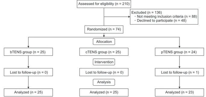
This research was designed as a single-blind study. The participants were not informed whether they were being given the main treatment or a placebo. Patients’ demographic data such as age, sex, body mass index, and occupation were recorded together with their clinical characteristics. Detailed histories were taken, and clinical examinations were performed. Existing laboratory and ra-diological tests were also assessed. Patients were evaluated immediately after treatment (postT) for short term effects, and the third month after treatment (postT3) for long term effects. The study CONSORT (Consolidated Standards of Reporting Trials) flow diagram is summarized in Fig. 1.
2. Assessment methods
1) Pain measurement
A VAS was employed to assess the severity of low back pain [21]. Patients were asked to mark a point on a 10-cm line, on which 0 represented no pain and 10 the worst possible pain, corresponding to the severity of their pain. Patients were asked about the mean pain level felt during the day and night-time pain before treatment, at the end of treatment, and at follow-ups in the third month. VAS was
applied in the early period, in contrast to the other assess-ment methods employed in the study, since neurological pain mechanisms are more complex, and we anticipated that a longer time would be required until significant im-provement occurred.
2) Neuropathic pain evaluation
The severity of neuropathic pain was assessed at the start of treatment and in postT3 using the Douleur Neu-ropathique 4 Questions (DN4) scale. DN4 consists of four questions using seven items related to symptoms, three of which are associated with neurological examination. Total scores were calculated, with scores above 4 indicating the presence of neuropathic pain [22].
3) Functional status evaluation
The Modified Oswestry Low Back Pain Disability Ques-tionnaire (MOS) consisting of 10 questions scored 0-5 was used to assess patients’ functional status. The scale in-vestigates intensity of pain, and degree of change in pain during personal care, lifting, walking, sitting, standing, sleeping, social life, and traveling. Patients’ MOS values were evaluated at the start of treatment and in postT3 [23].
4) Evaluation of symptoms of depression
The Beck Depression Inventory (BDI) was applied for the purpose of measuring physical, emotional, and cognitive signs encountered in depression, such as pessimism, ir-ritability, fatigue, and weight loss. BDI scores are
evalu-Fig. 1. CONSORT (Consolidated Standards of Reporting Trials) flow diagram of the patient inclusion process. TENS: transcutaneous electrical nerve stimulation, bTENS: burst TENS, cTENS: conventional TENS, pTENS: placebo TENS.
Assessed for eligibility (n = 210)
Randomized (n = 74)
Excluded (n = 136)
Not meeting inclusion criteria (n = 88) Declined to participate (n = 48)
Allocation
Intervention
Analysis
Analyzed (n = 25) Analyzed (n = 25) Analyzed (n = 23) bTENS group (n = 25) Lost to follow-up (n = 0) cTENS group (n = 25) Lost to follow-up (n = 0) pTENS group (n = 24) Lost to follow-up (n = 1)
ated from absence of symptoms to severe symptoms. Total scores range between 0 (no depression) to 63 (severe de-pression). Patients’ BDI scores were evaluated at the start of treatment and in postT3 [24].
5) Electromyographic evaluation
Conduction examinations and needle EMG were per-formed on all patients before treatment in order to dif-ferentiate peripheral neuropathy associated with other causes and potential peripheral nerve and root lesions. SSR was subsequently studied from the hands and feet, with amplitude and latency values being recorded. SSR evaluation was performed at a room temperature of 25°C, in a standardized, quiet, and semi-dark environment, at the Istanbul University Medical Faculty Neurology De-partment using an EMG device (Dantec Key Point® or
two-channel Keypoint v3.03; Medtronic, Skovlunde, Denmark) permitting computer software SSR measurements. SSR was measured at a room temperature of 25°C, with pa-tients at rest in a supine position. Six-millimeter silver disk electrodes were employed as recording electrodes. For SSR evaluation of the upper extremity, the active (–) electrode was attached to the palmar region, the reference electrode (+) to the dorsum of the hand, and the earth electrode to the forearm. Similarly in the lower extremities, the active electrode was attached to the sole of the foot, and the refer-ence electrode to the dorsum of the foot. SSR was obtained from the right upper and lower extremities through stimu-lation of the median nerve from the second finger or toe. Duration of stimulation was set to 0.1 msec and intensity to 15-20 mA. Stimuli were applied at varying intervals and with > 30 seconds between stimuli in order to avoid ha-bituation. A maximum of five unilateral stimulations were applied to each patient. SSR was regarded as unresponsive if no response was achieved to these five stimuli. Latency was defined as the time to the start of the first deflection from the start of the stimulus artefact (msec), and ampli-tude was measured from peak to peak (mV). The shortest latency and highest amplitude values were included in the analysis. SSR was re-evaluated postT3.
3. Treatment method
Patients were divided into three groups — cTENS, bTENS, and placebo TENS (pTENS). Randomization was per-formed with a sequential order list using the Microsoft Excel© 2003 (Microsoft, Redmond, WA) random number
production function.
TENS Intellect® Advanced (Chattanooga, Mouguerre,
France) device is used in our study. cTENS stimulation parameters are 60-80 Hz, with a pulse width of short
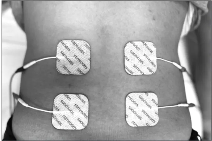
dura-tion (50-80 µs), 10-30 mA intensity and bTENS comprises a baseline low frequency current (1-4 Hz) together with high frequency trains (50-100 Hz). Patients were informed about the therapeutic method and potential side-effects before being taken for treatment. Two channels (four electrodes), one applied to the right and the other to the left, were used in treatment. The active electrode emerging from the dif-ferent channel was attached 1.5 cm laterally to the ver-tebrae at the L2-L4 level, while the passive electrode was placed 3 cm distally to the active electrode. TENS therapy was applied for 30 minutes in the placebo group, with no current being given to the TENS device, and at moderate intensity, depending on the patient’s tolerance, in such a way as not to cause pain or discomfort, in the individuals receiving burst and conventional therapy. Patients were not given any regular medical treatment, but were permit-ted to use tablets containing paracetamol if required, on the condition that the dosage did not exceed 1 g a day. Pain diaries were given to all patients, and they were asked to register their pain intensities and paracetamol consump-tion at home. Treatment was applied for a total of 15 ses-sions, five times a week for three weeks. The placement of the electrodes in the lumbar region is shown in Fig. 2.
4. Statistical analysis
Statistical analysis was conducted on SPSS version 21.0 (IBM Co., Armonk, NY) software. Mean, standard devia-tion, median, minimum, and maximum values were cal-culated for all parameters.
The Kolmogorov–Smirnov and Shapiro–Wilk tests were applied on commencement to evaluate normality of dis-tribution among the three groups. The Student t, Wilcoxon signed ranks, and Freidman tests were employed to com-pare intragroup changes. Intergroup comparisons were performed using the one-way ANOVA and Kruskal–Wallis
tests. For multiple comparisons, the Mann–Whitney U test with Bonferroni adjustment was used, where P < 0.05/ (number of comparsions) indicates a statistically signifi-cant difference between two groups. Numerical data were assessed using the chi-square test. Correlations between measurements were assessed using Spearman’s rho test and the Pearson test in the light of the character and distribution of variables. Results were analyzed at a 95% confidence interval, and P-values < 0.05 were regarded as statistically significant.
RESULTS
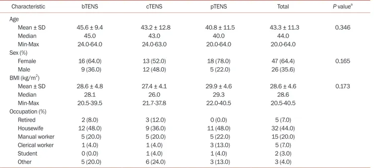
The 74 patients taking part in the study were assigned into three groups: bTENS (n = 25), cTENS (n = 25), and pTENS (n = 24). One patient from the placebo group was excluded from the analysis due to a failure to attend follow-ups, and the study was finally completed with 73 patients (25 bTENS, 25 cTENS, and 23 pTENS). Patients’ demographic data and clinical characteristics are summarized in Table 1. No TENS-associated side-effect developed in any patient. No patients reported using paracetamol. Demo-graphic and clinical characteristics were similar between Table 1. Patients’ demographic data and clinical characteristics by groups
Characteristic bTENS cTENS pTENS Total P valuea
Age Mean ± SD 45.6 ± 9.4 43.2 ± 12.8 40.8 ± 11.5 43.3 ± 11.3 0.346 Median 45.0 43.0 40.0 44.0 Min-Max 24.0-64.0 24.0-63.0 20.0-64.0 20.0-64.0 Sex (%) Female 16 (64.0) 13 (52.0) 18 (78.0) 47 (64.4) 0.165 Male 9 (36.0) 12 (48.0) 5 (22.0) 26 (35.6) BMI (kg/m2) Mean ± SD 28.6 ± 4.8 27.4 ± 4.1 29.9 ± 4.6 28.6 ± 4.6 0.173 Median 28.1 26.0 29.3 28.6 Min-Max 20.5-39.5 21.7-37.8 22.0-40.5 20.5-40.5 Occupation (%) Retired 2 (8.0) 3 (12.0) 0 (0.0) 5 (7.0) Housewife 12 (48.0) 9 (36.0) 11 (48.0) 32 (44.0) Manual worker 5 (20.0) 5 (20.0) 5 (22.0) 15 (20.0) Clerical worker 1 (4.0) 1 (4.0) 3 (13.0) 5 (7.0) Student 0 (0.0) 1 (4.0) 1 (4.0) 2 (3.0) Other 5 (20.0) 6 (24.0) 3 (13.0) 3 (4.0)
TENS: transcutaneous electrical nerve stimulation, bTENS: burst TENS, cTENS: conventional TENS, pTENS: placebo TENS, SD: standard deviation, Min: minimum, Max: maximum, BMI: body mass index.
aKruskal–Wallis test, analysis of variance test.
Table 2. Intragroup comparison of preT, postT, and postT3 VAS scores
VAS scores bTENS (n = 25) cTENS (n = 25) pTENS (n = 23)
Mean VAS scores
preT 6.8 ± 1.5 7 (4-10) 6.2 ± 1.7 6 (4-9) 6.6 ± 1.1 7 (5-8)
postT 2.8 ± 2.1 3 (0-9) 4.0 ± 2.3 4 (0-9) 4.5 ± 1.6 5 (1-8)
postT3 4.2 ± 2.9 4 (0-10) 3.7 ± 2.9 4 (0-10) 5.0 ± 1.9 5 (1-8)
P valuea < 0.001 < 0.001 0.016
Night-time VAS scores
preT 4.6 ± 3.0 5 (0-9) 4.4 ± 3.3 5 (0-9) 2.2 ± 2.5 6 (0-8)
postT 1.8 ± 2.4 0 (0-9) 2.7 ± 2.9 2 (0-9) 2.9 ± 3.0 3 (0-8)
postT3 2.2 ± 2.5 1 (0-10) 3.1 ± 2.3 2 (0-10) 3.6 ± 3.0 4 (0-8)
P valuea < 0.001 0.003 < 0.001
Values are presented as mean ± standard deviation or median (minimum-maximum).
preT: before treatment, postT: after treatment, postT3: third month after treatment, VAS: visual analogue scale, TENS: transcutaneous electrical nerve stimulation, bTENS: burst TENS, cTENS: conventional TENS, pTENS: placebo TENS.
the three groups (P > 0.05).
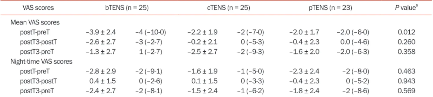
The preT, postT, and postT3 mean, standard devia-tion, median, and min-max values for the mean VAS and night-time VAS scores are shown in Table 2. Intragroup comparisons revealed significant improvement in mean and night-time pain severity measured using VAS in postT and postT3 mean values compared to preT (Table 2). PreT, postT, and postT3 VAS score changes are shown in Fig. 3. Intergroup comparison revealed that this significance was between postT and preT values (Table 3). A Mann–Whitney U test significance level with Bonferroni correction of P < 0.05/3 (0.0167) was adopted. Significant differences be-tween postT and preT values were observed bebe-tween the bTENS and pTENS groups (P = 0.007). Multiple
compari-sons of postT and preT VAS scores among the groups using the Mann–Whitney U test (with Bonferroni correction) revealed significant improvement in favor of bTENS (Table 4).
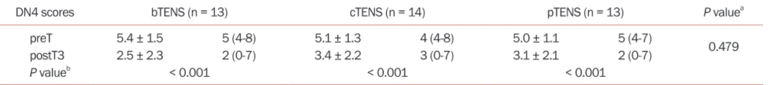
Neuropathic pain assessment was performed on 74 patients included in the study before treatment using the DN4 scale. The 40 patients subsequently diagnosed with neuropathic pain (DN4 > 4) were then subjected to subgroup analysis based on treatment groups. PreT and postT3 mean, standard deviation, median, and min-max DN4 values are shown in Table 5. Intragroup analysis re-vealed statistically significant improvement in all groups in DN4 measurements in postT3 values. However, no sta-tistically significant difference was observed in intergroup
preT postT postT3
10 8 6 4 2 Mean V A S scores Measurement time 0 * * bTENS cTENS pTENS TENS modality
preT postT postT3
10 8 6 4 2 Night-time V AS scores Measurement time 0 bTENS cTENS pTENS TENS modality
Fig. 3. Intergroup comparisons of mean and night-time visual analogue scale (VAS) scores. Asterisk (*) indicates statistically significant differences be-tween the time points within the group. For the lines in a box and whisker plot: error bars are the 95% confidence interval, the bottom and top of the box are the 25th and 75th percentiles, the line inside the box is the 50th percentile (median), and any outliers are shown as open circles or asteriks. TENS: transcutaneous electrical nerve stimulation, bTENS: burst TENS, cTENS: conventional TENS, pTENS: placebo TENS, preT: before treatment, postT: after treatment, postT3: third month after treatment.
Table 3. Intergroup comparison of preT, postT, and postT3 VAS scores
VAS scores bTENS (n = 25) cTENS (n = 25) pTENS (n = 23) P valuea
Mean VAS scores
postT-preT –3.9 ± 2.4 –4 (–10-0) –2.2 ± 1.9 –2 (–7-0) –2.0 ± 1.7 –2.0 (–6-0) 0.012 postT3-postT –2.6 ± 2.7 –3 (–2-7) –0.2 ± 2.1 0 (–5-3) –0.4 ± 2.3 0.0 (–4-6) 0.260 postT3-preT –1.3 ± 2.7 1 (–2-7) –2.5 ± 2.7 –2 (–9-3) –1.6 ± 2.0 –2.0 (–6-3) 0.358 Night-time VAS scores
postT-preT –2.8 ± 2.9 –2 (–9-1) –1.6 ± 1.9 –1 (–5-0) –2.3 ± 2.4 –2 (–8-0) 0.463 postT3-postT 0.4 ± 1.5 0 (–2-6) 0.1 ± 1.5 0 (–3-3) –0.4 ± 2.3 0 (–5-2) 0.943 postT3-preT –2.4 ± 2.7 –2 (–8-1) –1.5 ± 2.4 –1 (–6-2) –1.8 ± 2.4 –2 (–8-6) 0.569 Values are presented as mean ± standard deviation or median (minimum-maximum).
preT: before treatment, postT: after treatment, postT3: third month after treatment, VAS: visual analogue scale, TENS: transcutaneous electrical nerve stimulation, bTENS: burst TENS, cTENS: conventional TENS, pTENS: placebo TENS.
comparisons.
Third-month intragroup analysis of MOS values re-vealed significant improvement in all the treatment groups, but no statistically significant difference was ob-served between them (Table 6). No statistically significant improvement was observed in BDI depressive symptoms at postT3 at intragroup analyses, and no significant differ-ence was also determined among the three groups (Table 6).
Intragroup comparisons revealed no statistically
sig-nificant difference at postT3 in terms of SSR latency and amplitude values obtained from the upper extremity, or of SSR latency values recorded from the lower extremity. Similarly, there was no statistically significant difference between the groups. Intragroup analysis of SSR amplitude values recorded from the lower extremity increased signif-icantly in the bTENS and pTENS groups, but no significant difference was observed between the three study groups
(Table 7).
DISCUSSION
This study evaluated the efficacy of TENS on pain and neuropathic pain in chronic back pain in a placebo-con-trolled manner. The primary outcome of this study was that bTENS treatment was significantly more effective in short-term pain control compared to cTENS and pTENS in terms of pain severity capable of measurement using VAS scores. The secondary outcome was that bTENS and cTENS treatments were no different from the placebo in terms of neuropathic pain control.
Although data exist for the short-term effectiveness of TENS therapy, which has been employed in clinical prac-Table 4. Intergroup multiple comparison analysis of postT and preT VAS
scores
postT-preT VAS Mann–Whitney U test (Bonferroni correction)
Z P value
bTENS-cTENS –2.375 0.018
bTENS-pTENS –2.685 0.007a
cTENS-pTENS –0.348 0.728
preT: before treatment, postT: after treatment, VAS: visual analogue scale, TENS: transcutaneous electrical nerve stimulation, bTENS: burst TENS, cTENS: conventional TENS, pTENS: placebo TENS.
aP values indicate a statistical significance at P < 0.0167, after
Bonfer-onni adjustment.
Table 5. Intra- and intergroup comparison of preT and postT3 DN4 scores
DN4 scores bTENS (n = 13) cTENS (n = 14) pTENS (n = 13) P valuea
preT 5.4 ± 1.5 5 (4-8) 5.1 ± 1.3 4 (4-8) 5.0 ± 1.1 5 (4-7)
0.479
postT3 2.5 ± 2.3 2 (0-7) 3.4 ± 2.2 3 (0-7) 3.1 ± 2.1 2 (0-7)
P valueb < 0.001 < 0.001 < 0.001
Values are presented as mean ± standard deviation or median (minimum-maximum).
preT: before treatment, postT3: third month after treatment, DN4: Douleur Neuropathique 4 Questions, TENS: transcutaneous electrical nerve stimula-tion, bTENS: burst TENS, cTENS: conventional TENS, pTENS: placebo TENS.
aKruskal–Wallis test. bt-test.
Table 6. Intra- and intergroup comparison of preT and postT3 MOS and BDI values MOS and BDI
values bTENS (n = 25) cTENS (n = 25) pTENS (n = 23) P valuea
MOS preT 32.0 ± 6.3 31 (18-45) 30.6 ± 8.2 32 (14-47) 32.6 ± 6.3 33 (18-44) 0.476 postT3 25.4 ± 9.4 25 (11-45) 26.0 ± 10.6 28 (10-48) 28.3 ± 7.9 29 (14-43) P valueb 0.001 0.003 0.002 BDI preT 15.4 ± 7.3 16 (3-29) 10.0 ± 6.1 9 (0-26) 10.7 ± 5.5 10 (1-23) 0.283 postT3 14.2 ± 8.5 14 (1-29) 10.7 ± 7.7 11 (0-30) 9.3 ± 6.1 8 (1-26) P valueb 0.428 0.594 0.160
Values are presented as mean ± standard deviation or median (minimum-maximum).
preT: before treatment, postT3: third month after treatment, MOS: Modified Oswestry Low Back Pain Disability Questionnaire, BDI: Beck Depression In-ventory, TENS: transcutaneous electrical nerve stimulation, bTENS: burst TENS, cTENS: conventional TENS, pTENS: placebo TENS.
tice for many years, the data for long-term effectiveness are inconsistent [14,25-27]. Marchand et al. [28] compared the effectiveness of cTENS and pTENS in chronic low back pain. Those authors reported that cTENS application was significantly more effective than placebo in the control of early period pain, but that no difference was detected between the two therapies at the third and sixth months after treatment. Jarzem et al. [29] reported that cTENS was superior to a placebo in the control of short-term pain. Topuz et al. [20] also concluded that cTENS was more ef-fective in the control of short-term pain than a placebo in chronic low back pain.
The superiority of cTENS over a placebo reported in these studies shows that active TENS application is effec-tive in the control of short-term pain. Although the aceffec-tive TENS treatment bTENS was superior to placebo in short-term pain control, cTENS was not superior to placebo in the control of short-term pain in the present study. This discrepancy between the present study and previous re-search may derive from methodological differences. The cTENS method stimulates group II (Aβb) fibers and pro-vides analgesia through spinal segmental mechanisms providing inhibition of type IV (C) fibers and type III (Aδd) fibers. The bTENS method stimulates group III (Aδd) no-ciceptive fibers to release endogenous opiate at the spinal and supraspinal levels [30]. bTENS provides spinal analge-sia, and may also provide stronger analgesia by binding to opioid receptors in the brain by releasing the body’s own
endorphins. While the effectiveness of TENS in chronic low back pain has previously been evaluated in conven-tional or acupuncture mode, to the best of our knowledge there have been no studies investigating burst, and the present research is thus the first from that perspective.
The great majority of studies involving TENS have fo-cused on healing in the nociceptive component of pain, and very few studies have examined its effectiveness on the neuropathic component. The majority of those studies that have been conducted have considered diabetic pe-ripheral neuropathies and trigeminal neuralgias, and have reported that TENS is effective in controlling pain in these diseases [31-35]. Forst et al. [36] compared active TENS and a placebo in the treatment of diabetes-related neuropathic pain and reported that active TENS was superior to the placebo at 6 and 12 weeks. Active TENS may have emerged as superior to the placebo on neuropathic pain scores, in contrast to the present study, due to the use in that study of a questionnaire, a subjective evaluation tool. In another study, Jin et al. [32] reported that active TENS adminis-tered to patients with diabetic peripheral neuropathy resulted in a significantly greater improvement in short-term neuropathic pain scores compared to a placebo, but observed no statistically significant difference between the groups in the third month. The long-term results of the present research are consistent with these previous stud-ies. In addition, due to different pathophysiology, clinical findings and healing processes involved in neuropathic Table 7. Intra- and intergroup comparison of preT and postT3 SSR values
SSR values bTENS (n = 25) cTENS (n = 25) pTENS (n = 23) P valuea
Upper extremity Latency (ms) preT 1.4 ± 0.2 1.4 (1.2-1.8) 1.3 ± 0.2 1.3 (0.9-1.6) 1.3 ± 0.2 1.4 (0.8-1.8) 0.913 postT3 1.4 ± 0.2 1.4 (0.7-1.8) 1.3 ± 0.1 1.3 (0.9-1.6) 1.4 ± 0.1 1.4 (1.2-1.5) P valueb 0.993 0.542 0.821 Amplitude (mV) preT 2.7 ± 2.6 2.0 (0.8-10.9) 2.9 ± 1.7 1.7 (0.8-7.4) 2.7 ± 1.6 2.4 (0.4-5.2) 0.660 postT3 2.7 ± 1.9 1.9 (0.9-8.0) 2.8 ± 1.4 2.6 (1.2-4.9) 3.6 ± 2.1 3.0 (1.2-8.4) P valuec 0.937 0.575 0.508 Lower extremity Latency (ms) preT 2.1 ± 0.3 2.0 (1.7-2.7) 1.7 ± 0.2 1.8 (1.2-2.0) 1.8 ± 0.3 1.9 (1.1-2.1) 0.903 postT3 1.9 ± 0.4 1.8 (1.2-2.6) 1.7 ± 0.2 1.7 (1.2-2.1) 1.6 ± 0.2 1.7 (1.2-2.0) P valuec 0.367 0.484 0.314 Amplitude (mV) preT 0.9 ± 0.4 0.8 (0.3-1.7) 1.1 ± 0.6 1.0 (0.3-2.0) 1.5 ± 1.3 0.9 (0.4-4.8) 0.275 postT3 2.0 ± 1.6 1.3 (0.5-5.9) 1.2 ± 0.9 0.8 (0.4-2.9) 2.3 ± 1.4 1.8 (0.8-4.9) P valuec 0.016 0.575 0.037
Values are presented as mean ± standard deviation or median (minimum-maximum).
preT: before treatment, postT3: third month after treatment, SSR: sympathetic skin response, ms: millisecond, mV: millivolt, TENS: transcutaneous elec-trical nerve stimulation, bTENS: burst TENS, cTENS: conventional TENS, pTENS: placebo TENS.
pains, the treatment results for neuropathic pain develop-ing in association with diabetes and low back pain are not suitable for comparison.
Of the 74 patients included in the present study, 40 had neuropathic pain detected using the DN4 scale. Epidemio-logical studies show that the incidence of neuropathic pain in patients with low back pain ranges between 17% and 54% [10]. The neuropathic pain rates for each group in the present study were thus consistent with the previous lit-erature. The effect of TENS treatment on neuropathic pain being no different from that of the placebo in this study may be due to the insufficient number of patients. In addi-tion, improvement may not have been adequately defined due to the broad spectrum of neuropathic pain symptoms and the relatively limited content of the DN4 scale com-pared to other scales evaluating neuropathic pain. To the best of our knowledge, very few studies have investigated the effectiveness of TENS in terms of the neuropathic pain component of low back pain [37], and the present research is the first such extensive placebo-controlled study. The absence of any neurological or systemic disease or vitamin deficiency in our patients, and the use of objective EMG examinations for assessment purposes all enhance the re-liability of this study.
Numerous studies have assessed pain together with functional status, but no statistically significant improve-ment has been determined following TENS therapies [20,29]. Similarly, in the present patient group, active TENS had no statistically significant effect on functional status. Pain is known to be correlated with functional restriction [38]. The lack of a significant decrease in pain at the third month controls in the present study may have resulted in the lack of significant improvement in functional restric-tion.
Psychological and social dimensions may be affected in addition to the presence of physical symptoms in patients with chronic pain. The reported prevalence of major de-pression in patients with pain is 13%-85% [39]. The pres-ence of anxiety, stress, and somatization symptoms in patients with chronic pain has been linked to pain devel-opment [40,41]. It should be remembered that while pain may be a symptom of depression, depressive disorders triggered by pain may also be encountered [42]. One sys-tematic review detected no difference between the effects of a placebo and TENS applied for chronic pain control on depression scales [14]. This is also consistent with the find-ings of the present study. The lack of any major change in BDI values after treatment may be due to the fact that pa-tients diagnosed with major depression were not included in the study. In addition, the exclusion of patients with major depression also made it possible to exclude pain emerging in association with depression.
Electrophysiological reflections of diseases associated with neurological involvements are highly important in terms of diagnosis, prognosis and treatment. In addition to nerve conduction studies and needle EMG, SSR assessing the autonomous nervous system reveals momentary po-tential changes reflecting sudomotor changes on the skin surface [43]. Although SSR has been evaluated in numer-ous diseases, few studies have examined the relationship between chronic low back pain and SSR. Some studies have determined abnormal sympathetic system find-ings in SSR measurements in patients with lumbosacral radiculopathy [44,45], and these impairments have been reported to be capable of contributing to pain exhaustion and chronicization [45].
The number of studies evaluating neurophysiological responses, in terms of treatment methods employed in low back pain, is limited. Perry et al. [46] reported that ac-tive manipulation therapies directed toward the lumbar region affected simultaneous SSR measurements and in-creased sympathoexcitatory activities, although another study reported that active exercise and manipulation re-sulted in significant changes in SSR [47]. One study of the effect on SSR of active TENS and sham TENS in healthy individuals reported no change in post-application latency values, while amplitude values increased significantly in the active TENS group [48]. Similarly in the present study, while no significant change was observed after treatment in the bTENS group, a significant increase was recorded in amplitude values. In addition, although the SSR am-plitude changes were detected in pTENS, no statistically significant difference was determined between active and placebo applications. Although this shows that placebo administration may be effective, the wide variability in SSR amplitudes in normal individuals means that this measurement is not always regarded as a reliable param-eter [49]. The fact that, in contrast to other studies, the present research evaluated long-term results may account for the inconsistency between the findings. Moreover, no statistically significant change in SSR values may be due to the absence of a significant improvement in pain and neu-ropathic pain scores at third month measurements.
The principal limitation of this study is that DN4, MOS, BDI, and SSR values were not assessed at the time when significant improvement was achieved in terms of VAS scores. Although the effects of TENS methods were no different from those of the placebo on neuropathic pain scores, the low sample size means that it is not possible to conclude that TENS treatment is not effective in neu-ropathic pain control. In addition, patients with lumbar discopathy or degenerative disc disease were included in the study, and, therefore, a homogeneous patient group was not established. It was also not possible to evaluate
disease-related SSR pathologies due to the absence of a healthy control group.
In conclusion, bTENS treatment aimed at the lumbar regions in patients with chronic mechanical low back pain is an effective and safe method capable of use in early nociceptive pain control. We think that it will be useful to combine TENS therapies with other physiotherapeutic modalities in order to achieve long-term effects. We also think that further studies involving larger case numbers and permitting analysis of specific diagnostic groups for chronic low back pain should now be designed to investi-gate the effectiveness of TENS in neuropathic pain deriv-ing from chronic low back pain.
ACKNOWLEDGMENTS
We would like to thank Scientific Research Projects Coor-dination Unit of Istanbul University for funding this study.
CONFLICT OF INTEREST
No potential conflict of interest relevant to this article was reported.
FUNDING
This study was funded by Scientific Research Projects Co-ordination Unit of Istanbul University (Project number: 28997).
ORCID
Elif Yakşi, https://orcid.org/0000-0003-1534-8205 Ayşegül Ketenci, https://orcid.org/0000-0003-3165-7358 Mehmet Barış Baslo, https://orcid.org/0000-0003-1551-0559 Elif Kocasoy Orhan, https://orcid.org/0000-0002-2110-4832
REFERENCES
1. Freburger JK, Holmes GM, Agans RP, Jackman AM, Darter JD, Wallace AS, et al. The rising prevalence of chronic low back pain. Arch Intern Med 2009; 169: 251-8.
2. Hoy D, Brooks P, Blyth F, Buchbinder R. The epidemiology of low back pain. Best Pract Res Clin Rheumatol 2010; 24: 769-81.
3. Weiner SS, Nordin M. Prevention and management of chron-ic back pain. Best Pract Res Clin Rheumatol 2010; 24: 267-79.
4. Kamper SJ, Apeldoorn AT, Chiarotto A, Smeets RJ, Ostelo RW, Guzman J, et al. Multidisciplinary biopsychosocial rehabili-tation for chronic low back pain: Cochrane systematic review and meta-analysis. BMJ 2015; 350: h444.
5. Will JS, Bury DC, Miller JA. Mechanical low back pain. Am Fam Physician 2018; 98: 421-8.
6. Freynhagen R, Baron R. The evaluation of neuropathic com-ponents in low back pain. Curr Pain Headache Rep 2009; 13: 185-90.
7. Morlion B. Pharmacotherapy of low back pain: targeting no-ciceptive and neuropathic pain components. Curr Med Res Opin 2011; 27: 11-33.
8. Treede RD, Jensen TS, Campbell JN, Cruccu G, Dostrovsky JO, Griffin JW, et al. Neuropathic pain: redefinition and a grading system for clinical and research purposes. Neurol-ogy 2008; 70: 1630-5.
9. Jensen TS, Baron R, Haanpää M, Kalso E, Loeser JD, Rice ASC, et al. A new definition of neuropathic pain. Pain 2011; 152: 2204-5.
10. Kaki AM, El-Yaski AZ, Youseif E. Identifying neuropathic pain among patients with chronic low-back pain: use of the Leeds Assessment of Neuropathic Symptoms and Signs pain scale. Reg Anesth Pain Med 2005; 30: 422-8.
11. Mehra M, Hill K, Nicholl D, Schadrack J. The burden of chronic low back pain with and without a neuropathic com-ponent: a healthcare resource use and cost analysis. J Med Econ 2012; 15: 245-52.
12. Koes BW, van Tulder MW, Thomas S. Diagnosis and treat-ment of low back pain. BMJ 2006; 332: 1430-4.
13. Chou R, Deyo R, Friedly J, Skelly A, Hashimoto R, Weimer M, et al. Nonpharmacologic therapies for low back pain: a sys-tematic review for an American College of Physicians Clini-cal Practice Guideline. Ann Intern Med 2017; 166: 493-505. 14. Khadilkar A, Odebiyi DO, Brosseau L, Wells GA.
Transcuta-neous electrical nerve stimulation (TENS) versus placebo for chronic low-back pain. Cochrane Database Syst Rev 2008; 2008: CD003008.
15. Vance CG, Dailey DL, Rakel BA, Sluka KA. Using TENS for pain control: the state of the evidence. Pain Manag 2014; 4: 197-209.
16. Melzack R, Wall PD. Pain mechanisms: a new theory. Science 1965;150:971-9.
17. Sluka KA, Walsh D. Transcutaneous electrical nerve stimula-tion: basic science mechanisms and clinical effectiveness. J Pain 2003; 4: 109-21.
18. Moran F, Leonard T, Hawthorne S, Hughes CM, McCrum-Gardner E, Johnson MI, et al. Hypoalgesia in response to transcutaneous electrical nerve stimulation (TENS) depends on stimulation intensity. J Pain 2011; 12: 929-35.
19. Johnson M. Transcutaneous electrical nerve stimulation: review of effectiveness. Nurs Stand 2014; 28: 44-53.
trans-cutaneous electrical nerve stimulation and pertrans-cutaneous neuromodulation therapy in chronic low back pain. J Back Musculoskelet Rehabil 2004; 17: 127-33.
21. Price DD, McGrath PA, Rafii A, Buckingham B. The valida-tion of visual analogue scales as ratio scale measures for chronic and experimental pain. Pain 1983; 17: 45-56.
22. Bouhassira D, Attal N, Alchaar H, Boureau F, Brochet B, Bruxelle J, et al. Comparison of pain syndromes associated with nervous or somatic lesions and development of a new neuropathic pain diagnostic questionnaire (DN4). Pain 2005; 114: 29-36.
23. Hudson-Cook N, Tomes-Nicholson K, Breen A. A revised Oswestry disability questionnaire. In: Back pain: new ap-proaches to rehabilitation and education. Edited by Roland MO, Jenner JR. Manchester, Manchester University Press. 1989, pp 187-204.
24. Hisli N. [A study on the validity of Beck depression inven-tory]. Psikol Derg 1988; 6: 118-22. Turkish.
25. Milne S, Welch V, Brosseau L, Saginur M, Shea B, Tugwell P, et al. Transcutaneous electrical nerve stimulation (TENS) for chronic low back pain. Cochrane Database Syst Rev 2001; 2: CD003008.
26. Brosseau L, Milne S, Robinson V, Marchand S, Shea B, Wells G, et al. Efficacy of the transcutaneous electrical nerve stim-ulation for the treatment of chronic low back pain: a meta-analysis. Spine (Phila Pa 1976) 2002; 27: 596-603.
27. Khadilkar A, Milne S, Brosseau L, Robinson V, Saginur M, Shea B, et al. Transcutaneous electrical nerve stimulation (TENS) for chronic low-back pain. Cochrane Database Syst Rev 2005; 3: CD003008.
28. Marchand S, Charest J, Li J, Chenard JR, Lavignolle B, Lau-rencelle L. Is TENS purely a placebo effect? A controlled study on chronic low back pain. Pain 1993; 54: 99-106. 29. Jarzem PF, Harvey EJ, Arcaro N, Kaczorowski J.
Transcutane-ous electrical nerve stimulation [TENS] for short-term treat-ment of low back pain–randomized double blind crossover study of sham versus conventional TENS. J Musculoskelet Pain 2005; 13: 11-7.
30. van der Spank JT, Cambier DC, De Paepe HM, Danneels LA, Witvrouw EE, Beerens L. Pain relief in labour by transcu-taneous electrical nerve stimulation (TENS). Arch Gynecol Obstet 2000; 264: 131-6.
31. Dubinsky RM, Miyasaki J. Assessment: efficacy of transcuta-neous electric nerve stimulation in the treatment of pain in neurologic disorders (an evidence-based review): report of the Therapeutics and Technology Assessment Subcommittee of the American Academy of Neurology. Neurology 2010; 74: 173-6.
32. Jin DM, Xu Y, Geng DF, Yan TB. Effect of transcutaneous electrical nerve stimulation on symptomatic diabetic pe-ripheral neuropathy: a meta-analysis of randomized con-trolled trials. Diabetes Res Clin Pract 2010; 89: 10-5.
33. Pieber K, Herceg M, Paternostro-Sluga T. Electrotherapy for the treatment of painful diabetic peripheral neuropathy: a review. J Rehabil Med 2010; 42: 289-95.
34. Singla S, Prabhakar V, Singla RK. Role of transcutaneous electric nerve stimulation in the management of trigeminal neuralgia. J Neurosci Rural Pract 2011; 2: 150-2.
35. Yameen F, Shahbaz NN, Hasan Y, Fauz R, Abdullah M. Ef-ficacy of transcutaneous electrical nerve stimulation and its different modes in patients with trigeminal neuralgia. J Pak Med Assoc 2011; 61: 437-9.
36. Forst T, Nguyen M, Forst S, Disselhoff B, Pohlmann T, Pfüt-zner A. Impact of low frequency transcutaneous electrical nerve stimulation on symptomatic diabetic neuropathy us-ing the new Salutaris device. Diabetes Nutr Metab 2004; 17: 163-8.
37. Ghoname EA, White PF, Ahmed HE, Hamza MA, Craig WF, Noe CE. Percutaneous electrical nerve stimulation: an alter-native to TENS in the management of sciatica. Pain 1999; 83: 193-9.
38. Pengel LH, Herbert RD, Maher CG, Refshauge KM. Acute low back pain: systematic review of its prognosis. BMJ 2003; 327: 323.
39. Tsang A, Von Korff M, Lee S, Alonso J, Karam E, Angermeyer MC, et al. Common chronic pain conditions in developed and developing countries: gender and age differences and comorbidity with depression-anxiety disorders. J Pain 2008; 9: 883-91.
40. Andersson GB. Epidemiological features of chronic low-back pain. Lancet 1999; 354: 581-5.
41. Oksuz E. Prevalence, risk factors, and preference-based health states of low back pain in a Turkish population. Spine (Phila Pa 1976) 2006; 31: E968-72.
42. Kara H, Abay E. [Psychiatric approach to chronic pain]. An-adolu Psikiyatri Derg 2000; 1: 89-99. Turkish.
43. Vetrugno R, Liguori R, Cortelli P, Montagna P. Sympathetic skin response: basic mechanisms and clinical applications. Clin Auton Res 2003; 13: 256-70.
44. Erdem Tilki H, Coşkun M, Unal Akdemir N, Incesu L. Axon count and sympathetic skin responses in lumbosacral ra-diculopathy. J Clin Neurol 2014; 10: 10-6.
45. El-Badawy MA, El Mikkawy DM. Sympathetic dysfunction in patients with chronic low back pain and failed back surgery syndrome. Clin J Pain 2016; 32: 226-31.
46. Perry J, Green A, Singh S, Watson P. A randomised, indepen-dent groups study investigating the sympathetic nervous system responses to two manual therapy treatments in pa-tients with LBP. Man Ther 2015; 20: 861-7.
47. Perry J, Green A. A longitudinal observational clinical study of neurophysiological and patient-reported responses to a program of physiotherapy for acute and subacute low back pain. J Manipulative Physiol Ther 2018; 41: 456-66.
Mansuroğlu A. Does transcutaneous nerve stimulation have effect on sympathetic skin response? J Clin Neurosci 2018; 47: 160-2.
49. Aramaki S, Kira Y, Hirasawa Y. A study of the normal values and habituation phenomenon of sympathetic skin response. Am J Phys Med Rehabil 1997; 76: 2-7.