서 론
이물질의 섭취 및 흡인은 임상에서 발생하는 우발적 사 고로 소아환자들에게 흔히 발생한다(Choi et al. 2014). 많은 소아들이 이물질의 섭취 및 흡인하는 행동은 만 1세까지 외 부 환경을 이해하려는 자연스러운 것이며, 4세 까지는 작은 물질들을 입안에 넣어 확인 또는 탐구하려는 경향이 있으 며, 이러한 이물질 섭취 및 흡인은 소아 성장과정의 자연스 러운 행동이기도 하지만 비의도적으로 이물질을 섭취하는 경향이 발생하기도 한다(Cha et al. 2013). 기도(airway)에 이물질이 흡인된 경우의 소아환자는 호 흡기계에 주원인이 되며 급성 호흡 부전이나 만성 폐질환을 유발하여 사망할 수도 있어 조기 검사 및 진단으로 적절한 치료가 필요하다. 이러한 소아환자 중 80%는 3세 미만이며 10%는 1세 미만의 영아에서 주로 발생하고 있다(Shubba et al. 2009). 이러한 이물질은 효과적인 방법으로 내시경적으 로 제거가 가능하지만 일부에서는 수술적 방법을 시행하는 경우도 있다(Webb 1995). 유아 및 소아의 경우에는 배터리, 견과류, 동전, 장난감, 크레용, 안전핀, 바둑돌 등으로 삼킬 수 있는 물체로 종류가다양하다(Webb 1995; Kim et al. 2009). 소아에서 이물질의
이물질 섭취 및 흡인 소아환자의 엑스선 영상 평가
권 대 철1· 최 지 원2,*
1신한대학교 바이오생태보건대학 방사선학과, 2전주대학교 방사선학과
Evaluation of Radiography of Ingested and
Aspirated Foreign Bodies in Pediatric Patients
Dae Cheol Kweon
1and Jiwon Choi
2,*
1Department of Radiological Science, College Bioecological Health, Shinhan University,
Uijeongbu 11644, Republic of Korea
2Department of Radiological Science, Jeonju University, Jeonju 56079, Republic of Korea
Abstract - The purpose of this study was to introduce the radiography for the natural course and clinical diagnosis of foreign body ingestion and aspiration, to help diagnosis and treatment, to evaluate the accuracy of radiographic images of pediatric patients. A 2 to 7 year-old patient who ingested a foreign body was ingested and aspirated with foreign substances such as coin, cloth pin, earring, baduk stone, and hairpins, and chest and abdomen of the plain radiography. The pediatric patient who ingested and aspirated the foreign body of the coins, the clothespins, the earrings, the stones, and the hairpins were examined by chest and abdomen of the plain radiography and fluoroscopic images. The radiography examination can be combined to effectively cope with the treatment and the treatment of the foreign substance removal. It can be applied to the diagnosis of foreign body in pediatric patient's clinic and appropriate treatment and treatment direction.
Key words : Aspiration, Foreign body, Ingestion, Radiography
─ 291 ─ Technical Paper
* Corresponding author: Jiwon Choi, Tel. +82-63-220-3260, Fax. +82-63-220-2054, E-mail. [email protected]
이에 소아에서 발생하는 이물질의 섭취에 대한 엑스선 영상 을 보고하여 적절한 치료 및 시술에 기초적 자료로 적용할 수 있도록 보고하고자 한다. 연구의 목적은 이물질 섭취 및 흡인 환자의 자연경과 및 임상적 진단을 위한 엑스선 영상을 소개하여 진단 및 치료 에 대한 고려에 일조하고 소아환자 방사선 영상의 정확성을 평가하여 임상에 적절한 시술 및 치료 방향을 제시하여 적 용하는데 목적이 있다.
대상 및 방법
이물질을 섭취 및 흡인한 2세에서 7세의 남아 소아환자로 동전, 옷핀, 귀걸이, 바둑돌, 머리핀의 이물질을 단순 엑스선 의 흉부 및 복부를 투시 촬영하여 이물질 종류별로 구분하 였다. 소아환자의 방사선 검사는 보호자의 동의 및 기관연 소아환자가 섭취한 이물질은 동전, 옷핀, 귀걸이, 바둑돌, 머리핀이었고 이에 따른 연동운동과 이동시간 및 영상의 결 과를 획득하였다. 1. Coins 동전은 어린이들에게 이물질 섭취가 가장 흔한 유형으로 동전은 다행히도 날카로운 모서리가 없고 일반적으로 무독 성으로 위장에 도달한 동전은 보수적으로 관리할 수 있다 (Wyllie 2006). 그러나 Fig. 1과 같이 증상을 일으키고 식도 또는 위장에 있는 섭취된 동전은 24시간 후에 식도를 벗어Table 1. General characteristics of foreign bodies aspirated of pe-diatrics patient
Foreign
bodies Sex Year Time Procedure Coin Male 7 Not applicable
Male 7 Not applicable
Safety pin Male 1 27 hour Evacuation Earing Male 2 Not applicable
Baduk stone Male 3 Not applicable Hairpin Male 2 Not applicable Ring Male 2 Not applicable
Fig. 1. Chest radiographs of a 7-year-old male who was reported to have aspirated a radiopaque foreign body. (a) Chest PA of the chest shows a foreign body projecting over the upper mediastinum. (b) Lateral radiograph of the chest confirms the esophageal location and shows that the foreign body.
(a)
(b)
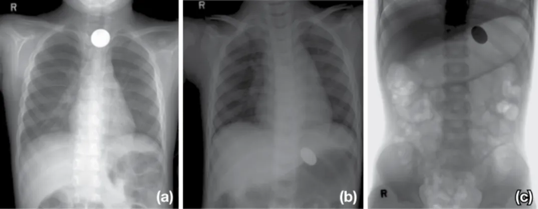
Fig. 2. Chest radiograph of a 6-year-old male showed of aspirated foreign body in chest PA radiograph and 2 hour delayed abdominal fluo-roscopy spot image(b, c).
나는 증상을 일으키지 않는 동전 또는 4주 후에 위를 벗어 나지 증상을 일으키지 않는 동전은 대개 내시경 제거가 필 요하다. 2. Safety Pin 옷핀의 이물질은 소화기관에 존재하고 있으며 장기관의 연동운동으로 시간이 지나서 배출하였다. 1세 소화환자가
처음 엑스선을 촬영(Fig. 3a, b), 3시간(Fig. 3c), 5시간(Fig. 3d), 9시간(Fig. 3e), 12시간(Fig. 3f), 21시간,(Fig. 3g), 27시
간(Fig. 3h)의 복부 영상이고 옷핀이 제거된 영상이다. 3. Earring 귀걸이 이물질은 2세의 소아환자로 엑스선 영상에서 원 형으로 위장에 위치하고 있으며 복부를 누운 자세(Fig. 5a) 와 선 자세(Fig. 5b)로 촬영한 영상이다. 4. Baduk Stone 바둑돌 이물질은 3세의 소아환자로 엑스선 영상에서 원 형으로 위장에 위치하고 있으며 복부를 누운 자세(Fig. 5a) 와 선 자세(Fig. 5b)로 촬영하였다. 5. Hairpin 머리핀 이물질은 2세의 소아환자로 엑스선 영상에서 위
Fig. 3. Chest radiograph of a 1-year-old male showed of aspirated foreign body in abdominal radiograph(a and b). Foreign body delayed of 3 hour(c), 5 hour(d), 9 hour(e), 12 hour(f) 21 hour(g), 27 hour(h) and clear foreign body(i) abdomi-nal radiograph.
(a) (b)
(c) (d)
(e) (f)
(g) (h)
(i)
Fig. 4. Chest radiograph of pectus excavatum post operation state a 2-year-old male showed of aspirated foreign body in chest radiograph AP(a) lateral image(b).
(a) (b)
Fig. 5. Abdominal radiograph supine(a) and erect(b) of a 3-year-old male showed of aspirated baduk stone foreign body in the stomach.
장에 위치하고 있으며 복부를 누운 자세(Fig. 6a)와 선 자세 (Fig. 6b)로 촬영하였다.
6. Ring
반지 이물질은 2세의 소아환자로 엑스선 영상에서 위장
에 위치하고 있으며 흉부(Fig. 7a), 시상면(Fig. 7a), 복부를 누운 자세(Fig. 7c)와 선 자세(Fig. 7d)로 촬영하여 반지가
방사선 불투과성으로 보이고 있다.
은 방사선 노출의 위험이 있고, 위장관 내시경, 기관지 내시
경은 침습적이다(Spector et al. 2008; Kim et al. 2009). 이물질의 섭취 및 흡인 소아에서 발생하며 이물질은 대 부분은 합병증 없이 소화기관으로 자연적으로 배출되나, 장 내에 고정되어 합병증을 일으킬 수 있기 때문에 상황에 따 른 적절한 검사와 진단으로 처치가 필요하다는 보고도 있다 (Panieri et al. 1995). 이물질의 섭취 및 흡인은 주로 6개월에서 3세 소아에서 발생하며, 남아에서 더 많이 발생한다(Wagoner 1997). 본 연구에서는 2세부터 7세의 이물질 섭취 및 흡인에 대한 방 사선 촬영에 따른 흉부 및 복부 영상이며, 모두 남아에서 발 생한 종류별로 이물질의 사례를 보고하였다. 또한 이물질을 섭취 및 흡인 환자의 호흡에 대한 이동과정 및 엑스선 영상, 내시경 영상, CT 영상을 보고하였고, 이러한 이물질의 영상 에 대한 묘출 및 평가를 위한 임상적인 유의성과 시술 및 사례를 보고하였다(Butterworth et al. 2007; Pugmire et al. 2015). 소화기관의 이물질을 직접 삼키어 위석과 같이 음식물이 나 기타 물질로 구성되어 소화기관 위 안에서 종괴를 형성 하기도 한다. 유아 및 소아에서 많이 발생하며, 귀걸이, 칫 솔, 견과류, 동전, 장난감, 돌, 뼈조각 등으로 종류는 시대 및 문화 환경에 따라 다양하게 나타날 수 있으며, 종류가 매우
다양하다(Webb 1995; Kim et al. 2009).
소아환자에서 기도에 있는 이물질의 이물의 종류는 대다 수의 논문에서 땅콩 등의 견과류가 가장 많았으며(Kim et al. 2009). 그러나 땅콩, 옥수수, 과일 등 유기물(organic)의 경우 금속, 플라스틱과 같은 무기물(inorganic)보다 진단 및 치료가 지연되기도 하고 장기 합병증이 빈번히 발생하는 경 향도 있다(Karakoç et al. 2002). 유기물은 방사선 비투과성 (radiolucent)으로 단순 흉부 촬영에서 초기에 진단하기 어 려운 점과 무기물보다 염증성 반응이 잘 일어나기 때문이 다. 본 연구에서는 무기물의 이물질을 보고하여 유기물을 섭취 및 흡인한 환자의 단순 엑스선 영상은 없었다. 이물질의 섭취 및 흡인의 종류에 따라 날카로운 이물질은 내시경으로 제거하거나 어려운 경우도 있으며 제거하는 과 정에서 출혈과 천공과 같은 위험한 합병증이 발생할 수 있 다. 이러한 이물질의 영상은 소아환자의 섭취 및 흡입된 이 물질의 진단 및 관리에 중요한 역할을 한다. 여러 종류의 일 반적으로 섭취된 이물질 제거가 필요하기 때문에 섭취된 이 물질의 신속한 식별은 적절한 치료에 필수적이다. 방사선
Fig. 6. Abdominal radiograph supine(a) and erect(b) of a 2-year-old male showed of aspirated hairpin foreign body in the stomach.
(a) (b)
Fig. 7. Radiograph of a 2-year-old male showed of aspirated for-eign body in chest AP(a), lateral(b) and abdominal radio-graph of supine(c) and erect(d).
(a) (b)
촬영은 섭취되거나 흡입된 이물질의 평가에서 가장 중요한 검사이며 다양한 검사방법인 투시와 CT 검사와 같은 부수 적인 검사가 이물질 제거를 위해 부수적인 역할을 할 수 있 다(Pugmire et al. 2015). 본 연구의 제한점으로는 소아환자의 다양한 환자 및 이 물질 섭취에 대한 사례의 부족으로 방사선 영상에는 한계 가 있어 추후 연구에서는 후향적 연구로 섭취 및 흡인 이후 의 방사선 검사 및 이물질 제거에 따른 시술 및 치료에 대 한 결과를 후향적으로 연구하여 보고할 필요가 있겠다.
결 론
이물질을 섭취 및 흡인한 소아환자에서 동전, 옷핀, 귀걸 이, 바둑돌, 머리핀의 이물질을 단순 엑스선의 흉부 및 복부 및 투시 촬영하여 이물질 종류별로 구분하여 확인하였다. 방사선 검사를 종합하여 이물질 제거의 시술 및 치료에 효 과적으로 대응할 수 있다. 소아환자의 임상에서 이물질 진 단 및 적절한 시술 및 치료 방향에 유용하게 적용할 수 있 다.참 고 문 헌
Butterworth J and Feltis B. 2007. Toy magnet ingestion in chil-dren: revising the algorithm. J. Pediatr. Surg. 42(12):e3-e5. Cha KM, Kim SW, Kim JH, Oh SH, Choi SM, Choi KH and
Cho SP. 2014. The characteristics and outcomes of foreign body aspiration and ingestion in pediatric patients who vis-it an emergency department. J. Korean Soc. Emerg. Med.
25(1):79-83.
Choi ES, Lee HG, Choi JN and Chun SY. 2014. Clinical Anal-ysis of Foreign Bodies in Gastrointestinal Tract in
Chil-dren. J. Korean Assoc. Pediatr. Surg. 20(1):12-16.
Karakoç F, Karadağ B, Akbenlioğlu C, Ersu R, Yildizeli B, Yüksel M and Dağli E. 2002. Foreign body aspiration: what is the outcome? Pediatr. Pulmonol. 34(1):30-36. Kim HY, Kong SG and Park HJ. 2009. Foreign body aspiration
in children: 30-years experience in a single institution. Pe-diat. Allergy Respir. Dis. 19(4):383-391.
Panieri E and Bass DH. 1995. The management of ingested foreign bodies in children a review of 663 cases. Eur. J. Emerg. Med. 2(2):83-87.
Pugmire BS, Lim R and Avery LL. Review of ingested and aspirated foreign bodies in children and their clinical sig-nificance for radiologists. 2015. Radiographics: a review publication of the Radiological Society of North America, Inc. 35(5):1528-1538.
Shubha AM and Das K. 2009. Tracheobronchial foreign bodies in infants. Int. J. Pediatr. Otorhinolaryngol. 73(10):1385-1389.
Spector J and Fernadez WG. 2008. Chemical, thermal, and bi-ological ocular exposures. Emerg. Med. Clin. North Am.
26(1):125-136.
Uyemura MC. 2005. Foreign body ingestion in children. Am. Fam. Physician. 72(2):287-291.
Wagoner MD. 1997. Chemical injuries of the eye: Current con-cepts in pathophysiology and therapy. Surv. Ophthalmol.
41:275-313.
Webb WA. 1995. Management of foreign bodies of the up-per gastrointestinal tract: update. Gastrointest. Endosc.
41(1):39-51.
Wyllie R. 2006. Foreign bodies in the gastrointestinal tract. Curr. Opin. Pediatr. 18(5):563–564.
Received: 3 September 2018 Revised: 27 September 2018 Revision accepted: 29 October 2018