1
Introduction
In the emergency department (ED), abdominal pain is a common complaint among children. Although most children visiting the ED with abdominal pain have nonsurgical illnesses, appendicitis is one of the most common causes of acute abdominal pain that requires surgery in pediatric patients (Tseng, 2008; Larson, 2011). History and physical examination in children with suspected appendicitis are highly variable that requires physicians to use imaging (McCollough, 2006 ; Shah, 2008).
While ultrasonography (US) has the benefit of being fast without ionizing radiation, it is highly operator-dependent and may not be achieved anytime depending on the circumstances (van Randen, 2008; Burr, 2011). On the other hand, CT is less operator-dependent. With its easy visualization of various anatomies, CT has been considered as the gold standard for the diagnosis of appendicitis with high sensitivity and specificity (Doria, 2006; Newman, 2011; Aspelund, 2014). For this reason there has been a dramatic increase (from 0.9% in 1998 to 15.4% in 2008) of pediatric CT use for abdominal pain (Fahimi et al, 2012). Although CT is helpful for physicians to diagnose or exclude acute appendicitis, radiation exposure has attracted increased attention in the medical community and the general public. Cancer-related mortality risk due to radiation exposure among children is estimated to be approximately one in 1,500 for a head CT scan and one in 550 for an abdominal CT scan (Brenner et al, 2001).
To reduce the risk of radiation exposure, many researchers have suggested to use reduced radiation dose or single phase CT scan without unenhanced phase for evaluating abdominal pain in children (Donnelly, 2001; Slovis, 2002; Strauss, 2010). However, the practice of using single phase CT scan without unenhanced phase has not been accepted universally. In clinical practice, it is common to take a CT scan with both unenhanced and contrast-enhanced phases in children. We performed a preliminary telephone survey to identify whether the unenhanced phase was used to diagnose suspected appendicitis in pediatric patients in teaching hospitals. Surprisingly, 22 (52.4%) of 42 teaching hospitals performed CT scan with both phases (unenhanced and contrast-enhanced). According to the result of our telephone survey, it was unclear whether a CT scan without an unenhanced phase could be used for the diagnosis of appendicitis in pediatric patients visiting ED with acute non-traumatic right lower abdominal pain.
Therefore, the purpose of this study was to evaluate the usefulness of abdominal CT scan without the unenhanced phase for the diagnosing acute appendicitis in pediatric patients visiting ED.
2
Materials and Methods
This study protocol was approved by our institutional review board.
We retrospectively reviewed abdominal CT scans of children with right lower abdominal pain who visited the ED of Ajou University Hospital from January to December 2013. The ages of children ranged from 5 to 10 years old. Patients whose abdominal pain was due to physical injury were excluded from this study. Total 233 CT scans were found. They consisted of 105 ‘normal’, 65 ‘appendicitis’ and 63 ‘the others’ CT scans that were confirmed by pediatric radiologists. One researcher among us randomly collected one hundred abdominal CT scans. For minimizing the selective bias, that researcher reviewed the reports read by radiologists without checking CT images. These samples consisted of final diagnosis with ‘normal’ or ‘appendicitis’. Half of the study samples (50) were randomly chosen from 105 ‘normal’ CT scans. The other half of the study samples (50) were randomly chosen from 65 ‘appendicitis’ CT scans. Each CT scan was reviewed twice. On the first occasion, contrast images alone were presented without unenhanced ones (protocol A). After two weeks, the same CT scans were again presented with unenhanced phase (protocol B). The contrast-enhanced coronal images were included in both protocols, which can be reconstructed without additional radiation exposure and are performed routinely in most hospitals. The order of the 100 study samples was randomly assigned into each protocol.
All of abdominal CT scans was performed transversely in the supine position. SOMATOM sensation16TM (Siemens, Erlangen, Germany) was used to take a CT scan in accordance with a pediatric protocol of radiology department in our institution. The conditions of 60mAs and 120kVp were used for 3-6 year old, whereas 80mAs and 120kVp were used for 6-10 year old. The slice width of 4mm with 1-2ml/kg IV contrast was used for CT scans.
The participants in this study were comprised of thirty emergency physicians (25 residents and 5 specialists) from two university hospitals. To assess the differences of CT interpretation depending on experiences, we categorized participants into two groups: Junior and Senior. Junior group consisted of first and second year residents. Senior group consisted of specialists and residents with three or more years of experience. They separately reviewed all CT images twice (protocol A and protocol B). We provided the information that the patients were between ages of 5 and 10 who had non-traumatic right lower abdominal pain. All participants were required to choose a single diagnosis of either ‘normal’ or ‘appendicitis’. We also provided the following criteria of acute appendicitis based on literature report (Tintinalli, 2010): dilated appendix >
3
6mm with thickened wall, stranding of periappendiceal inflammation, and potential visualization of an appendicolith or abscess. At the end of this study, we conducted a survey to evaluate how helpful the following phase of the CT scan was in making a diagnosis of appendicitis: 1) unenhanced-transverse; 2) enhanced-transverse; and 3) contrast-enhanced-coronal. The 3-item questionnaire was composed of Likert-type scale. All participants were asked to pick up a number between 1 and 9, with number 1 indicating not helpful at all whereas number 9 indicating the most helpful.
The sensitivity and specificity of protocol A and B for diagnosing suspected appendicitis was calculated using interpretation provided by pediatric radiologist as the standard reference. We also compared the sensitivity and specificity between the Junior group and the Senior group.
Intraobserver agreement between results from the two protocols (protocol A and B) for each participant was measured. Interobserver agreement for the two protocols was also measured as compared with the result from each participant.
All data in this study were analyzed using Microsoft Excel 2010 (Microsoft, Redmond, WA) and SPSS ver. 21(SPSS Inc., Chicago, IL). A t-test was used to determine whether the means of populations were statistically different from each other and to compare the average scores of questionnaire from participants. To evaluate the Intraobserver and interoobserver agreements, kappa values were calculated. As suggested by Landis and Koch (Landis, 1977; Kundel, 2003), the strength of the agreement was classified in the following categories: κ<0, poor; 0-0.20 slight; 0.21-0.40, fair; 0.41-0.60, moderate; 0.61-0.80, substantial; and 0.81-1.00, almost perfect. Statistical significance was defied as p-value < 0.05.
Results
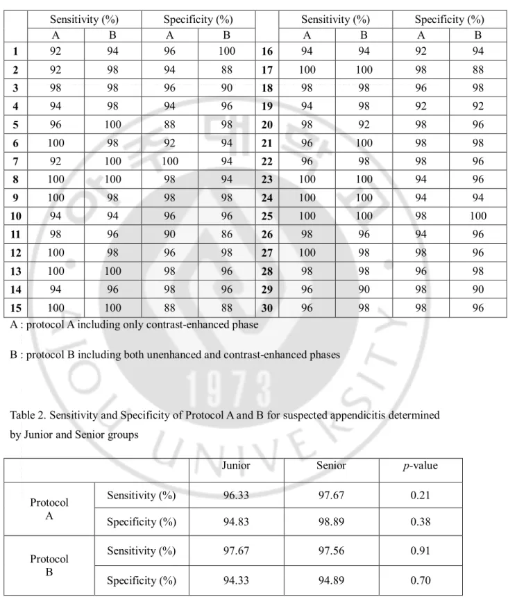
The sensitivity and specificity of protocol A and B for diagnosing suspected acute appendicitis were shown in Table 1. The mean sensitivities for protocol A and B were 97.13% [95% Confidence interval = 96.13-98.14] and 97.60% [96.67-98.53], respectively. The mean specificities for protocol A and B were 95.47% [94.34-96.59] and 94.67% [93.33-96.00], respectively. No statistically significant difference on the sensitivity and specificity was found between the two protocols (p-value = 0.51 for sensitivity, p-value = 0.37 for specificity). The mean sensitivity and specificity of the Junior and Senior group were shown in Table 2. No statistical difference on the sensitivity and specificity was observed between the two groups.
4
Table 1. Sensitivity and Specificity of Protocol A and B for suspected appendicitis, using interpretation of CT scan read by radiologist as standard reference
Sensitivity (%) Specificity (%) Sensitivity (%) Specificity (%)
A B A B A B A B 1 92 94 96 100 16 94 94 92 94 2 92 98 94 88 17 100 100 98 88 3 98 98 96 90 18 98 98 96 98 4 94 98 94 96 19 94 98 92 92 5 96 100 88 98 20 98 92 98 96 6 100 98 92 94 21 96 100 98 98 7 92 100 100 94 22 96 98 98 96 8 100 100 98 94 23 100 100 94 96 9 100 98 98 98 24 100 100 94 94 10 94 94 96 96 25 100 100 98 100 11 98 96 90 86 26 98 96 94 96 12 100 98 96 98 27 100 98 98 96 13 100 100 98 96 28 98 98 96 98 14 94 96 98 96 29 96 90 98 90 15 100 100 88 88 30 96 98 98 96
A : protocol A including only contrast-enhanced phase
B : protocol B including both unenhanced and contrast-enhanced phases
Table 2. Sensitivity and Specificity of Protocol A and B for suspected appendicitis determined by Junior and Senior groups
Junior Senior p-value
Protocol A Sensitivity (%) 96.33 97.67 0.21 Specificity (%) 94.83 98.89 0.38 Protocol B Sensitivity (%) 97.67 97.56 0.91 Specificity (%) 94.33 94.89 0.70
5
Table 3. Intraobserver agreement of 30 participants between results obtained from the two protocols κ [95% CI ] κ [95% CI ] κ [95% CI ] 1 0.860 [0.663-1.055] 11 0.778 [0.582-0.974] 21 0.960 [0.764-1.156] 2 0.720 [0.523-0.915] 12 0.960 [0.763-1.155] 22 0.960 [0.764-1.156] 3 0.799 [0.603-0.995] 13 0.980 [0.783-1.175] 23 0.940 [0.743-1.135] 4 0.820 [0.623-1.015] 14 0.840 [0.643-1.035] 24 1.000 [0.804-1.196] 5 0.798 [0.601-0.993] 15 1.000 [0.804-1.196] 25 0.980 [0.783-1.175] 6 0.880 [0.683-1.075] 16 0.820 [0.623-1.015] 26 0.960 [0.763-1.155] 7 0.860 [0.663-1.055] 17 0.900 [0.703-1.095] 27 0.960 [0.763-1.155] 8 0.960 [0.763-1.155] 18 0.980 [0.783-1.175] 28 0.980 [0.783-1.175] 9 0.980 [0.783-1.175] 19 0.840 [0.643-1.035] 29 0.800 [0.604-0.996] 10 1.000 [0.804-1.196] 20 0.880 [0.683-1.075] 30 0.960 [0.764-1.156] κ : kappa value, CI : confidence interval
The interobserver agreement for protocol A was almost perfect (κ=0.90 [0.89-0.91]), and the interobserver agreement for protocol B was also almost perfect (κ=0.87 [0.86-0.88]). There was no statistically significant difference in the interobserver agreement for either protocol.
The inraobserver agreement between the results from protocol A and B were shown in Table 3. The mean kappa value for intraobserver agreements was also almost perfect (κ=0.91 [0.88-0.93]).
Based on the survey results using the questionnaire to rate the usefulness of each phase of CT, the average scores of unenhanced-transverse, enhanced-transverse, and contrast-enhanced-coronal scans were 3.13, 8.57, and 7.13, respectively. The scores of the three categories were significantly different (p < 0.001).
Discussion
To our knowledge, our study was the first attempt to compare pediatric abdominal CT protocols in the diagnosis of suspected appendicitis in the ED. Our study revealed that the unenhanced images were not helpful in revealing the presence of appendicitis in children.
It is well accepted that US is a useful modality for the diagnosis of suspected appendicitis in children without the exposure to ionizing radiation. However, CT scan has been favored over
6
US because it is less operator-dependent. In addition, CT scan provides more readily recognizable anatomy and appreciated abnormality than US (Newman, 2011).Because of its short scanning time, less need for sedation and high quality image, CT scan has become an essential tool for rapidly diagnostic evaluation of children in the ED (Pappas, 2000; Donnelly, 2005; Broder, 2007).There has been a steep rise in the usage of CT scan in pediatric patients visiting the ED with abdominal pain (Broder, 2007; Larson, 2011; Larson, 2011; Newman, 2011),with the greatest increase of usage for children between 4 and 18 years old (Fahimi et al, 2012).Although CT studies typically represent less than 11% of all imaging studies performed at most university-based hospitals, it can generate as much as 70% of the overall radiation dose generated in radiology department (Brenner, 2007; Smith-Bindman, 2008).There has been increasing awareness of radiation exposure because of the potential cancer risk caused by CT use (Brenner and Hall, 2007). This is particularly true in pediatric patients, because they have higher susceptibility to radiation than adults and a longer remaining life expectancy in which cancer may form (Brenner, 2002).
Since the multidisciplinary conference organized by radiologists organized in August 2001 to discuss radiation doses used in pediatric CT (as low as reasonable achievable, or ALARA, Concept in pediatric CT-Intelligent dose reduction), a lot of efforts have been made to reduce the radiation dose and the overall number of CT scans performed in children (Slovis, 2002). Strauss et al. (Strauss et al, 2010) have suggested ten steps, concrete ways to lower CT radiation dose while maintaining image quality in children, which is known as the “Image Gently campaign”. The utilization of CT scan for abdominal pain has increased much more than in other illness. Abdominal CT is typically associated with effective doses of radiation of up to seven times of that used in head CT (Smith-Bindman et al, 2009). Each phase of the CT scan contributes to the total dose of radiation. When both unenhanced and contrast-enhanced abdominal CT are used in pediatric patients, its radiation dose will be twice of the radiation dose used for a single phase CT (da Costa e Silva et al, 2007). Many efforts such as the ALARAconcept and the Image gently campaign have been attempted to reduce radiation exposure from CT scan in children (Slovis, 2002; Strauss, 2010). It has been recommended to take a single contrast-enhanced phase scan in children with acute abdominal pain (Donnelly, 2001; Slovis, 2002; Strauss, 2010). However, over half of the teaching hospitals have a pediatric abdominal CT protocol that includes the unenhanced phase CT scan based on our preliminary telephone survey. Therefore, efforts should be made to reduce the unnecessary radiation dose in pediatric abdominal CT. The teaching hospitals participated our telephone survey included 10 pediatric emergency centers. Eight (80%) of them performed only single phase of abdominal CT to diagnose appendicitis. It shows that pediatric emergency centers have more concerns and make efforts to reduce the radiation dose in children compared to
7 general emergency centers.
In this study, we found that it was unnecessary to perform the unenhanced phase of CT scan for the diagnosis or exclusion of appendicitis in children visiting the ED. Based on our results, it is recommended to take only contrast enhanced CT for diagnosis of pediatric appendicitis. In addition, we should follow the ALARA principle and ‘ten steps’ from Image Gently campaign to optimize the image quality and lower the radiation dose in CT scans for pediatric patients, especially in the pediatric emergency centers (Slovis, 2002;; Strauss, 2010) .
US and CT share the same specificity (94%) in diagnosing suspected appendicitis in children. However, the sensitivity of US (up to 88%) alone is lower than that of CT (95%) (Doria, 2006; Newman, 2011). According to our study, the sensitivity and specificity for protocol B as a standard for abdominal CT were 97.60% and 94.67%, suggesting that all participants are excellent in evaluating appendicitis. The sensitivity and specificity of protocol A were as good as those of protocol B or previous studies reported in the literature. No statistical difference in sensitivity or specificity was observed between Junior and Senior groups, suggesting that more experienced physicians do not necessarily have higher accuracy of diagnosis than the less experienced ones. For each emergency physician, no significant difference was found between the results obtained from the two protocols. The almost perfect kappa values of the intraobserver and interobserver agreements strongly suggest that it is unnecessary to take an unenhanced CT scan for evaluating suspected appendicitis in children with abdominal pain visiting the ED.
Based on our survey on the usefulness of different phase CT for the diagnosis of appendicitis, the unenhanced-transverse phase had the lowest score of 3.13, whereas the contrast-enhanced phase had the highest score of 8.57. There was statistically significant difference between the two results. It was not surprising that all participants chose contrast-enhanced phase as the most helpful CT scan, because most conditions met for criteria of acute appendicitis were revealed by contrast-enhanced phase CT (Tintinalli, 2010). It was believed that unenhanced phase is useful for detecting calcification. However, da Costa e Sivan and Silva found that contrast-enhanced phase CT has good sensitivity for detection of calcification compared to unenhanced one (da Costa e Silva et al, 2007). Therefore, it is easy to find appendicolith in contrast images, although appendicolith is valuable but unnecessary for the diagnosis of appendicitis.
This study has several limitations. The limitations include the small size of participants and the narrow age range of patients. In addition, our investigation was only focused on one cause
8
of abdominal pain - appendicitis. Therefore, additional studies using CT without unenhanced phase in other applications should be performed. Lastly, theses samples of CT scans were selected retrospectively.Therefore, there is a possibility of biased study such as easy cases were chosen, despite our efforts to select samples randomly. Further research on the CT utilization in pediatric-focused EDs versus non-pediatric-focused EDs will be helpful to identify the best way to use CT scan to reduce the dose of radiation.
Conclusion
We observed that unenhanced images of CT scan have no advantage over contrast-enhanced CT scan for the evaluation of suspected acute appendicitis in children visiting the ED with abdominal pain. Consequently we concluded that a CT protocol without unenhanced phase is a feasible alternative for the diagnosis of acute appendicitis in pediatric patients visiting the ED. .References
1. Aspelund G, Fingeret A, Gross E, Kessler D, Keung C, Thirumoorthi A, Oh PS, Behr G, Chen S, Lampl B, Middlesworth W, Kandel J, Ruzal-Shapiro C:
Ultrasonography/MRI versus CT for diagnosing appendicitis. Pediatrics 133: 586-593, 2014
2. Brenner D, Elliston C, Hall E, Berdon W: Estimated risks of radiation-induced fatal cancer from pediatric CT. AJR Am J Roentgenol 176: 289-296, 2001
3. Brenner DJ, Hall EJ: Computed tomography--an increasing source of radiation exposure. N Engl J Med 357: 2277-2284, 2007
4. Brenner DJ: Estimating cancer risks from pediatric CT: going from the qualitative to the quantitative. Pediatr Radiol 32: 228-221; discussion 242-224, 2002
5. Broder J, Fordham LA, Warshauer DM: Increasing utilization of computed tomography in the pediatric emergency department, 2000-2006. Emerg Radiol 14: 227-232, 2007
6. Burr A, Renaud EJ, Manno M, Makris J, Cooley E, DeRoss A, Hirsh M: Glowing in the dark: time of day as a determinant of radiographic imaging in the evaluation of abdominal pain in children. J Pediatr Surg 46: 188-191, 2011
7. Donnelly LF, Emery KH, Brody AS, Laor T, Gylys-Morin VM, Anton CG, Thomas SR, Frush DP: Minimizing radiation dose for pediatric body applications of single-detector helical CT: strategies at a large Children's Hospital. AJR Am J Roentgenol 176: 303-306, 2001
9
8. Donnelly LF: Reducing radiation dose associated with pediatric CT by decreasing unnecessary examinations. AJR Am J Roentgenol 184: 655-657, 2005
9. Doria AS, Moineddin R, Kellenberger CJ, Epelman M, Beyene J, Schuh S, Babyn PS, Dick PT: US or CT for Diagnosis of Appendicitis in Children and Adults? A Meta-Analysis. Radiology 241: 83-94, 2006
10. Fahimi J, Herring A, Harries A, Gonzales R, Alter H: Computed tomography use among children presenting to emergency departments with abdominal pain. Pediatrics 130: e1069-1075, 2012
11. Kundel HL, Polansky M: Measurement of observer agreement. Radiology 228: 303-308, 2003
12. Landis JR, Koch GG: The measurement of observer agreement for categorical data. Biometrics 33: 159-174, 1977
13. Larson DB, Johnson LW, Schnell BM, Salisbury SR, Forman HP: National trends in CT use in the emergency department: 1995-2007. Radiology 258: 164-173, 2011 14. Larson DB, Johnson LW, Schnell BM, Goske MJ, Salisbury SR, Forman HP: Rising
use of CT in child visits to the emergency department in the United States, 1995-2008. Radiology 259: 793-801, 2011
15. McCollough M, Sharieff GQ: Abdominal pain in children. Pediatr Clin North Am 53: 107-137, vi, 2006
16. Newman B: Ultrasound body applications in children. Pediatr Radiol 41 Suppl 2: 555-561, 2011
17. Pappas JN, Donnelly LF, Frush DP: Reduced frequency of sedation of young children with multisection helical CT. Radiology 215: 897-899, 2000
18. Shah NB, Platt SL: ALARA: is there a cause for alarm? Reducing radiation risks from computed tomography scanning in children. Curr Opin Pediatr 20: 243-247, 2008
19. Slovis TL: The ALARA concept in pediatric CT: myth or reality? Radiology 223: 5-6, 2002
20. Smith-Bindman R, Lipson J, Marcus R, Kim KP, Mahesh M, Gould R, Berrington de Gonzalez A, Miglioretti DL: Radiation dose associated with common computed tomography examinations and the associated lifetime attributable risk of cancer. Arch Intern Med 169: 2078-2086, 2009
21. Smith-Bindman R, Miglioretti DL, Larson EB: Rising use of diagnostic medical imaging in a large integrated health system. Health Aff (Millwood) 27: 1491-1502,
10 2008
22. Strauss KJ, Goske MJ, Kaste SC, Bulas D, Frush DP, Butler P, Morrison G, Callahan MJ, Applegate KE: Image gently: Ten steps you can take to optimize image quality and lower CT dose for pediatric patients. AJR Am J Roentgenol 194: 868-873, 2010 23. Tintinalli JE: Tintinalli's Emergency Medicine: A Comprehensive Study Guide. 7
edition ed., McGraw-Hill Professional, pp.574-577, 2010
24. Tseng YC, Lee MS, Chang YJ, Wu HP: Acute abdomen in pediatric patients admitted to the pediatric emergency department. Pediatr Neonatol 49: 126-134, 2008
25. van Randen A, Bipat S, Zwinderman AH, Ubbink DT, Stoker J, Boermeester MA: Acute appendicitis: meta-analysis of diagnostic performance of CT and graded compression US related to prevalence of disease. Radiology 249: 97-106, 2008
26. da Costa e Silva EJ, da Silva GA: Eliminating unenhanced CT when evaluating abdominal neoplasms in children. AJR Am J Roentgenol 189: 1211-1214, 2007
11 국문요약
-소아 급성 충수염의 진단에 조영 전 CT가 필요한가 ?
본 연구의 목적은,급성 비외상성 우하복부 통증을 주소로 응급실을 방문한 소아에 서 급성 충수염 여부를 가리기 위해 조영 전 스캔이 없는 CT 프로토콜이 유용한가 를 알아보는 데에 있다. 우리는 후향적으로 응급실에서 촬영된 100개의 소아 복부 CT 샘플들을 모았으며, 이것들은 모두 소아 영상의학 전문의에 의해 공식 판독되어진 것이었다.30명의 응 급의학과 의사들 각각에게 100개의 샘플들을 두 차례에 걸쳐 판독해 줄 것을 요청 하였다. 첫 번째 판독은 조영 전 스캔을 포함하지 않고 있으며 이를 protocolA 라 고 한다.두 번째 판독은 조영 전 스캔 및 조영 증강 스캔 모두를 포함하며 이를 protocolB라고 한다.급성 충수염을 진단하기 위한 각 protocol의 민감도와 특이도를 계산하였으며,kappa값을 이용하여 intraobserver및 interobserver일치도를 측 정하였다.
두 protocol에 대한 평균 민감도와 특이도는 서로 비슷한 값을 보였다. protocolA
에 대한 민감도는 97.13%[95%신뢰구간=96.13-98.14]였으며,protocolB에 대한 민감 도는 97.60% [96.67-98.53]이었다.protocolA와 B에 대한 특이도는 각각 95.47% [94.34-96.59]와 94.67% [93.33-96.00]이었다.Intraobserver일치도에 대한 평균 kappa 값은 0.91 [0.88-0.93] 이었다.Interobserver 일치도에 대한 kappa 값은 protocol A에 대한 것이0.90 [0.89-0.91] 이었고 protocol B에 대한 것이 0.87[0.86-0.88]이었다.
급성 복통을 주소로 응급실을 방문한 소아에서,조영 전 스캔을 촬영하지 않는 복
![Table 3. Intraobserver agreement of 30 participants between results obtained from the two protocols κ [95% CI ] κ [95% CI ] κ [95% CI ] 1 0.860 [0.663-1.055] 11 0.778 [0.582-0.974] 21 0.960 [0.764-1.156] 2 0.720 [0.523-0.915] 12 0.960 [0.76](https://thumb-ap.123doks.com/thumbv2/123dokinfo/4676475.1544/5.892.121.796.222.616/table-intraobserver-agreement-participants-results-obtained-protocols-ci.webp)
