High-Flexion Total Knee Arthroplasty:
Survivorship and Prevalence of Osteolysis
Results After a Minimum of Ten Years of Follow-up
Young-Hoo Kim, MD, Jang-Won Park, MD, and Jun-Shik Kim, MDInvestigation performed at the Joint Replacement Center, Ewha Womans University School of Medicine, Seoul, South Korea
Background: We are aware of no information about the mid-term performance of the high-flexion total knee arthroplasty, although early results have been reported. The purpose of this study was to evaluate the mid-term results of high-flexion and conventional knee prostheses.
Methods: We prospectively compared the results of 100 patients with osteoarthritis who had received a NexGen Legacy Posterior Stabilized (NexGen LPS) prosthesis in one knee and a NexGen Legacy Posterior Stabilized-Flex (NexGen LPS-Flex) prosthesis in the other. Seventy-five patients (150 knees) were women and twenty-five (fifty knees) were men. The mean age was sixty-five years (range, forty-eight to eighty-five years) at the time of the index procedure. The mean duration of follow-up was 10.3 years (range, ten to 10.6 years). The patients were assessed with radiographs, with the rating system of the Knee Society, and with the Western Ontario and McMaster Universities Osteoarthritis Index (WOMAC) score at three months, one year, and annually thereafter.
Results: Total knee scores, knee function scores, pain scores, WOMAC scores, knee motion, and activity scores did not differ significantly between the two designs of the implants, on the basis of the numbers studied, either preoperatively or at the time of final follow-up. One knee in the NexGen LPS-Flex group was revised because of recurrent infection. No knee in either group had aseptic loosening of the components. The Kaplan-Meier survivorship at ten years postoperatively, with revision defined as the end point, was 100% (95% confidence interval, 94 to 100) for the NexGen LPS prosthesis and 99% (95% confidence interval, 93 to 100) for the NexGen LPS-Flex prosthesis.
Conclusions: After a minimum duration of follow-up of ten years, there were no significant differences between the two groups with regard to implant survivorship, functional outcome, knee motion, or prevalence of osteolysis.
Level of Evidence: Therapeutic Level I. See Instructions for Authors for a complete description of levels of evidence.
T
he high-flexion total knee arthroplasty system was intro-duced to increase the articular surface area at high flexion angles and increase posterior femoral translation and knee motion. It was postulated that improved tibiofemoral contact at high flexion could decrease contact surface stress and subsequently decrease osteolysis and wear of the tibial polyethylene component. There are conflicting reports of the early results of the high-flexion total knee prosthesis1-10. Han et al.1
reported a disturbingly high prevalence of early femoral component loosening after high-flexion total knee arthroplasty. However, authors of other recent follow-up studies reported excellent early results following
high-flexion total knee arthroplasty, even when they had used the same or a similar implant type2-10
. Although there is some information about the early performance of high-flexion total knee arthro-plasty, there is none, to our knowledge, about the mid-term per-formance of this prosthesis. The question thus arises as to whether, in the longer term, use of a high-flexion total knee prosthesis improves implant longevity, knee function, and knee motion while decreasing polyethylene wear and osteolysis compared with the same parameters after use of a conventional prosthesis.
The purpose of this study was to evaluate the mid-term results of high-flexion and conventional knee prostheses, with a Disclosure: None of the authors received payments or services, either directly or indirectly (i.e., via his or her institution), from a third party in support of any aspect of this work. None of the authors, or their institution(s), have had any financial relationship, in the thirty-six months prior to submission of this work, with any entity in the biomedical arena that could be perceived to influence or have the potential to influence what is written in this work. Also, no author has had any other relationships, or has engaged in any other activities, that could be perceived to influence or have the potential to influence what is written in this work. The complete Disclosures of Potential Conflicts of Interest submitted by authors are always provided with the online version of the article.
particular emphasis on implant survivorship, knee function and motion, and prevalence of osteolysis.
Materials and Methods
Demographics
T
he senior author (Y.-H.K.) performed 111 consecutive primary bilateral total knee arthroplasties in 111 patients (222 knees). Five patients died from un-related causes and six were followed for less than two years postoperatively. Therefore, 100 patients (200 knees) were included in the study. The study was registered in ClinicalTrials.gov Protocol Registration System (trial number, NCT 01422642). The study was approved by our institutional review board, and all patients provided informed consent. Randomization between the use of a NexGen Legacy Posterior Stabilized-Flex (NexGen LPS-Flex; Zimmer, Warsaw, Indiana) or a NexGen Legacy Posterior Stabilized (NexGen LPS; Zimmer) prosthesis was determined from a sequential pool on the basis of a table of random numbers. Each of the 100 patients received a NexGen LPS-Flex prosthesis on one side and a NexGen LPS prosthesis on the contralateral side as treatment for knee osteo-arthritis. Seventy-five patients (150 knees) were women, and twenty-five patients (fifty knees) were men. The mean age of the patients at the time of the arthroplasty was sixty-five years (range, forty-eight to eighty-five years). The mean patient height (and standard deviation) was 159.2 ± 7.21 cm (range, 145 to 185 cm), and the mean weight was 65.8 ± 7.9 kg (range, 51 to 93 kg). The mean body mass index was 26 kg/m2(range, 24 to 27 kg/m2). Thirty-three patients (33%) had undergone bilateral arthroscopic debridement previously.Surgical Technique
All procedures were performed by the senior author. A bloodless field was ob-tained with use of a pneumatic tourniquet at a pressure of 250 mm Hg after exsanguination with an Esmarch bandage. In all knees, an anterior midline skin incision (10 to 12 cm in length) was used, followed by a medial parapatellar capsular incision, and the femur was prepared before the tibia. The anterior and posterior cruciate ligaments were excised. Ligament balancing was done, and an attempt was made to resect 10 mm of tibial bone to achieve a surface that was perpendicular to the shaft of the tibia in the coronal plane with 7° of posterior slope in the sagittal plane in both groups. The distal part of the femur was resected with an attempt to achieve valgus femorotibial alignment in the coronal plane in both groups. All patellae were resurfaced with use of a polyethylene patellar prosthesis. All implants were fixed with CMW cement (DePuy, Warsaw, Indiana) without antibiotics, after pulsed lavage, drying, and pressurization of the cement. Pain Management Protocol
All patients received intra-articular analgesic cocktail infiltration (200 mg of 0.5% bupivacaine, 10 mg of morphine, 40 mg of methylprednisolone acetate, and 300 mg of epinephrine [1:1000]), and all received epidural or patient-controlled intravenous analgesia (patient-patient-controlled anesthesia: a morphine bolus of 1.5 mg, a lockout time of six minutes, and a maximum of 15 mg per hour). The epidural catheter was left in place for continuous pain management for forty-eight hours after the surgery. Pethidine, tramadol, or oxycodone was administered after discontinuation of the epidural or patient-controlled intravenous anesthesia. Rehabilitation
Starting on the second postoperative day, the patients used a continuous passive motion machine for passive knee motion exercise twice daily for thirty minutes each time. On the same day, they started active knee motion exercises and began standing at the bedside or walking with crutches or a walker twice daily for thirty minutes each time under the supervision of a physical therapist. The patients used crutches or a walker with full weight-bearing for six weeks and used a cane when needed thereafter. Clinical Evaluation
Clinical evaluations were done at three months after the operation, at one year, and then yearly thereafter. The mean duration of follow-up was 10.3 years (range, ten to 10.6 years). All clinical data at the time of each follow-up were recorded and
compiled by a research associate who was not part of the operative team and was blinded to treatment allocation. We obtained the Knee Society knee score11and the Western Ontario and McMaster Universities Osteoarthritis Index (WOMAC) score12separately for each knee. We found that it was relatively easy for patients to distinguish the degree of pain in each knee. We inquired with regard to the degree of stiffness with use of the WOMAC instrument separately for each knee, and it was again relatively easy for patients to distinguish the degree of stiffness in each knee. We also asked about function separately for each knee. Although it was somewhat difficult for patients to distinguish the degree of impairment of the function of each knee, on careful questioning they were able to do so. For ex-ample, when they had difficulty in ascending or descending stairs, they were able to specify which knee bothered them more.
Active knee motion, both with the patient in the supine position and while weight-bearing, was determined with use of a standard (60-cm) clinical goniometer before the operation and at the time of the review. The patients were asked to extend the knees as much as possible while lying in a supine position to measure flexion contracture. They were told to bend the knees as much as possible while lying in a supine position and in a squatting position to measure the flexion angle. Knee motion was measured for all patients on two occasions by two of the authors (Y.-H.K. and J.-S.K.), both of whom were blinded to the type of implanted prosthesis. When the measured knee motion differed by >5° between the two observers, the values were averaged and that number was reported. Interobserver agreement regarding knee motion was 0.95 to 0.99. The knee motion was considered to be the arc of motion instead of the flexion angle13. The level of activity was assessed by using the Tegner and Lysholm activity score14. Radiographic Evaluation
Anteroposterior radiographs with the patient standing (including the hip and the ankle) and with the patient lying supine, lateral radiographs, and skyline patellar radiographs were made preoperatively and at each follow-up visit and were as-sessed for the alignment of the limb (tibiofemoral angle), the position of the components, the posterior slope of the tibial component, the level of the joint line, and the presence and location of radiolucent lines at the bone-cement interface according to the recommendations of the Knee Society11. Antero-posterior standing radiographs were used to determine any sequential change in the alignment of the limb as a result of polyethylene wear and/or loosening of the implant. Supine anteroposterior radiographs were used to determine the presence of a radiolucent line more precisely. Skyline patellar radiographs were examined for patellar tilt, subluxation, or dislocation. All radiographs were made under fluoroscopic guidance to control rotation of the knee. Radiographic data at the time of each follow-up were analyzed and recorded by a research associate who was not part of the operative team. However, this assessment was not blinded to allocation because the radiographic appearances of the two implants differ. Computed Tomography Scan Evaluation
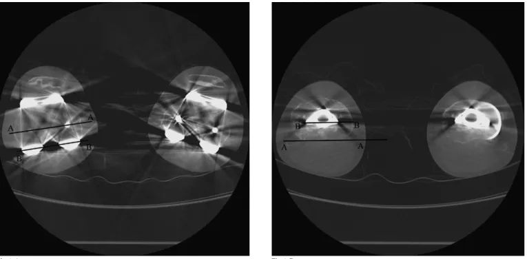
At the latest follow-up evaluation, all patients underwent computed tomog-raphy (CT) scanning with a multislice scanner (General Electric LightSpeed Plus; GE Healthcare, Milwaukee, Wisconsin) to determine the rotational alignment of the components and to assess for osteolysis. The scan sequence was between 10 cm above the superior pole of the patella and 10 cm below the tibial tuberosity, using contiguous 2.5-mm slices. Rotational alignment of the femoral component was determined by measuring the angle between a line joining the medial and lateral epicondyles of the femur and one joining the posterior margins of the femoral component (Fig. 1-A). Rotational alignment of the tibial component was assessed by measuring the angle between a line joining the posterior margins of the medial and lateral tibial plateaus and one joining the posterior margins of the tibial component (Fig. 1-B). Osteolysis was defined as any nonlinear region of periprosthetic cancellous bone loss with delineable margins. One author (Y.-H.K.) examined all CT scans. The intra-observer kappa statistic for the radiographic and CT examinations was 0.94. Statistical Analysis
An a priori power calculation was performed with use of a clinically relevant difference in range of motion of 5° and a standard deviation of 10°. Calculations
revealed that, for an effect size of 20% in functional outcome as measured with a validated instrument such as the linear analog scale assessment and with a = 0.05 and b = 0.80, ninety patients would be needed in each group. In addition to the required number of subjects, ten more patients were recruited to allow for possible attrition. The changes in the Knee Society knee scores, knee
mo-tion, WOMAC scores, and activity scores were evaluated with a paired t test. Complication rates and radiographic data were compared between the two groups with use of a paired t test as well. All statistical analyses were performed with a two-tailed t test. The level of significance was set at p < 0.05. Survivorship analysis was performed to determine the cumulative rate of survival of the
TABLE I Clinical Results
Preoperative Final Follow-up
NexGen LPS NexGen LPS-Flex P Value* NexGen LPS NexGen LPS-Flex P Value* Mean Knee Society knee
score (range) (points)
Total knee score 25 (4-47) 28 (4-44) 0.8431* 93 92 0.8681*
Function score 50.1 (27-68) 50.1 (28-68) 1.000* 85 82 0.8329*
Pain score 0 0 45 43 0.641†
Degree of pain (no. [%] of knees)
None — — 75 (75%) 71 (71%)
Mild — — 25 (25%) 28 (28%)
Moderate 10 (10%) 14 (14%) — 1 (1%)
Severe 90 (90%) 86 (86%) — —
Mean WOMAC score (points)
65.2 67.3 0.912* 28.9 32.3 0.931*
Mean activity score (points)
1.6 1.7 0.989* 5.7 5.9 0.972*
*Paired t test. †Chi-square test.
Fig. 1-A Fig. 1-B
Figs. 1-A and 1-B Postoperative CT scans of a fifty-five-year-old woman with osteoarthritis. Fig. 1-A Measurement of axial rotation of the femoral component in relation to the transepicondylar axis (AA) and the posterior margins of the femoral component (BB). Fig. 1-B Measurement of axial rotation of the tibial component in relation to the posterior margins of the medial and lateral tibial plateaus (AA) and one joining the posterior margins of the tibial component (BB).
implant during the period of the study15and reported with 95% confidence intervals (CI). The end point for the analysis was aseptic loosening and revision surgery for any reason.
Source of Funding
There was no external funding source for this study.
Results
Clinical Results Knee Score
T
he Knee Society total knee scores did not differ significantly between the two groups either preoperatively (p = 0.8431; paired t test) or postoperatively (p = 0.8681; paired t test). In the NexGen LPS group, the mean postoperative Knee Society totalknee score was 93 points (range, 75 to 100 points) and the Knee Society function score was 85 points (range, 60 to 100 points). In the NexGen LPS-Flex group, the mean postoperative Knee So-ciety total knee score was 92 points (range, 70 to 100 points) and the Knee Society function score was 82 points (range, 71 to 100 points) (Table I).
Pain
The postoperative Knee Society pain scores did not differ signif-icantly between the groups (p = 0.641) (Table I). Of the 100 knees treated with the NexGen LPS implant, seventy-five (75%) were not painful at the time of the latest follow-up, twenty-five (25%) were mildly painful, and none were moderately or severely painful.
TABLE II Total Arc of Knee Motion*
Preoperative One Year Five Years Ten Years
Non-Weight-Bearing Weight-Bearing Non-Weight-Bearing Weight-Bearing Non-Weight-Bearing Weight-Bearing Non-Weight-Bearing Weight-Bearing NexGen LPS 128 (75-150) 113 (70-135) 131 (90-145) 116 (75-130) 132 (90-140) 117 (75-130) 133 (90-140) 118 (75-130) NexGen LPS-Flex 125 (80-150) 110 (80-130) 132 (85-150) 119 (70-130) 133 (70-135) 120 (70-135) 135 (80-140) 121 (70-135) P value (paired t test) 0.141 0.831 0.231 0.265 0.516 0.567 0.191 0.182
*The values are given, in degrees, as the mean with the range in parentheses.
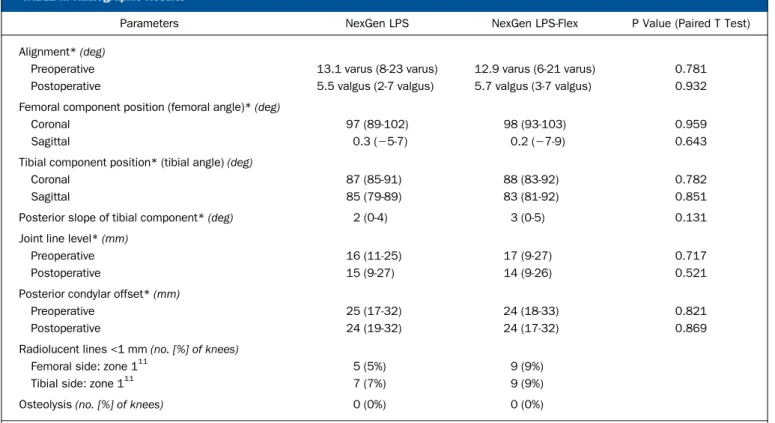
TABLE III Radiographic Results
Parameters NexGen LPS NexGen LPS-Flex P Value (Paired T Test)
Alignment* (deg)
Preoperative 13.1 varus (8-23 varus) 12.9 varus (6-21 varus) 0.781
Postoperative 5.5 valgus (2-7 valgus) 5.7 valgus (3-7 valgus) 0.932
Femoral component position (femoral angle)* (deg)
Coronal 97 (89-102) 98 (93-103) 0.959
Sagittal 0.3 (25-7) 0.2 (27-9) 0.643
Tibial component position* (tibial angle) (deg)
Coronal 87 (85-91) 88 (83-92) 0.782
Sagittal 85 (79-89) 83 (81-92) 0.851
Posterior slope of tibial component* (deg) 2 (0-4) 3 (0-5) 0.131
Joint line level* (mm)
Preoperative 16 (11-25) 17 (9-27) 0.717
Postoperative 15 (9-27) 14 (9-26) 0.521
Posterior condylar offset* (mm)
Preoperative 25 (17-32) 24 (18-33) 0.821
Postoperative 24 (19-32) 24 (17-32) 0.869
Radiolucent lines <1 mm (no. [%] of knees)
Femoral side: zone 111 5 (5%) 9 (9%)
Tibial side: zone 111 7 (7%) 9 (9%)
Osteolysis (no. [%] of knees) 0 (0%) 0 (0%)
Of the 100 knees treated with the NexGen LPS-Flex prosthesis, seventy-one (71%) were not painful, twenty-eight (28%) were mildly painful, one (1%) was moderately painful, and none were severely painful.
WOMAC Score
In both groups, the preoperative WOMAC scores (65.2 and 67.3 points) were found to be significantly improved at the time of latest follow-up (28.9 and 32.3 points) (Table I).
Activity Score
The preoperative activity scores of Tegner and Lysholm (1.6 and 1.7 points) were also improved significantly at the time of latest follow-up (5.7 and 5.9 points) in both groups (Table I).
Range of Motion
Preoperatively, the mean knee flexion contracture was 15° (range, 0° to 50°) in the NexGen LPS group and 14° (range, 0° to 50°) in the NexGen LPS-Flex group. Eighty-nine patients (89%) had a notable (10° or greater) flexion contracture preoperatively. At one year, no knee had a measurable flexion contracture. The mean ranges of flexion in supine and squatting positions pre-operatively, at one year, at five years, and at ten years did not differ significantly between the two groups (p = 0.381) (Table II). Satisfaction
Seventy-five patients (75%) were fully satisfied and twenty-five patients (25%) were satisfied with the outcome of the operation with the NexGen LPS prosthesis. Seventy-two patients (72%) were fully satisfied with the NexGen LPS-Flex prosthesis, twenty-seven patients (27%) were satisfied, and one (1%) was dissat-isfied because of constant moderate pain. Eighty-seven patients (87%) had no preference, seven patients (7%) preferred the NexGen LPS prosthesis, and six patients (6%) preferred the NexGen LPS-Flex prosthesis. Preference of one knee over the other knee was determined by stiffness of one knee. Radiographic Results
There were no significant differences between the two groups with regard to the alignment of the knee, the position of the femoral and tibial components in the coronal and sagittal planes, the posterior slope of the tibial component, the joint line, or the
Fig. 2-A
Figs. 2-A and 2-B Radiographs of both knees of a sixty-six-year-old woman with osteoarthritis made at ten years after surgery. Fig. 2-A Antero-posterior radiograph revealing that the NexGen LPS (right) and the NexGen LPS-Flex (left) prostheses are embedded rigidly. No radiolucent lines or osteolysis are demonstrated around the tibial components in either knee, and no gross wear of the polyethylene tibial insert is visu-alized in either knee.
Fig. 2-B
amount of the tibial surface area covered by the implants (tibial capping). The alignment of the knee was a mean of 5.5° of valgus in the NexGen LPS group and 5.7° of valgus in the NexGen LPS-Flex group. No knee in either group had tibial, femoral, or patellar osteolysis (Table III).
CT Evaluation
The CT scans showed no significant difference in external ro-tation of the femoral or tibial components between the two designs. The CT scans showed no tibial, femoral, or patellar osteolysis in either group.
Complications
The complication rate was low and was similar in the two groups. One knee in each group exhibited deep infection. Both knees were treated with open debridement, followed by intravenous antibiotics for six weeks. There was no recurrence of infection in the knee in the NexGen LPS group, but infection recurred in the knee in the NexGen LPS-Flex group; that knee was subsequently treated with a two-stage revision total knee arthroplasty. Survivorship Analysis
Kaplan-Meier survivorship15
analysis, with revision defined as the end point, showed a 100% implant-survival rate for the NexGen LPS prosthesis (95% CI, 94 to 100) and a 99% rate for the NexGen LPS-Flex prosthesis (95% CI, 93 to 100) at ten years postoperatively. With aseptic loosening as the end point, the survival rate was 100% (95% CI, 95 to 100) in both groups at ten years postoperatively (Fig. 2).
Discussion
T
he NexGen LPS-Flex total knee system was introduced to enhance knee flexion and to decrease wear of tibial poly-ethylene after total knee arthroplasty. Compared with the NexGen LPS prosthesis, the NexGen LPS-Flex total knee system includes an extension of the posterior condyle of the femoral component by 2 mm, a modification of the cam and tibial spine, and a re-duction of patellar impingement. The purpose of an extended posterior condyle of the femoral component is to extend the surface of the femoral component posteriorly to increase the ar-ticular contact area at high flexion angles and to increase posterior femoral translation and knee flexion. Furthermore, an improved tibiofemoral contact environment at high flexion angles could theoretically decrease the contact surface stress on the tibial polyethylene and potentially decrease wear of the tibial poly-ethylene. Moreover, the femoral cam design increases the contact surface between the cam and the tibial spine beyond that of the standard design at flexion angles of >130° and thereby may decrease the wear of the polyethylene tibial post and the rate of osteolysis. However, in this study, the survivorship, knee function, knee motion, and prevalence of osteolysis were similar between the two groups.Several authors1,16,17
reported that the high-flexion knee prosthesis had a higher risk of loosening at high flexion angles. In the current study, the ten-year survivorship of the NexGen LPS and NexGen LPS-Flex prostheses was excellent, but there
was no evidence proving the superiority of the NexGen LPS-Flex prosthesis over the NexGen LPS prosthesis. We believe that the excellent results of the NexGen LPS-Flex and NexGen LPS prostheses are attributable to adequate support of the anterior and posterior femoral condyles due to accurate bone cuts, rela-tively good bone quality, and good cementing technique.
There are inconsistencies regarding the reported differ-ences in the range of knee motion between patients with a high-flexion knee prosthesis and those with a conventional knee prosthesis. Several authors2,18,19
found that patients with a high-flexion knee prosthesis gained notably more knee motion than those with a conventional knee prosthesis. However, others3,10
did not find a difference in knee motion between the high-flexion and conventional knee components. In the current study, maximal flexion in the group with a NexGen LPS-Flex component was similar to that in the group with a NexGen LPS component. The NexGen LPS-Flex component did not dem-onstrate its theoretical advantage of providing a better range of motion of the knee. Therefore, we believe that other factors such as a good preoperative range of motion, flexion space bal-ancing, posterior tibiofemoral articular contact stability, limb characteristics (long and slender versus short and thick), and the patient’s motivation may have affected the range of knee motion. All of the patients in both groups in the current study had an improvement in knee function as assessed with the Knee Society knee function score and the WOMAC score.
Several authors have suggested that the articulating surface of the high-flexion femoral component is much more con-forming at a high flexion angle than is the conventional total knee design4,6,20
. Therefore, they suggested that the high-flexion total knee designs decrease stress concentration on the polyethylene surface and lead to decreased wear and less osteolysis in patients who attain high flexion. In contrast, the authors of another study21
reported that all high-flexion knee prostheses could result in almost point contact in an extremely high range of motion. The absence of osteolysis in both the NexGen LPS-Flex and the NexGen LPS group in our study may be related to a prepon-derance of female patients, the use of solid locking of the tibial baseplate to reduce backside wear of the insert, a compression-mold polyethylene tibial insert sterilized with gamma-radiation in inert gas, and the short shelf life of the tibial insert. It is possible that the follow-up was not sufficiently long to reveal osteolysis. The concept that a NexGen LPS-Flex prosthesis is associated with less wear and a lower prevalence of osteolysis than a NexGen LPS prosthesis remains to be proven in a longer-term follow-up study. There are several strengths of this study. First, evaluating patients treated by a single surgeon at a single center means that there was consistency in surgical technique and implant use in the study. Second, the follow-up was long enough to determine the functional outcome, survivorship, prevalence of osteolysis, and loosening. Finally, activity level data were collected and can be analyzed as a risk factor for failure.
This study also had some limitations. First, we did not assess interobserver variability in the radiographic and CT scan-ning measurements. However, we did determine the intraobserver agreement of the radiographic and CT scanning measurements22
Second, it is frequently difficult for a patient who has under-gone bilateral total knee arthroplasty to distinguish the func-tion of one knee from that of the other. Therefore, the WOMAC function scores should be interpreted with caution because it is difficult for patients to attribute functional status to a particular knee.
After a minimum duration of follow-up of ten years, we found no significant differences between these two groups with regard to survivorship, functional outcome, knee motion, or prevalence of osteolysis. Although the present study does not clearly direct the surgeon toward either arm of treatment, we believe that the extra bone that was removed from the posterior femoral condyles and intercondylar notch to allow for the thicker posterior femoral condyles and the femoral cam in the
NexGen LPS-Flex prosthesis (particularly in patients with small bones) may affect femoral component fixation and may be a concern should revision arthroplasty be necessary.n
Young-Hoo Kim, MD Jang-Won Park, MD Jun-Shik Kim, MD
The Joint Replacement Center,
Ewha Womans University MokDong Hospital, 911-1, MokDong, YangChun-Ku,
Seoul, South Korea 158-710.
E-mail address for Y.-H. Kim: [email protected]
References
1. Han HS, Kang SB, Yoon KS. High incidence of loosening of the femoral compo-nent in legacy posterior stabilised-flex total knee replacement. J Bone Joint Surg Br. 2007 Nov;89(11):1457-61.
2. Huang HT, Su JY, Wang GJ. The early results of high-flex total knee arthroplasty: a minimum of 2 years of follow-up. J Arthroplasty. 2005 Aug;20(5):674-9. 3. Kim YH, Sohn KS, Kim JS. Range of motion of standard and high-flexion posterior stabilized total knee prostheses. A prospective, randomized study. J Bone Joint Surg Am. 2005 Jul;87(7):1470-5.
4. Kim YH, Choi Y, Kim JS. Range of motion of standard and high-flexion posterior cruciate-retaining total knee prostheses a prospective randomized study. J Bone Joint Surg Am. 2009 Aug;91(8):1874-81.
5. Kim YH, Choi Y, Kwon OR, Kim JS. Functional outcome and range of motion of high-flexion posterior cruciate-retaining and high-flexion posterior cruciate-substituting total knee prostheses. A prospective, randomized study. J Bone Joint Surg Am. 2009 Apr;91(4):753-60.
6. Suggs JF, Kwon YM, Durbhakula SM, Hanson GR, Li G. In vivo flexion and kine-matics of the knee after TKA: comparison of a conventional and a high flexion cruciate-retaining TKA design. Knee Surg Sports Traumatol Arthrosc. 2009 Feb;17(2):150-6. Epub 2008 Oct 7.
7. Mehin R, Burnett RS, Brasher PM. Does the new generation of high-flex knee prostheses improve the post-operative range of movement?: a meta-analysis. J Bone Joint Surg Br. 2010 Oct;92(10):1429-34.
8. Nutton RW, van der Linden ML, Rowe PJ, Gaston P, Wade FA. A prospective randomised double-blind study of functional outcome and range of flexion following total knee replacement with the NexGen standard and high flexion components. J Bone Joint Surg Br. 2008 Jan;90(1):37-42.
9. Endres S, Wilke A. High flexion total knee arthroplasty - mid-term follow up of 5 years. Open Orthop J. 2011 Apr 14;5:138-42.
10. Choi WC, Lee S, Seong SC, Jung JH, Lee MC. Comparison between standard and high-flexion posterior-stabilized rotating-platform mobile-bearing total knee arthroplasties: a randomized controlled study. J Bone Joint Surg Am. 2010 Nov 17;92(16):2634-42. Epub 2010 Oct 15.
11. Insall JN, Dorr LD, Scott RD, Scott WN. Rationale of the Knee Society clinical rating system. Clin Orthop Relat Res. 1989 Nov;(248):13-4.
12. Bellamy N, Buchanan WW, Goldsmith CH, Campbell J, Stitt LW. Validation study of WOMAC: a health status instrument for measuring clinically impor-tant patient relevant outcomes to antirheumatic drug therapy in patients with osteoarthritis of the hip or knee. J Rheumatol. 1988 Dec;15(12): 1833-40.
13. Kim YH, Choi Y, Kwon OR, Kim JS. Functional outcome and range of motion of high-flexion posterior retaining and high-flexion posterior cruciate-substituting total knee prostheses. A prospective, randomized study. J Bone Joint Surg Am. 2009 Apr;91(4):753-60.
14. Tegner Y, Lysholm J. Rating systems in the evaluation of knee ligament injuries. Clin Orthop Relat Res. 1985 Sep;(198):43-9.
15. Kaplan EL, Meier P. Nonparametric estimation from incomplete observations. J Am Statist Assoc. 1958;53:457-81.
16. King TV, Scott RD. Femoral component loosening in total knee arthroplasty. Clin Orthop Relat Res. 1985 Apr;(194):285-90.
17. Zelle J, Janssen D, Van Eijden J, De Waal Malefijt M, Verdonschot N. Does high-flexion total knee arthroplasty promote early loosening of the femoral com-ponent? J Orthop Res. 2011 Jul;29(7):976-83. Epub 2011 Feb 9. doi:10.1002/ jor.21363.
18. Bin SI, Nam TS. Early results of high-flex total knee arthroplasty: comparison study at 1 year after surgery. Knee Surg Sports Traumatol Arthrosc. 2007 Apr;15(4):350-5. Epub 2006 Oct 28.
19. Gupta SK, Ranawat AS, Shah V, Zikria BA, Zikria JF, Ranawat CS. The P.F.C. sigma RP-F TKA designed for improved performance: a matched-pair study. Ortho-pedics. 2006 Sep;29(9 Suppl):S49-52.
20. Barink M, De Waal Malefijt M, Celada P, Vena P, Van Kampen A, Verdonschot N. A mechanical comparison of high-flexion and conventional total knee arthroplasty. Proc Inst Mech Eng H. 2008 Apr;222(3):297-307.
21. Shiramizu K, Vizesi F, Bruce W, Herrmann S, Walsh WR. Tibiofemoral contact areas and pressures in six high flexion knees. Int Orthop. 2009 Apr;33(2):403-6. Epub 2007 Nov 22.
22. Bach CM, Biedermann R, Goebel G, Mayer E, Rachbauer F. Reproducible assessment of radiolucent lines in total knee arthroplasty. Clin Orthop Relat Res. 2005 May;(434):183-8.