Reduction of CD16
-
CD56
bright
NK cell
subset Precedes NK Cell Dysfunction
in Prostate Cancer
Kyo Chul Koo
Department of Medicine
The Graduate School, Yonsei University
Reduction of CD16
-
CD56
bright
NK cell
subset Precedes NK Cell Dysfunction
in Prostate Cancer
Directed by Professor Sung Joon Hong
The Doctoral Dissertation submitted to the
Department of Medicine the Graduate School of
Yonsei University in partial fulfillment of the
requirements for the degree of Doctor of Philosophy
Kyo Chul Koo
This certifies that the
Doctoral Dissertation of Kyo Chul Koo
is approved.
---
Thesis Supervisor : Sung Joon Hong
---
Thesis Committee Member#1 : Kim Jong Sun
---
Thesis Committee Member#2 : Rha Koon Ho
---
Thesis Committee Member#3: Song Yun Seob
---
Thesis Committee Member#4: Yoon Ho Geun
The Graduate School
Yonsei University
ACKNOWLEDGEMENTS
I would like to express the deepest appreciation to my committee chair
Professor Sung Joon Hong, who continually and convincingly conveyed
a spirit of adventure in regard to research and scholarship. Without his
guidance and persistent help this dissertation would not have been
possible.
I would like to thank my committee members, Professor Yoon Ho
Geun, Professor Koon Ho Rha, Professor Yun Seob Song, and Professor
Jong Sun Kim, who embodied the figure of a kind professor, provided
maximum support and instilled enthusiasm in regards to my research.
Greatest thanks to Professor Jae Myun Lee, who kindly provided critical
revision of the manuscript for important intellectual content, material
support, and for introducing me to endearing Doctors; Doo Hee Shim,
Chang Mo Yang, Saet Byul Lee, and Shi Mun Kim who spent their late
hours for assisting me with experimentation. I also thank Tae Young
Shin and Kwang Hyun Kim for their assistance in preparing samples in
midst of bustling OR duty.
Finally, I express my deepest appreciation to my father and mother,
Dotory and Tory for providing me a reason.
<TABLE OF CONTENTS>
ABSTRACT………1
I. INTRODUCTION……….….3
II. MATERIALS AND METHODS………..6
1. Patients and healthy controls……….………... 6
2. Measurement of NK cell activity……….. 7
3. Measurement of NK cell subset distribution………. 7
A. Preparation of PBMCs………..7
B. Antibody staining………..7
C. Flow cytometry……….9
4. Prostate-specific antigen (PSA) detection……….……… 9
5. Tumor stage classification……….…9
6. Statistical analysis………. 9
III. RESULTS………. 10
1. Demographic data of investigated subjects………. 10
2. Frequency of total NK cells and distribution of CD56
dimand
CD56
brightNK cell subsets………...10
3. NK cell activity………... 12
4. Analysis by ROC curves………..13
5. NK cell activity and CD56dim to CD56bright cell ratio according
to clinicopathological variables……….…...15
IV. DISCUSSION………18
1. Preferential reduction of CD56bright NK cells in PCa patients…..20
2. Alteration of CD56bright NK cells as a response mechanism to
tumor microenvironment………..21
3. NK cell dysfunction as a consequence to reduction of CD56
brightNK cells………23
4. NK cell activity, a supportive diagnostic marker to PSA…………25
V. CONCLUSION………...27
REFERENCES………29
ABSTRACT (IN KOREAN)………...35
LIST OF FIGURES
Figure 1(A) Representative flow cytometric data of the distribution
of total NK cell population represented by CD3
-CD56
+cells………...11
Figure 1(B) Representative flow cytometric data of two major NK
cell subsets detected in peripheral blood…..…………11
Figure 2(A) Flow cytometric distribution results of total NK cell
population %...12
Figure 2(B) Flow cytometric distribution results of CD56
dimand
CD56
brightsubset distributions within total NK cells..14
Figure 2(C) CD56
dim-to-CD56
brightratio measurements between
controls and patients, according to cancer stage…….14
Figure 3 NK cell activity between controls and patients, grouped
according to cancer stage………..……...15
Figure 4(A) ROC curves comparing performances of NK cell
activity and CD56
dim-to-CD56
brightratio ...16
Figure 4(B) ROC curves comparing the performances of NK cell
LIST OF TABLES
Table 1. Demographic data of clinicopathological characteristics of
PCa patients and controls………8
Table 2. Comparisons of NK cell activity, % total NK cell
population, distribution of CD56
dimand CD56
brightsubsets,
and the CD56
dimto CD56
brightratio between patients and
controls………..13
Table 3. Comparisons of sensitivity and specificity of NK cell
activity and CD56
dimto CD56
brightratio to detect PCa...16
Table 4. Correlation of NK cell activity and CD56
dim-to-CD56
brightratio between clinicopathological variables………..18
Table 5. Comparisons of NK activity and the ratio of
CD56dim-to-CD56bright between controls and patients,
1
ABSTRACT
Reduction of CD16
-CD56
brightNK cell subset Precedes
NK Cell Dysfunction in Prostate Cancer
Kyo Chul Koo
Department of Medicine
The Graduate School, Yonsei University
Directed by Professor Sung Joon Hong
Introduction: Natural cytotoxicity, mediated by natural killer (NK) cells is
known to play an important role in inhibition and elimination of malignant tumor cells. In order to investigate the immunoregulatory role of NK cells and potential as a diagnostic tool, NK cell activity of prostate cancer (PCa) patients were analyzed with particular focus on NK cell subset distribution.
Materials and Methods: The study was based on a prospective cross-sectional
analysis of 51 patients initially diagnosed with PCa and 54 healthy controls. NK cell activity and NK cell subset distribution patterns were analyzed. NK cell activity was represented by levels of IFN-γ after stimulation of peripheral blood with proinflammatory cytokines. For distribution of NK cell subsets, PBMCs
were stained with FITC-anti-CD3, Alexa Fluor® 647-anti-CD16, and
PE-anti-CD56 monoclonal antibodies. Then, CD16+CD56dim and
CD16-CD56bright cells gated on CD56+CD3- cells were analyzed by LSRII flow cytometer.
2
Results: Significant associations between NK cell-related factors and
clinicopathological data were evident. NK cell activity was significantly lower in patients compared to controls (430.9 pg/ml vs. 975.2 pg/ml; p < 0.001). Moreover, patients with higher stages tended to show greater reduction in NK cell activity (stages II-IV: 546.1 pg/ml, 427.8 pg/ml, and 194.5 pg/ml; p for trend = 0.001). The proportion of CD56bright cells was lower in patients (2.48 % vs. 4.13 %; p<0.001). According to stage progression, CD56bright cells tended to gradually reduce (2.74 %, 2.21 %, and 1.72 %; p for trend < 0.001) in relation to CD56dim cells, confirmed by an increase in CD56dim to CD56bright cell ratio (p for trend = 0.001). The sensitivity and specificity of NK cell activity regarding PCa detection were 72% and 74%, respectively (best cut-off value at 530.9 pg/ml, AUC = 0.786 ± 0.051).
Conclusions: Results of the present study implicate that reduction of CD56bright
cells may precede NK cell dysfunction, leading to impaired cytolytic activity against PCa cells. These observations may explain one of the mechanisms behind immunoregulatory function of NK cells, and lend further support to potential immunotherapeutic strategies for tumor.
---
Key words : Cytotoxicity; Immunity; Killer cell, Natural; Prostate cancer
3
Reduction of CD16
-CD56
brightNK cell subset precedes
NK cell dysfunction in prostate cancer
Kyo Chul Koo
Department of Medicine
The Graduate School, Yonsei University
Directed by Professor Sung Joon Hong
I. INTRODUCTION
Natural killer (NK) cells serve a major role in innate and adaptive immune responses against tumor transformation or pathogen-infected cells.1,2 Without prior sensitization or class I MHC restriction, NK cells exert natural cytotoxic activity in eliminating malignant cells.3,4 Furthermore, NK cells stimulate the adaptive immune response by secreting proinflammatory cytokines such as IFN-γ to counteract the escape mechanisms promoted by tumor cells.5,6 Much progress has been made in understanding the biology of NK cells regarding cytolytic activity and subset distribution; nonetheless, considering the importance of NK cells in tumor control and elimination of blood-borne metastases, further clarification remains to be determined.
4
CD3 expression.7 According to surface membrane densities of CD56 and CD16,
NK cells are classified into CD16+CD56dim and CD16-CD56bright subsets which possess distinct distribution and function.8 The majority of NK cells are CD56dim cells, which mainly exert potent cytotoxicity.9 CD56dim NK cells, constituting approximately 90% of total NK cells, recognize tumor cells by antibody-dependent cellular toxicity and exert strong cytotoxicity.9 In contrast, CD56bright cells constitutes approximately 10% of total NK cells, mediate low cytotoxicity, but upon activation by monokines, acquire greater cytolytic activity than CD56dim cells by releasing proinflammatory cytokines such as IFN-γ.10,11 The level of IFN-γ secretion, i.e., NK cell activity, is generally associated with oncologic outcome of cancers, which implies the essential role of differential NK cell subset expression in immune regulation of tumor cells.12 NK cell activity has been shown to serve an important role in immune surveillance and elimination of tumor cells.13,14 Both in vivo and in vitro studies have shown that low NK cell activity leads to high incidences of tumor occurrence and metastasis, and that the degree of impairment correlates with invasiveness of malignancy.15,16 On the contrary, high NK cell activity has been shown to correlate with good oncological prognosis.3,17
Prostate cancer (PCa) is the most common solid organ malignancy and the
second most common cause of cancer-related death among men in
industrialized nations.18 The exact cause of PCa is not yet clearly defined, although several mechanisms have been proposed; including hereditary, environmental factors such as diet or occupational exposure, age-related
remodeling, and immune response.19 Regarding immune response, suppression
5
distribution have been observed; however, the role of NK cell dysfunction is not yet fully understood.
There is accumulating evidence that impaired immune response is a crucial factor in the pathogenesis of prostate cancer (PCa).18,19 Recent developments of novel drugs based on immune response, namely, Sipuleucel-T (Provenge®), Prostvac-VF®, or Ipilimumab (Yervoy®), and their survival benefit in castration-resistant PCa, lend further support to the notion that immune response partly has role in development and progression of PCa. Moreover, such observations suggest that PCa has rationale targets for immunotherapy. NK cell dysfunction has been noticed in PCa along with a wide variety of tumors.20-22 Despite several proposed mechanisms including reduced number, immunosuppressive cytokines, activating and inhibitory receptor repertoire imbalance, the pathophysiology of NK cell dysfunction in the development and progression of PCa is not fully understood.8 Regarding the underlying role of NK cell activity in tumor regulation, harnessing the mechanisms behind NK cell biology is clearly an important component for a successful immunotherapy against PCa.
Prostate-specific antigen (PSA) is the most widely used serum marker that
has revolutionized early detection and management of PCa.23 Although PSA, as
an independent variable, is the best predictor of PCa, the relative lack of cancer-specificity, without upper or lower threshold value, is a major drawback which may lead to unnecessary risks and costs.24 Various factors are associated with false-elevations of PSA in noncancerous conditions, namely, benign prostatic hyperplasia, infection, trauma, or age, which not only cause unnecessary biopsies that lead to potential complications, but often mask
6
aggressive forms of cancers that may lead to substantial harm.23 Various attempts have been made to overcome these limitations, such as the utilizations of PSA density, percentage of free PSA, PSA velocity, age-specific PSA ranges, complex PSA, and novel genetic-based, serologic, and urinary biomarkers. However, none of these have clearly outweighed diagnostic benefits against drawbacks and achieved satisfactory results applicable to everyday clinical practice.
To address these issues, the level of NK cell activity and the distributions of CD56dim and CD56bright NK cell subsets were analyzed between PCa patients and healthy controls. Our findings indicate that immunoregulation in PCa is impaired due to a reduction in NK cell activity preceded by redistribution of NK cell subsets. Moreover, NK cell activity itself may be applied as a supportive diagnostic marker to patients whose PSA is indefinitely correlated to clinical findings.
II. MATERIALS AND METHODS 1. Patients and healthy controls
A prospective cross-sectional analysis involved 51 patients with newly diagnosed pathological PCa and 54 age-matched healthy controls from March to December, 2012. None of the patients had received prior treatment for PCa, were known of immunological or other malignant conditions, and were free of active infection or inflammation as shown by C-reactive protein < 1.0 mg/L and white blood cell count < 10,000 cells/ul (Table 1). All controls were also free from inflammatory conditions without prior exposure to immunosuppressive agents. Independent approval was obtained from Yonsei University Ethics
7
Committee (4-2011-0660), with blood samples collected from all individuals after obtaining informed consent.
2. Measurement of NK cell activity
The cytotoxic activity of NK cells were determined by NK Vue Kit® (ATgen,
Sungnam, Korea). Whole blood was collected by BD Vacutainer® heparinN1 tubes. 1 ml of whole blood was incubated at 37°C, 7.5% CO2 with the indicated
dose of recombinant IL-2 and 1 ml of RPMI 1640 media. The cell free supernatants were then harvested and IFN-γ levels were determined according to the manufacturer’s protocols.
3. Measurement of NK cell subset distribution A. Preparation of PBMCs
Three milliliters of heparinized venous blood was obtained and cell surface markers were analyzed within 5 hours of collection. PBMCs were isolated by density gradient centrifugation using CPT® cell preparation tubes (BD Vacutainer®) at 1600g for 20 min at 20°C. The collected PBMCs (1-2 x 10⁶ cells/ml) were washed and resuspended in 10% fetal bovine serum (FBS) + phosphate buffered saline (PBS).
B. Antibody staining
For the expression of CD3, CD16, and CD56 on NK cells, PBMCs were stained with FITC-anti-CD3, Alexa Fluor® 647-anti-CD16, and PE-anti-CD56 fluorochrome-conjugated monoclonal antibodies, all obtained from BD Biosciences. After staining for 30 min at 4°C, cells were washed extensively
8
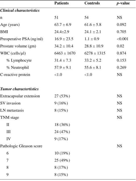
Table 1. Demographic data of clinicopathological characteristics of patients with PCa and healthy controls. All values are given as means ± SD.
Patients Controls p-value Clinical characteristics n 51 54 NS Age (years) 63.7 ± 6.9 61.6 ± 5.8 0.092 BMI 24.4±2.9 24.1 ± 2.1 0.705 Preoperative PSA (ng/ml) 16.9 ± 23.5 1.1 ± 0.9 <0.001 Prostate volume (gm) 34.2 ± 10.4 28.8 ± 10.9 0.02 WBC (cells/µl) 6463 ± 1670 6278 ± 1315 0.874 % Lymphocyte 31.4 ± 7.3 33.2 ± 5.2 0.153 % Neutrophil 57.9 ± 9.1 55.6 ± 8.1 0.269 C-reactive protein <1.0 <1.0 NS Tumor charactertistics Extracapsular extension 27 (53%) NS SV invasion 9 (16%) NS LN metastasis 8 (15%) NS TNM stage NS II 18 (36%) III 24 (47%) IV 9 (17%)
Pathologic Gleason score NS
6 10 (19%)
7 25 (49%)
8 8 (17%)
9
and fixed in 1% paraformaldehyde-PBS until assessment.
C. Flow cytometry
To determine the total percentage of NK cells gated on a CD3-CD56+ cell population, at least 10,000 target cells were acquired by LSRII flow cytometry (BD Biosciences). For the distribution of CD56dim and CD56bright NK cells, CD16+CD56dim NK cells and CD16-CD56bright NK cells gated on CD3-CD56+ cells were presented as the percentage of total NK cells. For each sample, data were further analyzed by FlowJo 8.1.1.1 (Tree Star, Inc, Ashland, OR, USA).
4. Prostate-specific antigen (PSA) detection
PSA levels of all patients and healthy controls enrolled in the study were detected by a photometric method using the Cobas C601 analyzer (ROCHE Diagnostic, USA).
5. Tumor stage classification
PCa staging was determined according to the 7th American Joint Committee
on Cancer (AJCC) TNM system. Stage distribution and pathological characteristics of the patients are shown in Table 1. All classifications were confirmed by a single pathologist.
6. Statistical analysis
Data are presented as mean ± standard deviation. Statistical analyses were performed using Student’s t-tests when comparing unpaired two group non-parametric data, and Kruskal-Wallis tests with Bonferroni post-hoc
10
correction when comparing more than two groups. The accuracy of NK cell activity and CD56dim to CD56bright ratio in detecting PCa was determined by the Receiver Operating Characteristic (ROC) and the corresponding area under a curve (AUC). The sensitivity and specificity were calculated at the best cut-off value of each ROC curve. Correlation analysis was used to evaluate the association between clinicopathological variables and measurements of NK cell activity and CD56dim to CD56bright ratio. Findings were considered significant when p-values were less than 0.05.
III. RESULTS
1. Demographic data of investigated subjects
All patients and controls were investigated clinically and pathologically with respect to factors shown in Table 1. Neither group of subjects showed significant differences in age, BMI, WBC count, % lymphocyte, % neutrophil, and C-reactive protein.
2. Frequency of total NK cells and distribution of CD56dim and CD56bright NK cell subsets
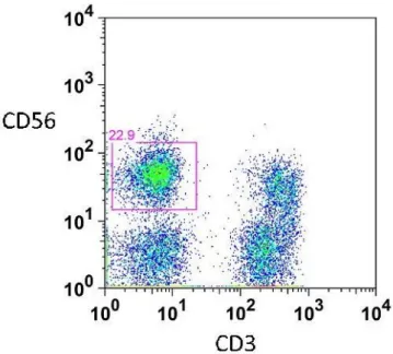
Representative flow cytometric data shows the distribution of total NK cell population represented as CD3-CD56+ cells (Fig. 1A) and two major subsets, CD16+CD56dim and CD16-CD56bright expressed as a percentage of total NK cells (Fig. 1B). Circulating frequencies of total NK cell population did not differ between patients and controls, or between stage groups (Fig. 2A; Table 2).
11
Fig. 1(A) Representative flow cytometric data of the distribution of total NK cell population represented by CD3-CD56+ cells
Fig. 1(B) Representative flow cytometric data of two major NK cell subsets detected in peripheral blood. Left, upper box: CD16+CD56dim NK cell subset. Right, lower box: CD16-CD56bright NK cell subset
12
Fig. 2(A) Boxplot diagrams showing flow cytometric distribution results of total NK cell population % between controls and patients, grouped according to cancer stage.
However, a preferential decrease in frequency of CD56bright cells was noted in patients compared to controls. Moreover, CD56bright cells tended to gradually reduce according to tumor stage progression (p for trend < 0.001) (Fig. 2B; Table 2). In order to evaluate the relative distributional shift between the two phenotypes, CD56dim to CD56bright NK cell ratio was calculated. A significantly higher ratio was observed in patients compared to controls. According to tumor progression, a gradual increase of the ratio was observed (p for trend = 0.001), implying a significant reduction of CD56bright cells in relation to alteration of CD56dim cells (Fig 2C; Table2).
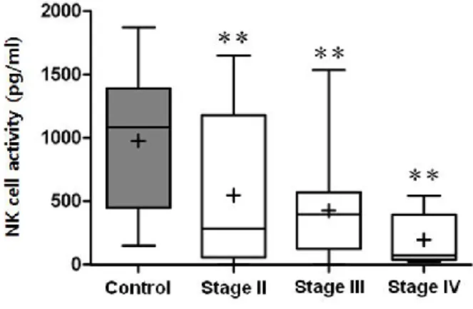
3. NK cell activity
NK cell activity was measured in culture supernatants derived from patients prior to treatment. Results obtained in studies on NK cell activity are presented
13
in Fig. 3 and Table 2. As indicated, patients showed significantly lower NK cell activity than controls. According to stage progression, those with higher stages tended to show greater reduction of NK cell activity (p for trend < 0.001).
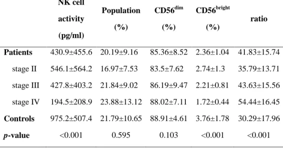
Table 2. Comparisons of NK cell activity, % total NK cell population, distribution of CD56dim and CD56bright subsets, and CD56dim to CD56bright ratio between patients and controls. All data represented as mean ± S.D.
NK cell activity (pg/ml) Population (%) CD56dim (%) CD56bright (%) ratio Patients 430.9±455.6 20.19±9.16 85.36±8.52 2.36±1.04 41.83±15.74 stage II 546.1±564.2 16.97±7.53 83.5±7.62 2.74±1.3 35.79±13.71 stage III 427.8±403.2 21.84±9.02 86.19±9.47 2.21±0.81 43.63±15.56 stage IV 194.5±208.9 23.88±13.12 88.02±7.11 1.72±0.44 54.44±16.45 Controls 975.2±507.4 21.79±10.65 88.91±4.61 3.76±1.78 30.29±17.96 p-value <0.001 0.595 0.103 <0.001 <0.001
4. Analysis by ROC curves
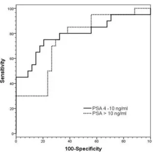
ROC curves and the best cut-off values were used to calculate the sensitivity and specificity of each NK cell-related parameter (Table 3). The sensitivity and specificity of NK cell activity levels with respect to PCa detection were 72% and 74%, whereas CD56dim to CD56bright cell ratio showed a sensitivity of 66% and a specificity of 71% (Fig. 4A). In further analysis, the sensitivity and specificity of NK cell activity were determined according to PSA values
14
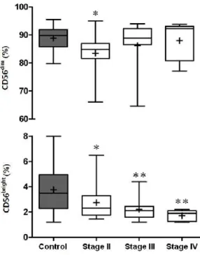
Fig. 2(B) Boxplot diagrams showing flow cytometric distribution results of CD56dim and CD56bright subset distributions within total NK cells between controls and patients. * p < 0.05, ** p < 0.01 in relation to controls
Fig. 2C Boxplot diagrams showing results of CD56dim-to-CD56bright ratio measurements between controls and patients. ** p < 0.01 in relation to controls
15
Fig. 3 Boxplot diagram comparing NK cell activity between controls and patients grouped according to cancer stage. ** p < 0.01 in relation to controls
grouped as; 4 to 10 ng/ml, i.e., critical diagnostic grey zone, and greater than 10 ng/ml. At a set specificity of 74%, NK cell activity for PSA values within the grey zone showed higher sensitivity (73% vs. 71%) and AUC (0.82 ± 0.06 vs. 0.76 ± 0.07) than for PSA values greater than 10 ng/ml (Fig. 4B).
5. NK cell activity and CD56dim to CD56bright cell ratio according to clinicopathological variables
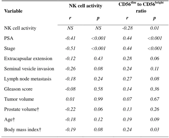
NK cell activity showed significant negative correlations with PSA (r =
-0.41), and with tumor stage (r = -0.51) (p < 0.001). Oppositely, CD56dim to CD56bright cell ratio showed positive correlations between and PSA (r = 0.44), and tumor stage (r = 0.44) (p < 0.001). Between NK activity and CD56dim to CD56bright cell ratio, a negative correlation was observed (r = -0.28, p = 0.01).
16
Table 3. Comparisons of sensitivity and specificity of NK cell activity and CD56dim to CD56bright ratio to detect PCa. The sensitivity of NK cell activity for corresponding PSA range is set at 74% specificity.
Diagnostic test AUC Sensitivity (95% CI) Specificity (95% CI) Cut-off value NK cell activity 0.79±0.05 72% 74% 530.9 pg/ml PSA 4-10 ng/ml 0.82±0.06 73% 74% PSA > 10 ng/ml 0.76±0.07 70% 74% CD56dim to CD56bright ratio 0.72±0.06 66% 71% 35.5
AUC = area under the curve
Fig. 4A ROC curves comparing the performances of NK cell activity and CD56dim-to-CD56bright ratio measurements. (AUC = Area Under the Curve)
17
Fig. 4B ROC curves comparing the performances of NK cell activity measurement according to PSA grouped as; 4 to 10 ng/ml and greater than 10 ng/ml. (AUC = Area Under the Curve)
However, there were no significant correlations noted between NK cell activity and total NK cell population, for the overall population or for the subgroups, i.e., controls and patients, and according to cancer stage category. In respect to other clinicopathological variates (e.g., Gleason score, extracapsular extension, seminal vesicle invasion, lymph node metastasis, prostate and tumor volume), analyses of NK cell activity and CD56dim to CD56bright ratio failed to demonstrate significant correlations (Table 4). NK activity and CD56dim to CD56bright ratio was compared between controls and patients grouped according to clinicopathological variables (Table 5). Although CD56dim to CD56bright ratio failed to discriminate patients with Gleason score <7 and those without
18
Table 4. Correlation of NK cell activity and CD56dim to CD56bright ratio between clinicopathological variables.
Variable
NK cell activity CD56dim to CD56bright ratio r p r p NK cell activity NS NS -0.28 0.01 PSA -0.41 <0.001 0.44 <0.001 Stage -0.51 <0.001 0.44 <0.001 Extracapsular extension -0.12 0.43 0.28 0.06
Seminal vesicle invasion -0.26 0.08 0.24 0.11
Lymph node metastasis -0.18 0.24 0.27 0.08
Gleason score -0.08 0.58 0.14 0.36
Tumor volume 0.01 0.99 0.07 0.67
Prostate volume† -0.22 0.06 0.13 0.26
Age† -0.18 0.12 0.19 0.09
Body mass index† -0.19 0.08 0.24 0.03
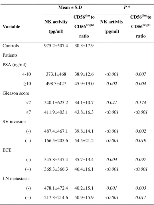
extracapsular extension, all other subgroups were distinguishable from controls by NK activity and CD56dim to CD56bright ratio. Analysis in-between patient subgroups revealed significantly higher CD56dim to CD56bright ratio in patients with pathologically confirmed LN metastasis (p = 0.043; data not shown).
IV. DISCUSSION
The aim of the present study was to clarify the role of NK cells in immune response against prostate cancer (PCa). Several mechanisms of PCa
19
Table 5. Comparisons of NK activity and CD56dim to CD56bright ratio between controls and patients grouped according to clinicopathological variables.
Mean ± S.D P * Variable NK activity (pg/ml) CD56dim to CD56bright ratio NK activity (pg/ml) CD56dim to CD56bright ratio Controls 975.2±507.4 30.3±17.9 Patients PSA (ng/ml) 4-10 373.1±468 38.9±12.6 <0.001 0.007 ≥10 498.3±427 45.9±19.0 0.002 0.004 Gleason score <7 540.1±625.2 34.1±10.7 0.041 0.174 ≥7 411.9±403.1 43.8±16.3 <0.001 <0.001 SV invasion (-) 487.4±467.1 39.8±14.1 <0.001 0.002 (+) 166.5±205.6 54.5±21.2 <0.001 0.019 ECE (-) 545.8±547.4 35.7±13.4 0.004 0.097 (+) 365.3±366.3 46.4±16.1 <0.001 <0.001 LN metastasis (-) 478.1±472.4 40.2±15.1 0.001 0.003 (+) 217.3±214.6 50.9±15.9 <0.001 0.011 ECE=Extracapsular extension SV=Seminal vesicle
20
development and progression have been proposed, e.g., age-related tissue
remodeling, hormonal, metabolic alterations, and immune response.9,25
Regarding immune response, there are accumulating evidences that host cell-mediated immune activity is suppressed in PCa,18,26-29 and that different lymphocyte populations are involved in its occurrence and progression.25 However, there is limited information regarding the functional role of NK cells in the immune response to PCa. To address this issue, we investigated levels of IFN-γ, i.e., NK cell activity, and distribution of NK cell subsets in PBMCs of PCa patients. The results of our study indicate that impaired NK cell activity is presumably preceded by a reduction of CD56bright NK cells, and that the level of NK cell activity could be potentially utilized as a supportive diagnostic marker to PSA.
1. Preferential reduction of CD56bright NK cells in PCa patients
NK cells are functionally classified into CD56dim and CD56bright NK subsets
according to surface expressions of CD56 and CD16. CD16+CD56dim NK cells
are effector cells with high quantities of cytolytic granules which express potent cytotoxicity against tumor cells.6,30 CD16-CD56bright NK cells release proinflammatory cytokines such as IFN-γ upon activation, which drives cellular and molecular inflammatory mechanisms that regulate tumor initiation, immunoevasion, survival, and outgrowth.31,32 Interesting discoveries on the importance of CD56bright NK cells have been made in recent years that it constitutes the majority of NK cells in lymphoid tissues and that it is not just a minor subpopulation among total NK cells, but are immature precursors of CD56dim NK cells.33 This article focuses on this particular subset, regarding its
21
importance in regulation of NK cell-mediated adaptive response mechanisms against tumor cells.
Investigation on distributional patterns of NK cell subsets revealed that the proportion of CD56bright NK cells of patients was lower than that of controls. Moreover, a tendency of gradual reduction of CD56bright NK cells was observed according to tumor progression, i.e., extracapsular extension, regional node or adjacent organ metastasis. The significant reduction of CD56bright NK cells in
relation to CD56dim NK cells was confirmed by an upward shift of the CD56dim
to CD56bright NK cell ratio. Previous studies on various tumor-bearing hosts have reported distinct interrelationships between CD56dim and CD56bright NK subsets. In contrast to our results, reduction of CD56dim NK cells without alteration of CD56bright NK cells were noted in gastric and esophageal cancers.34 On the other hand, reduction of CD56bright NK cells observed in breast, head, and neck cancers and equal distribution of CD56dim cells in PCa were observations that are consistent to the present study.10,25 The latter results imply that alteration of the CD56bright NK subset may be associated with immune response against various tumors.
2. Alteration of CD56bright NK cells as a response mechanism to tumor microenvironment
The significance of our study was to primarily observe a preferential reduction of CD56bright NK cells without alteration of CD56dim NK cells. Although the underlying cause has not yet been clearly defined, three possible explanations can be raised with regard to the 1) maturation process, 2) recruitment process, and 3) effect of regulatory cytokines on NK cells.
22
As mentioned, CD56bright NK cells are accepted as precursor cells of CD56dim NK cells, i.e., each subset represents distinct maturation stages of NK cells.35 Possibly, an excessive demand for effector cells in response to tumor may have provocated a rapid peripheral transition of immature CD56bright NK cells into CD56dim NK cells. A similar explanation has been proposed for reduction of CD56bright cells in patients with head and neck cancers.10 However, this presupposes a concomitant increase of CD56dim NK cells, which was not observed in the present study.
An alternative explanation without demonstration, is that peripheral CD56bright NK cells may have been recruited to lymphoid tissue sites, e.g., metastatic LNs, in order to acquire cytotoxicity. This postulation was raised by previous observations that CD56bright NK cells preferentially accumulate in the T cell area of LNs through selective expressions of CCR7 and CD62L, until being activated to produce abundant proinflammatory cytokines.10,31,33 Moreover, the
observation that CD56bright NK cells isolated from human LNs become strongly
cytotoxic upon stimulation by IL-2, suggests NK cells that are recruited to LNs might represent an immature pool of effector cells.36
A significantly higher CD56dim to CD56bright NK cell ratio in patients with pathologically confirmed LN metastasis was observed in our study, implying that these circulating cells may have been recruited to pathologic or secondary LNs in response to tumor. This is of relevance because draining LNs are usually the primary metastatic sites of cancers and CD56bright NK cells are the primary subset found in LNs that obviously counteracts metastatic cells at these sites.37 To confirm this issue, it would be interesting to examine whether CD56bright NK cells are excessively accumulated in metastatic LN specimens of patients who
23 receive radical prostatectomy.
Immunoregulatory cytokines may have a role in preferential reduction of peripheral CD56bright NK cells in PCa. An in vitro study demonstrated that culture of peripheral NK cells with transforming growth factor-β (TGF-β) induces transition of CD16+CD56dim NK cells into CD16-CD56bright NK cells.38 Although data not shown, level of peripheral TGF-β was observed to be significantly lower in our patients compared to controls. This implies that the down-regulation of TGF-β may have compromised the transition of CD56dim NK cells into CD56bright NK cells.
3. NK cell dysfunction as a consequence to reduction of CD56bright NK cells
NK cell activity was investigated to determine the influence reduction of CD56bright NK cells has on cytolytic activity against PCa tumor cells. Parallel to observations with CD56bright NK cells, NK cell activity was observed to be lower in patients along with a tendency to gradually decrease according to stage progression. These findings are consistent to previous reports that NK cell activity is compromised in a broad spectrum of tumors, including those of hematological origin and solid tumors such as bladder, esophageal, gastric, and laryngeal carcinomas.5,12,15,39-41 Several mechanisms of compromised NK cell activity in tumor microenvironment have been proposed, such as decreased number of tumor-infiltrating NK cells,34 increased NK cell surface receptors for immune suppressor factors,42 and inactivation of NK effector cells.15
Correlations observed between NK cell activity and the distribution of CD56bright NK cells may be of direct relevance to suggest an additional mechanism that weak NK cell activity, i.e., reduced secretion of IFN-γ, is a
24
consequence to reduced CD56bright NK cells. The major cytokines secreted by CD56bright NK cells are tumor necrosis factor-α, granulocyte-macrophage colony-stimulating factor, IL-10, IL-13, and IFN-γ.7 Focusing on IFN-γ, it has been reported that NK cells, particular the CD56bright NK cell subset is one of major sources of this cytokine.33 Secretion of IFN-γ seems to directly depend on CD56bright NK cells, as observed in in vitro studies that CD56bright NK cells preferentially proliferate in coculture with immature dendritic cells and lipopolysaccharides to produce IFN-γ.43 Also, stimulation of CD56bright NK cells with transduced carcinoma cells resulted with enhanced ability to produce IFN-γ and gain high cytotoxicity.9 Further to this notion, in vivo studies have observed production of IFN-γ by tonsillar CD56bright NK cells before maturation into effector cells.36 Reversely, reduction of CD56bright NK cells was observed to induce impaired secretion of IFN-γ in patients with allergic rhinitis.44
As of tumors, IFN-γ stimulates the adaptive immune response in the polarization of T-helper type 1 cells to counteract the escape mechanisms promoted by tumor cells.5 In practice, the level of IFN-γ is generally accepted to predict oncologic outcome, suggesting the importance of this particular subset of NK cells in adaptive immunity.12 Our findings that NK cell activity did not correlate to total NK cell population, implies that cytotoxicity exerted by NK cells, does not merely depend on the number of NK cells. This lends further support to the notion that the activity of CD56bright NK cells on cytokine secretion, relative to that of CD56dim NK cells, is the crucial factor that regulates NK cell activity.
Considering these supportive findings, we prefer to hypothesize that reduction of CD56bright NK cells is a potential mechanism involved in reduced
25
secretion of IFN-γ, i.e., low NK cell activity, which leads to impaired cytotoxicity against PCa cells.
4. NK cell activity, a supportive diagnostic marker to PSA
The area under a curve (AUC) for NK cell-related parameters in diagnosing PCa, implies that NK cell activity may serve as a supportive marker to PSA. It is clear that PSA provides the highest diagnostic value for PCa and that its application to clinical practice has revolutionized management of this disease.45 However, the major drawback is its relative lack of specificity, especially in the critical diagnostic grey zone of 4 to 10 ng/ml where only 25% of biopsied patients will demonstrate cancer.23,46 False-elevations in noncancerous conditions not only cause unnecessary biopsies that lead to potential complications, but often mask aggressive forms of cancer that may lead to substantial harm.19,24 Although this ongoing clinical challenge has aroused scientific challenges to evaluate novel methods of PCa screening, none have clearly outweighed diagnostic benefits against drawbacks.19 The American Urological Association best practice guideline for PSA screening has recommended screening to be offered in all healthy men over the age of 40. However, due to such limitations, in 2012 the United States Preventive Service
Task Force accumulated solid evidences based on prospective
randomized-controlled studies to recommend against routine PSA screening. This paper does not seek to settle these concerns, but raise the possibility that NK cell-related factors such as NK cell activity may be utilized in combination with PSA to provide additional diagnostic value for preventing underdiagnosis and overdiagnosis; as in our results, especially for those within
26 the diagnostic grey zone.
There are several limitations of this study that are worth mentioning. First, the current study was based on healthy controls versus patients diagnosed of PCa due to elevated PSA on routine health examinations. Therefore, the absence of PCa patients with normal PSA (< 4 ng/ml) was the major limitation which hampered a direct comparison of diagnostic yield between PSA and NK cell activity. Although this group of patients would most probably present clinically insignificant cancer and represent candidates for active surveillance, clearly an extended population study is needed to confirm our preliminary findings and to assess economic impact.
Second, although PCa patients were age-matched to healthy controls, and there were no differences in CRP or WBC counts confirming the absence of active infection or immunosuppression, the possibility of disparities in immune response may have existed. To confirm the absence of confounders related to immunoregulation that may affect the level of NK cell activity or the distribution in NK cell subsets, additional investigation with surrogate markers should have been performed.
Third, the indirect assay based on IFN-γ measurements after stimulation of the peripheral blood with IL-2 could be considered insufficient, and rather a direct evidence such as NK cell killing assay to confirm alterations in NK cell cytotoxicity is needed. However, the scopes of the current study was to utilize and validate the usefulness of a measurement that is reproducible, simple, less time-consuming, and less costly which is applicable to everyday clinical practice. The conventional widely-used measurements, such as the 51Cr release assay, has limitations regarding quantification and standardization as it utilizes
27
K-562 cell lines as target cells and PBMCs as effector cells. The NK Vue Kit used in this study is a measurement of indirectly stimulated IFN-γ levels; however, by monitoring IFN- γ levels after stimulation of the peripheral blood with IL-2, it may be assumed that this may provide an actual quantitation of NK cell cytotoxicity exerted on tumor cells. Indeed, the cytokines secreted by NK cells, most importantly IFN-γ, is known to be generally associated with oncologic outcome of cancers, which is the main scope of our study.
Fourth, NK cell activity was characterized in this study as the level of IFN-γ secretion in the supernatant following ex vivo activation of the total blood by IL-2. As IL-2 could also activate the secretion of IFN-γ by T cells, there is potential that the diminution of IFN-γ was not specific to NK cells, and intracellular flow cytometric staining or purification of NK cells from total blood should have performed. However, in a set of our unpublished data which utilized intracellular staining protocols, IL-2 was observed to mainly stimulate IFN-γ from NK cells, and not from T cells. Moreover, the concentration of IL-2 in the NK Vue kit was set at 8 ng/ml; a cut-off concentration that was insufficient to stimulate IFN-γ from T cells, but only from NK cells.
Further studies regarding activating and inhibitory receptor profiles and degranulation assays in a prospective well-designed, randomized control trial are warranted to confirm our results and utilize the biology of NK cell as a immunotherapeutic strategy against PCa development and progression.
V. CONCLUSION
The present study provided novel findings that the CD56bright NK cell subset is not only a minor subpopulation among total NK cells, but serves an important
28
role in adaptive response against PCa cells. This notion lends further support that longitudinal studies regarding NK cell immunosurveillance clearly deserves additional research, which could potentially lead to novel immunotherapeutic strategies for enhancing oncological outcomes of PCa.
29
REFERENCES
1. Cerwenka A, Lanier LL. Natural killer cells, viruses and cancer. Nat Rev Immunol 2001;1:41-9.
2. Szkaradkiewicz A, Karpinski TM, Drews M, Borejsza-Wysocki M, Majewski P, Andrzejewska E. Natural killer cell cytotoxicity and immunosuppressive cytokines (IL-10, TGF-beta1) in patients with gastric cancer. J Biomed Biotechnol 2010;2010:901564.
3. Imai K, Matsuyama S, Miyake S, Suga K, Nakachi K. Natural cytotoxic activity of peripheral-blood lymphocytes and cancer incidence: an 11-year follow-up study of a general population. Lancet 2000;356:1795-9.
4. Trinchieri G. Biology of natural killer cells. Adv Immunol 1989;47:187-376. 5. Dunn GP, Koebel CM, Schreiber RD. Interferons, immunity and cancer
immunoediting. Nat Rev Immunol 2006;6:836-48.
6. Takahashi E, Kuranaga N, Satoh K, et al. Induction of CD16+ CD56bright NK cells with antitumour cytotoxicity not only from CD16- CD56bright NK Cells but also from CD16- CD56dim NK cells. Scand J Immunol 2007;65:126-38.
7. Cooper MA, Caligiuri MA. Isolation and characterization of human natural killer cell subsets. Curr Protoc Immunol 2004;Chapter 7:Unit 7 34.
8. Watanabe M, Kono K, Kawaguchi Y, et al. NK cell dysfunction with down-regulated CD16 and up-regulated CD56 molecules in patients with esophageal squamous cell carcinoma. Dis Esophagus 2010;23:675-81.
9. Dowell AC, Oldham KA, Bhatt RI, Lee SP, Searle PF. Long-term proliferation of functional human NK cells, with conversion of CD56(dim) NK cells to a CD56 (bright) phenotype, induced by carcinoma cells
30
co-expressing 4-1BBL and IL-12. Cancer Immunol Immunother
2012;61:615-28.
10. Bauernhofer T, Kuss I, Henderson B, Baum AS, Whiteside TL. Preferential apoptosis of CD56dim natural killer cell subset in patients with cancer. Eur J Immunol 2003;33:119-24.
11. Soiffer RJ, Murray C, Shapiro C, et al. Expansion and manipulation of natural killer cells in patients with metastatic cancer by low-dose continuous infusion and intermittent bolus administration of interleukin 2. Clin Cancer Res 1996;2:493-9.
12. Lin CT, Yu MT, Li C, et al. Dysfunction of natural killer cells in patients with transitional cell carcinoma. Cancer Lett 2010;291:39-45.
13. Whiteside TL, Herberman RB. The role of natural killer cells in immune surveillance of cancer. Curr Opin Immunol 1995;7:704-10.
14. Kim S, Iizuka K, Aguila HL, Weissman IL, Yokoyama WM. In vivo natural killer cell activities revealed by natural killer cell-deficient mice. Proc Natl Acad Sci U S A 2000;97:2731-6.
15. Takeuchi H, Maehara Y, Tokunaga E, Koga T, Kakeji Y, Sugimachi K. Prognostic significance of natural killer cell activity in patients with gastric carcinoma: a multivariate analysis. Am J Gastroenterol 2001;96:574-8. 16. Talmadge JE, Meyers KM, Prieur DJ, Starkey JR. Role of NK cells in
tumour growth and metastasis in beige mice. Nature 1980;284:622-4.
17. Schleypen JS, Baur N, Kammerer R, et al. Cytotoxic markers and frequency predict functional capacity of natural killer cells infiltrating renal cell carcinoma. Clin Cancer Res 2006;12:718-25.
31 Opin Urol 2008;18:315-9.
19. Basch E, Oliver TK, Vickers A, et al. Screening for prostate cancer with prostate-specific antigen testing: American Society of Clinical Oncology Provisional Clinical Opinion. J Clin Oncol 2012;30:3020-5.
20. Lahat N, Alexander B, Levin DR, Moskovitz B. The relationship between clinical stage, natural killer activity and related immunological parameters in
adenocarcinoma of the prostate. Cancer Immunol Immunother
1989;28:208-12.
21. Oikawa T, Kawai K, Ishiwata I, Ohno T, Akaza H. Induction of potent antitumour natural-killer cells from peripheral blood of patients with advanced prostate cancer. BJU Int 2003;92:1009-15.
22. Kastelan M, Kraljic I, Tarle M. NK cell activity in treated prostate cancer patients as a probe for circulating tumor cells: hormone regulatory effects in vivo. Prostate 1992;21:111-20.
23. Lumen N, Fonteyne V, De Meerleert G, et al. Population screening for prostate cancer: an overview of available studies and meta-analysis. Int J Urol 2012;19:100-8.
24. O'Rourke ME. The prostate-specific antigen screening conundrum: examining the evidence. Semin Oncol Nurs 2011;27:251-9.
25. Sotosek S, Sotosek Tokmadzic V, Mrakovcic-Sutic I, et al. Comparative study of frequency of different lymphocytes subpopulation in peripheral blood of patients with prostate cancer and benign prostatic hyperplasia. Wien Klin Wochenschr 2011;123:718-25.
26. Aalamian M, Pirtskhalaishvili G, Nunez A, et al. Human prostate cancer regulates generation and maturation of monocyte-derived dendritic cells.
32 Prostate 2001;46:68-75.
27. Ebelt K, Babaryka G, Figel AM, et al. Dominance of CD4+ lymphocytic infiltrates with disturbed effector cell characteristics in the tumor microenvironment of prostate carcinoma. Prostate 2008;68:1-10.
28. Thomas JW, Jerkins G, Cox C, Lieberman P. Defective cell-mediated immunity in carcinoma of the prostate. Invest Urol 1976;14:72-5.
29. Wagenlehner FM, Elkahwaji JE, Algaba F, et al. The role of inflammation and infection in the pathogenesis of prostate carcinoma. BJU Int 2007;100:733-7.
30. Cooper MA, Fehniger TA, Caligiuri MA. The biology of human natural killer-cell subsets. Trends Immunol 2001;22:633-40.
31. Harlin H, Hanson M, Johansson CC, et al. The CD16- CD56(bright) NK cell subset is resistant to reactive oxygen species produced by activated granulocytes and has higher antioxidative capacity than the CD16+ CD56(dim) subset. J Immunol 2007;179:4513-9.
32. Zaidi MR, Merlino G. The two faces of interferon-gamma in cancer. Clin Cancer Res 2011;17:6118-24.
33. Poli A, Michel T, Theresine M, Andres E, Hentges F, Zimmer J. CD56bright natural killer (NK) cells: an important NK cell subset. Immunology 2009;126:458-65.
34. Izawa S, Kono K, Mimura K, et al. H(2)O(2) production within tumor microenvironment inversely correlated with infiltration of CD56(dim) NK cells in gastric and esophageal cancer: possible mechanisms of NK cell dysfunction. Cancer Immunol Immunother 2011;60:1801-10.
33
differentiate into CD56dim cells: role of contact with peripheral fibroblasts. J Immunol 2007;179:89-94.
36. Ferlazzo G, Munz C. NK cell compartments and their activation by dendritic cells. J Immunol 2004;172:1333-9.
37. Sheikhi A, Saadati K, Salmani R, Yahaghi N, Siemens DR. In vitro modulation of natural killer activity of human peripheral blood mononuclear cells against prostate tumor cell line. Immunopharmacol Immunotoxicol 2011;33:700-8.
38. Keskin DB, Allan DS, Rybalov B, et al. TGFbeta promotes conversion of CD16+ peripheral blood NK cells into CD16- NK cells with similarities to decidual NK cells. Proc Natl Acad Sci U S A 2007;104:3378-83.
39. Schantz SP, Campbell BH, Guillamondegui OM. Pharyngeal carcinoma and natural killer cell activity. Am J Surg 1986;152:467-74.
40. Rabinovich GA, Gabrilovich D, Sotomayor EM. Immunosuppressive strategies that are mediated by tumor cells. Annu Rev Immunol 2007;25:267-96.
41. Gonzalez FM, Vargas JA, Lopez-Cortijo C, et al. Prognostic significance of natural killer cell activity in patients with laryngeal carcinoma. Arch Otolaryngol Head Neck Surg 1998;124:852-6.
42. Yamaguchi Y, Takashima I, Funakoshi M, Kawami H, Toge T. Defective natural killer activity in gastric cancer patients: possible involvement of suppressor factor receptor. In Vivo 1994;8:279-83.
43. Vitale M, Della Chiesa M, Carlomagno S, et al. The small subset of CD56brightCD16- natural killer cells is selectively responsible for both cell proliferation and interferon-gamma production upon interaction with
34
dendritic cells. Eur J Immunol 2004;34:1715-22.
44. Scordamaglia F, Balsamo M, Scordamaglia A, et al. Perturbations of natural killer cell regulatory functions in respiratory allergic diseases. J Allergy Clin Immunol 2008;121:479-85.
45. Schroder FH. Landmarks in prostate cancer screening. BJU Int 2012;110 Suppl 1:3-7.
46. Khan MA, Partin AW, Rittenhouse HG, et al. Evaluation of proprostate specific antigen for early detection of prostate cancer in men with a total prostate specific antigen range of 4.0 to 10.0 ng/ml. J Urol 2003;170:723-6.
35 ABSTRACT 전립선암에서 관찰되는 자연살해세포 활성장애 기전으로써 CD16-CD56bright subset의 감소 지도교수 홍 성 준 연세대학교 대학원 의학과 구 교 철 I. 서론 자연살해세포에 의한 세포독성은 종양세포의 억제와 제거 기전에 있어 주요한 역할을 한다. 본 연구는 전립선암의 발생 및 진행의 면역요인에 있어서 자연살해세포의 역할에 대해 알아보기 위해 전립선암 환자의 말초혈액에서 자연살해세포의 subset에 초점을 맞춰 자연살해세포 활성도를 분석하고, 전립선암 진단 표지자로써 이들의 가치에 대해 알아보았다. II. 재료 및 방법 전립선암으로 확진되어 수술을 앞둔 51명의 환자들과 나이를
36 보정한 건강대조군 54명을 대상으로 자연살해세포 활성도와 자연살해세포 subset의 분포를 전향적으로 분석하였다. 자연살해세포 활성도는 말초혈액을 IL-2로 자극시킨 후 배양액의 상층액에서 검출된 IFN-γ치를 측정값으로 사용하였다. 자연살해세포 subset 분포를 분석하기 위해 말초혈액을 단일 클론 형광색소 항체로 염색 후 세포분석기를 이용하여 측정하였으며, 자연살해세포를 구성하는
CD56+CD3- 세포 중 CD16+CD56dim subset과 CD16-CD56bright subset의
세포수를 측정하여 전체 자연살해세포 수 중에서 차지하는 비율로써 분석하였다. III. 결과 자연살해세포 활성도와 CD56bright subset 세포수는 대조군에 비해 환자군에서 유의하게 낮았다(430.9 pg/ml 대 975.2 pg/ml, 2.3% 대 3.8%; p < 0.001). 암병기가 증가할수록 두 인자 모두 감소하는 유의한
경향이 관찰되었다 (p for trend = 0.001). CD56dim
대 CD56bright
subset 비율은 환자군에서 유의하게 높았으며 (41.8 대 30.3; p < 0.001) 병기가 높을수록 증가하는 유의한 경향을 보임으로써 (p for trend =
0.001), CD56dim subset 세포수의 변화에 비해 CD56bright subset 세포수가 유의하게 상대적으로 감소했음을 확인할 수 있었다. 전립선암 진단에 있어서 자연살해세포 활성도의 예민도와 특이도는 각각 72%와 74%로 나타났다.
37 IV. 결론 CD56bright subset의 세포수 감소는 자연살해세포 활성장애에 선행하는 것으로 생각되며, 이로 인하여 전립선암 세포에 대한 세포독성장애가 발생하는 것으로 보인다. 본 관찰 결과는 전립선암의 미세종양환경에서 자연살해세포 독성장애의 기전을 제시하며, 향후 면역기반 치료의 개발에 응용될 수 있으리라 기대한다.
---
핵심되는 말 : 세포독성; 자연살해세포; 전립선암
38
PUBLICATION LIST
Koo KC, Shim DH, Yang CM, et al. Reduction of the CD16-CD56bright NK Cell
Subset Precedes NK Cell Dysfunction in Prostate Cancer. PLoS ONE 2013;8:e78049. doi:10.1371/journal.pone.0078049