저작자표시-비영리-변경금지 2.0 대한민국 이용자는 아래의 조건을 따르는 경우에 한하여 자유롭게 l 이 저작물을 복제, 배포, 전송, 전시, 공연 및 방송할 수 있습니다. 다음과 같은 조건을 따라야 합니다: l 귀하는, 이 저작물의 재이용이나 배포의 경우, 이 저작물에 적용된 이용허락조건 을 명확하게 나타내어야 합니다. l 저작권자로부터 별도의 허가를 받으면 이러한 조건들은 적용되지 않습니다. 저작권법에 따른 이용자의 권리는 위의 내용에 의하여 영향을 받지 않습니다. 이것은 이용허락규약(Legal Code)을 이해하기 쉽게 요약한 것입니다. Disclaimer 저작자표시. 귀하는 원저작자를 표시하여야 합니다. 비영리. 귀하는 이 저작물을 영리 목적으로 이용할 수 없습니다. 변경금지. 귀하는 이 저작물을 개작, 변형 또는 가공할 수 없습니다.
The role of activated leukocyte cell
adhesion molecule (ALCAM/CD166)
in the pathogenesis of atopic dermatitis
Mi Seon Oh
Department of Medical Science
The Graduate School, Yonsei University
The role of activated leukocyte cell
adhesion molecule (ALCAM/CD166)
in the pathogenesis of atopic dermatitis
Directed by Professor Myung Hyun Sohn
The Master’s Thesis submitted to
The Department of Medical Science,
The Graduate School of Yonsei University
In partial fulfillment of the requirements for the degree of
Master of Medical Science
Mi Seon Oh
December 2016
This certifies that the Master’s Thesis of
Mi Seon Oh is approved.
Thesis Supervisor : Myung Hyun Sohn
Thesis Committee Member #1 : Kyung Won Kim
Thesis Committee Member #2 : Jae Myun Lee
The Graduate School
Yonsei University
감사의 글
대학원 진학을 위해 작성하는 학업계획서에 ‘생명의 메커니즘을 밝히 는 전문가가 되고 싶습니다’라는 거창한 문장을 서슴지 않고 적어 내리 는 철부지였던 제가, 지금은 석사과정을 마무리하는 단계에 있다는 것이 감개무량합니다. 연구실에 들어와서 실험뿐만 아니라 제가 삶을 살아가 는 데 도움이 될 귀한 경험들을 배운 것 같습니다. 배움이 많은 이 곳에 서, 저의 부족함을 깨닫고 스스로 많이 힘든 시간도 있었습니다. 하지만 많은 분들의 도움 덕분에 어려움을 극복하고, 학위 논문이라는 결실을 맺게 되었습니다. 저의 학문적인 부족함을 늘 세심한 조언으로 채워주시고, 제가 연구에 능동적으로 임할 수 있게 옆에서 지도해주신 손명현 교수님께 깊은 감사 드립니다. 그리고 반복되는 실험으로 심신이 지쳤을 때, 따듯한 조언과 배려로 응원해주셨던 김규언 교수님께 감사 드립니다. 제가 부진할 때마 다 안타까워하시며 제가 큰 성과를 이룰 수 있도록 옆에서 다독여주셨던 김경원 교수님, 제 논문을 더 풍성하게 채워갈 수 있도록 귀한 조언을 주셨던 이재면 교수님께도 감사 드립니다. 엉뚱한 저에게 늘 귀 기울여주시고 제가 이 곳에서도 소중한 존재라는 것을 느끼게 해주신 이경은 선생님 정말 감사합니다. 제 연구의 시작과 마무리까지 늘 관심 가져주시고 저의 많은 질문에도 항상 자상하게 알려 주셨던 홍정연 선생님, 연구자로서의 제 단점도 장점으로 승화시켜주시 며 긍정적으로 연구실 생활을 할 수 있게 도와주신 김미나 선생님께 감 사 드립니다. 더 좋은 연구 결과를 넣을 수 있도록 바쁘신 상황에서도저를 위해 시간을 내어주신 설인숙 선생님, 김수연 선생님께도 감사 드 립니다. 그리고 힘든 석사 과정을 함께 하면서 저의 부족함을 채워주던 친구, 윤선이에게 미안함과 고마움을 함께 전합니다. 지루한 삶에서 활력소가 되어준 유진, 진아, 민지. 나의 짜증도 유쾌 하게 받아주었던 희재, 영재. 연구실에서 깜짝선물과 같았던 보라, 호균 오빠, 지수오빠. 그리고 나의 어떤 모습도 이해해주고. 잘 될 거라며 자 신감을 북돋아주었던 상만이. 변함없는 마음으로 제게 큰 힘과 사랑을 주고, 끝까지 노력할 수 있도록 응원해준 친구들에게도 감사합니다. 마지막으로 오랫동안 공부하는 막내딸을 한결같이 응원해주시고 지금 껏 뒷바라지 하시느라 고생하신 부모님, 학업으로 가족에 소홀해진 저를 대신해 집을 항상 화기애애하게 해주고 저를 자랑스러운 동생으로 생각 해준 언니, 나에게 웃음을 선사하는 폭스, 그리고 나의 곁에 오랜 시간 함께 함으로 마음의 안정을 주었던 소중한 비다. 사랑하는 가족들이 있 었기에 포기하지 않을 수 있었습니다. 정말 감사합니다. 다시 한번, 제 인생에 이처럼 영광스러운 순간을 마련해주신 모든 분 들께 깊은 감사의 마음을 전합니다. 2016년 12월 오미선
TABLE OF CONTENTS
ABSTRACT··· 1
I. INTRODUCTION ··· 3
II. MATERIALS AND METHODS ··· 6
1. Mice and Induction of Atopic Dermatitis ··· 6
2. Hematoxylin and Eosin Staining ··· 7
3. Clinical Score ··· 7
4. Histological Score ··· 7
5. Enzyme-linked Immunosorbent Assay ··· 8
6. Real-time Polymerase Chain Reaction ··· 8
7. Electron Microscopy ··· 9
8. Flow Cytometry ··· 10
9. Subjects ··· 11
10. Statistical Analysis of Data ··· 12
III. RESULTS ··· 13
1. ALCAM is increased in OVA-induced AD mice ··· 13
2. ALCAM deficiency attenuates Th2 inflammatory responses ··· 15
3. ALCAM deficiency suppresses the activation of T cell ··· 18
4. ALCAM deficiency diminishes the skin barrier dysfunction ··· 21
5. ALCAM deficiency attenuates Th2 inflammatory responses in
OXA-induced AD mice ··· 24
IV. DISCUSSION ··· 30
V. CONCLUSION ··· 33
REFERENCES ··· 34
LISTS OF FIGURES
Figure 1. Increase of ALCAM in wild type mice induced to atopic
dermatitis by ovalbumin ··· 14
Figure 2. Attenuation of ovalbumin-induced atopic dermatitis in
ALCAM deficient mice ··· 16
Figure 3. Suppression of T cell activation in ALCAM deficient
mice ··· 19
Figure 4. Inhibition of skin barrier dysfunction in ALCAM
deficient mice ··· 22
Figure 5. Attenuation of oxazolone-induced atopic dermatitis in
ALCAM deficient mice ··· 25
Figure 6. Increase of ALCAM in pediatric atopic dermatitis
LISTS OF TABLES
ABSTRACT
The role of activated leukocyte cell adhesion molecule (ALCAM/CD166)
in the pathogenesis of atopic dermatitis
Mi Seon Oh
Department of Medical Science
The Graduate School, Yonsei University
(Directed by Professor Myung Hyun Sohn)
Activated leukocyte cell adhesion molecule (ALCAM/CD166) is a transmembrane protein expressed highly on dendritic cells (DC). ALCAM binds heterophilically to CD6 expressed on resting T cells. It has been reported that ALCAM-CD6 interaction is involved in the formation of immunological synapse. The interaction mediates the stable contact between DC and T cells. Moreover, it is associated with T cell activation, proliferation and maturation. Atopic dermatitis (AD) is known to T cell-mediated allergic skin diseases. The disease is characterized by skin barrier dysfunctions and Th2 inflammatory immune reactions. However, the role of ALCAM has not been fully identified in the allergic diseases. Therefore, this study determined the possibilities that ALCAM mediates the immunologic responses and affects to the function of skin barrier in the pathogenesis of AD.
To explore the contribution of ALCAM in allergic skin inflammation, wild-type (WT) mice were induced to AD, using ovalbumin (OVA). As a different allergen, oxazolone (OXA) was applied to induce AD murine model. ALCAM deficient (ALCAM-/-) mice were used to analyze the effects of the immunologic
response and barrier dysfunction in the pathogenesis of AD.
In this study, ALCAM mRNA expression was up-regulated in the skin of WT AD mice, comparing with a sham group. Although mice were induced to AD with OVA or OXA respectively, ALCAM deficiency was less worsened the skin barrier dysfunction than WT AD mice. In addition, T cell activation was inhibited by ALCAM deficiency. Serum IgE and Th2 type cytokines were down-regulated in ALCAM-/-AD mice
Therefore, these findings indicate that ALCAM mediates not only immunologic dysregulation but also impaired barrier function in the pathogenesis of AD.
KEY WORDS: atopic dermatitis, ALCAM, CD166, ovalbumin, oxazolone,
Th2 dominant immune response, skin barrier
The role of activated leukocyte cell adhesion molecule (ALCAM/CD166)
in the pathogenesis of atopic dermatitis
Mi Seon Oh
Department of Medical Science
The Graduate School, Yonsei University
(Directed by Professor Myung Hyun Sohn)
I. INTRODUCTION
Atopic dermatitis (AD) is associated with skin barrier defects and T cell-mediated chronic inflammation. AD symptoms are accompanied by dry, itchy skin and relapsing eczema.1-3 Dendritic cell (DC) and type 2 helper T cells (Th2) contribute to development of allergic skin inflammation. Th2 cells are classically dominant to elicit AD, producing the cytokines such as interleukin-4 (IL-4), interleukin-5 (IL-5) and interleukin-13 (IL-13). DC is found in the stratified epithelium of the skin and mucosa.3-6 An antigen penetrated through the impaired epithelial barrier, is uptaken by DC. DC is acquired to initiate atopic dermatitis after contact with an antigen. The activated DC migrates to the T cell zone of skin draining lymph node and promotes Th2 differentiation.7 As an antigen presenting cell (APC), DC gains the capacity to differentiate naïve CD4+ T cells into effector Th2 cells.8 T cell activation requires two receptor signals expressed on the T cells and DC respectively to initiate an immune response. The first signal is an interaction of T cell receptor (TCR) and MHC-peptide complexes of dendritic cell (DC), forming an immune synapse. The second signal is an interaction of
co-stimulatory molecules. The co-co-stimulatory molecules are expressed typically on T cell (CD28 and CTLA-4) and DC (CD80/B7.1 and CD86/B7.2).8-10 As an another co-stimulatory molecule, CD6 is a surface glycoprotein found on mature T cells, immature B cells. It binds to ALCAM (Activated leucocyte cell adhesion molecule / CD166) through their extracellular domains.11
ALCAM is a transmembrane protein of immunoglobulin superfamily.12 The molecule is structurally characterized by the presence of five extracellular immunoglobulin (Ig) domains including two membrane-distal variable (V) type and three membrane-proximal constant (C2) type Ig folds.13 ALCAM is expressed on diverse cell types, such as monocytes, keratinocytes, thymic epithelium, nervous ends, langerhans cells. Its expression is particularly high on DC. ALCAM has been reported to a critical role in tumor progression, metastasis, cell migration and immune response.14-16 ALCAM-CD6 interaction is important to mediate immunologic process, including initiation and sustaining of DC-T cell contact called ‘immune synapse’. In addition ALCAM-CD6 has a pivotal role in T cell differentiation, proliferation and maturation through enhancement of CD3 proliferation and increase of CD25 molecules.11,16
Meanwhile, an abnormal skin barrier is also a hallmark of AD. Epidermis is the outer layer of the skin and provides a barrier against the outside. It is composed of four layers, stratum corneum (SC), stratum granulosum (SG), stratum spinosum (SS) and stratum basale (SB). SC normally is composed with proteins, lipid rich peripheral envelope and lamellar body (LB) located in SG. The main function of the proteins and envelope is to prevent transepidermal water loss (TEWL) and to provide a cutaneous permeability barrier against environmental allergen. In AD patients, however, the proteins related with skin barrier and the lipids are deficient significantly. Moreover, allergic skin inflammatory response demonstrates Th2 dominant cytokine profile through the suppressed Th1 and enhanced Th2 cytokines.3,5,17-19 Impaired skin barrier might also provide an initial entry point for food allergens, potentially leading to development of eosinophilic esophagitis (EoE)
and food anaphylaxis by modulation of IL-33.20,21 Thus, compromised epithelial barrier can be a risk factor for AD as well as other allergic diseases.
Taken together, it is suggested that ALCAM is needed for T cell-mediated allergic inflammatory diseases. However, ALCAM is still unclear in pathogenesis of AD. Thus, the present study identified the role of ALCAM in immune response and skin barrier function, using ALCAM deficient and wild type mice.
II. MATERIALS AND METHODS
1. Mice and Induction of Atopic DermatitisWT C57BL/6 mice, aged 7 - 10 wk old, were purchased from Orient Bio (Sungnam, Korea). ALCAM-/- mice with C57BL/6 background were purchased from Jackson Laboratory (Bar Harbor, ME, USA). All mice were kept under controlled humidity, temperature and pathogen-free conditions.
To induce atopic dermatitis on the dorsal skin of mice by egg protein ovalbumin (OVA / Grade V; Sigma-Aldrich, St. Louis, MO, USA), Mice were anesthetized with ioflurane (Hana pharm, Sungnam, Korea) inhalation. The dorsal skin of mice was also shaved with an electric clipper and then tape stripped five times by tegaderm (1624W; 3M Health Care, St Paul, MN, USA). Then 100 μg of OVA in 100 μl of PBS was placed on a patch of sterile gauze (1 × 1 cm2). It was secured to the dorsal skin with tegaderm. The patch was kept on the skin for 1 wk. Thus, mice have a total of three 1 wk OVA exposures with 2 wk intervals between each exposure week. During the experiment periods, transepidermal water loss (TEWL) was assessed on the skin by Vapometer (Delfin, Kuopio, Finland) at controlled humidity and temperature conditions. On day 50, the dorsal skin and blood were collected after mice were sacrificed.22
To induce atopic dermatitis on the ear of mice by oxazolone (OXA / 4-Ethoxymethylene-2-phenyl-2-oxazolin-5-one; Sigma-Aldrich, St. Louis, MO, USA), Mice were sensitized with 50 μl of 3 % OXA in the vehicle mixed acetone and olive oil in the ratio of 4:1 at day 0. After 5 days, mice were challenged with 20 μl of 0.6 % OXA in vehicles every other day until day 13. Mice were measured to TEWL and ear thickness with a micrometer (Mitutoyo, Kanagawa, Japan) during the experiment. On the day 14, the ear and blood were collected after mice were sacrificed.4
2. Hematoxylin and Eosin Staining
Skin samples of mice were fixed in 10% buffered formalin at room temperature for 24 hr. And then, the samples were embedded in paraffin. The sections were cut at 4 μm for hematoxylin and eosin staining. Briefly, the sections were deparaffinized by xylene (Sigma-Aldrich, St. Louis, MO, USA) and rehydrated by ethanol (Duksan, Ansan, Korea). Deparaffinized sections were stained with hematoxylin and eosin.23 Stained sections were analyzed by light microscopy at × 100 or × 200.
3. Clinical Score
During the experimental periods, mice were assessed to AD symptoms by clinical observation. Clinical score was graded as follows: 0 (none), 1 (mild), 2 (moderate), 3 (severe) for the symptoms of erythema, edema, erosion, dryness in the skin lesions of mice.24,25 Clinical score was expressed by the average of each symptom in each group. Thus maximum score of each group was 3 points.
4. Histological Score
The hematoxylin and eosin stained sections were analyzed to histological characteristics. The sections were graded as follows: 0 (none), 1 (minimal), 2 (mild), 3 (moderate), 4 (marked) representing the histological responses of inflammation, edema, epithelial hyperplasia and immune cell infiltration under light microscopy at × 200.24 Histological score was expressed by the sum of each response in each group. Thus maximum score of each group was 16 points.
5. Enzyme-linked Immunosorbent Assay (ELISA)
After mice were sacrificed, blood was collected by cardiac puncture. Then, blood was centrifuged (20 min, 4 ℃, 4,000 rpm) to obtain the serum. IgE level was measured from the serum using mouse IgE ELISA assay kit (BD biosicience, San Diego, CA, USA). Briefly, 96-well plates were coated and incubated overnight at 4 ℃ with anti-IgE antibody in coating buffer (0.1 M sodium carbonate in PBS, pH 9.5). The plates were blocked with assay diluent (10 % FBS in PBS) and incubated for 1 hr at room temperature. The samples and diluted standards were added and incubated for 2 hr at room temperature. Then, working detector (detection antibody and SAv-HRP reagent) was added to the plate for 1 hr at room temperature. After each step was finished, the plates were washed with PBS containing 0.05% tween (Sigma-Aldrich, St. Louis, MO, USA). Substrate solution (TMB / KPL, Gaithersburg, MD, USA) was used to develop the reaction of serum and standards and incubated for 30 min at room temperature in the dark. The reaction of the samples was stopped by 1 % of sulfuric acid. The plates were read at absorbance 570 nm from absorbance 450 nm.
ALCAM levels of human serum were also measured by ELISA using human ALCAM/CD166 ELISA assay kit (BD biosicience, San Diego, CA, USA). The protocol was generally same with mouse IgE. However, ALCAM/CD166 ELISA assay kit was required to use PBS as coating buffer. The assay diluent was consisted of 1 % BSA in PBS. The concentration of standards was applied half as much again in the plates. Moreover, steps to detect and to use streptavidin-HRP were conducted respectively unlike mouse IgE ELISA assay kit.
6. Real-time Polymerase Chain Reaction (Real-time PCR)
Total RNA was isolated from the skin with TRIzol reagent (Invitrogen, Charlsabad, CA, USA). The isolated RNA was dissolved in RNase-free water and quantified. 2 μg of total RNA was used to synthesize complementary DNA (cDNA)
using 3 μg of random hexamer, 1 μl of 10 mM dNTP Mix (2’-deoxynucleoside 5’-triphosphate) and 200 U of Superscript II reverse transcriptase (Invitrogen, Charlsabad, CA, USA) in 20 μl volumes at 42 ℃ for 60 min and subsequently at 70 ℃ for 15 min. Real-time PCR was performed using an Exicycler96 (Bioneer, Daejeon, Korea) and target gene levels were quantified with an AccuPower® Greenstar qPCR PreMix (Bioneer, Daejeon, Korea) according to the manufacturer’s instructions. Primers of target genes were synthesized by integrated DNA technologies (Coralville, IA, USA) and were followed: ALCAM: 5’-TGG TAC ACT GTC AAC TCA GCA-3’ (forward primer) and 5’-ACC CAT CGG GCT TTT CAT ATT TC-3’ (reverse primer); IL-4: 5’-GGT CTC AAC CCC CAG CTA GT-3’ (forward primer) and 5’-GCC GAT GAT CTC TCT CAA GTG AT-3’ (reverse primer); IL-5: 5’-CTC TGT TGA CAA GCA ATG AGA CG-3’ (forward primer) and 5’-TCT TCA GTA TGT CTA GCC CCT G-3’ (reverse primer); IL-13: 5’-CCT GGC TCT TGC TTG CCT T-3’ (forward primer) and 5’-GGT CTT GTG TGA TGT TGC TCA-3’ (reverse primer); Filaggrin: 5’-CAC TGA GCA AAG AAG AGC TGA A-3’ (forward primer) and 5’-CGA TGT CTT GGT CAT CTG GA-3’ (reverse primer); Loricrin: 5’-TCC TTC CCT CAC TCA TCT TCC-3’ (forward primer) and 5’-CTC CTC CAC CAG AGG TCT TT-3’ (reverse primer); Involucrin: 5’-CTC CTG TGA GTT TGT TTG GTC T-3’ (forward primer) and 5’-GGA TGT GGA GTT GGT TGC TT-3’ (reverse primer); β-actin: 5’-GGC TGT ATT CCC CTC CAT CG-3’ (forward primer) and 5’-CCA GTT GGT AAC AAT GCC ATG T-3’ (reverse primer). The results of mRNA expression were quantified relative to the housekeeping gene, β-actin.
7. Electron Microscopy (EM)
Skin samples were pre-fixed in Karnovsky’s fixative solutions (2 % of glautaraldehyde-paraformaldehyde in 0.1 M phosephate buffer, pH 7.4) for 1 wk. After washing the samples with phosphate buffer saline (PBS), 1 % of osmium
tetroxide (OsO4) was added for post-fixation and then ethanol was used to dehydrate. To analyze the samples with transmission electron microscope (TEM), the samples were infiltrated with propylene oxide and embedded by Ply/Bed 812 kit (Polysciences Inc., Warrington, PA, USA). 70 nm of thin sections were cut from the embedded samples and stained with 7 % of uranyl acetate and lead citrate for contrast staining. Then, the thin section was observed by JEM-1011 TEM (JEOL, Tokyo, Japan). Images of the section were obtained by Mega View III camera (Olympus, Tokyo, Japan).
8. Flow Cytometry
Cell suspensions were isolated from skin and skin draining (dLN / axillary, brachial) lymph nodes.
Skin was detached from the body or ears and floated on RPMI 1640 (Hyclone, Thermo Ficher Scientific, Waltham, MA, USA) containing 1 mg/ml of dispase II (Sigma-Aldrich, St. Louis, MO, USA) for 60 min. Whole skin was cut into small pieces and incubated RPMI containing 10 % inactivated fatal bovine serum (FBS / Hyclone, Thermo Ficher Scientific, Waltham, MA, USA), 0.8 mg/ml of collagenase type II (Worthington Biochemical Corp. Lakewood, NJ, USA) and 50 μg/ml of DNaseI (Roche, Freehold, NJ, USA) at 37 ℃ for 90 min. Digested skin was filtered through a 40 μm cell strainer (BD Falcon, Corning, NY, USA), using a syringe plunger to obtain single cell suspension.26,27 On the other hands, skin draining lymph nodes were floated on PBS and passed through the 40 μm cell strainer, using the syringe plunger without the process of digestion. Red blood cells were lysed using ACK lysis buffer (150 mM of NH4Cl, 10 mM of KHCO3, 0.1 mM of Na2EDTA, pH 7.2-7.4) at room temperature for 5 min. Then, cells were centrifuged (10 min, 4 ℃, 400 g). Cell pallets were washed with PBS and counted by a light microscopy.
of FACS buffer (0.5 % FBS in PBS) and simultaneously stained with optimal concentration (1/100) of mouse antibodies (mAbs) specific for CD3 (clone: 17A2), CD4 (clone: GK1.5), CD44 (clone: IM7), CD62L (clone: MEL-14). mAbs of CD3 and CD4 were purchased from eBioscience Inc. (San Diego, CA, USA). mAbs of CD44 and CD62L were obtained from BD Biosciences (San Jose, CA, USA). The cells were stained for 1 hr and tapped slightly for blend every 15 min. After the process of staining, the suspensions were washed with FACS buffer. The samples were run on a FACS LSR II flow cytometer (BD Biosciences, San Jose, CA, USA). The data were analyzed by Flow Jo Software (Tree Star, Ashland, OR, USA).
9. Subjects
ALCAM level was evaluated from serum of children who visited the allergy clinic for AD or for a general health workup at Severance Children’s Hospital between June 2010 and November 2014. Children who were enrolled had no symptoms of other allergic disease such as asthma or allergic rhinitis besides AD. Children with AD fulfilled the revised Hanifin and Rajka criteria.28 Children with AD were subdivided into three groups, mild AD (SCORAD score ≤ 25), moderate AD (25 < SCORAD score ≤ 50) or severe AD (SCORAD score > 50) according to disease severity based on the SCORing Atopic Dermatitis (SCORAD) index.29,30 Healthy controls had no history of AD or other allergic diseases such as asthma, allergic rhinitis or inflammatory disease. At first visit, blood samples were obtained from all the children. The number of eosinophil was determined by using a hematology analyzer (NE-8000; Sysmex, Kobe, Japan). IgE was measured by the Pharmacia CAP assay (Uppsala, Sweden). This study was approved by the institutional review board of Severance Hospital, and written informed consent was obtained from participants or their parents (IRB No. 4-2004-0036).
10. Statistical Analysis of Data
The results of allergen-induced AD mice were expressed as mean ± standard deviations (SDs). Significant differences between each group were estimated with unpaired Student’s t-test.
In human serum analysis, continuous data were also reported as mean ± SDs. The Student’s t-test and one-way analysis of variance were used for analyzing continuous variables. Categorical data were presented as counts and percentages, and the Chi-square test was used for comparisons. Correlations between ALCAM levels and SCORAD scores were evaluated by using the Pearson correlation analysis.
All statistical analysis was performed by using SPSS 20 (SPSS Inc., Chicago, IL, USA). A result was deemed significant at P-value < 0.05.
III. RESULTS
1. ALCAM is increased in OVA-induced AD mice
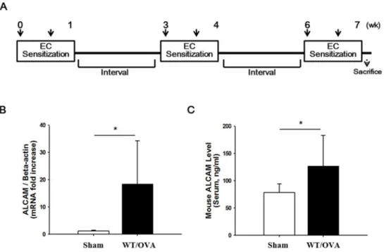
To identify the effect of ALCAM in the pathogenesis of AD, WT mice were sensitized by OVA, conducting the protocol (Fig. 1. A). This study investigated the level of ALCAM at skin lesion and serum respectively. OVA-induced AD WT mice were significantly increased to mRNA expression of ALCAM on the AD skin lesion, comparing with a sham (Fig. 1. B). ALCAM level of serum also was significantly increased in WT mice induced to AD (Fig. 1. C). Thus, the study supports a hypothesis that ALCAM is involved in AD.
Figure 1. Increase of ALCAM in wild type mice induced to atopic dermatitis by ovalbumin (A) Experimental protocol for ovalbumin-induced atopic dermatitis
model. (B) The mRNA expression of ALCAM in the skin lesion. (C) The level of ALCAM in the serum. The data represent means ± SD. * P < 0.05, n = 3 - 7 mice / group.
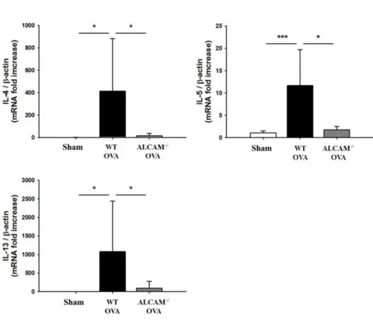
2. ALCAM deficiency attenuates Th2 inflammatory responses
To determine the role of ALCAM in Th2 dominant AD, the study used with ALCAM-/-mice, comparing WT mice. AD symptom was measured by clinical score and histological score. Clinical score was included some symptoms such as erythema, edema, erosion, dryness. Histological score was assessed by inflammation degree, edema, epithelial hyperplasia, immune cell infiltration. After induction of AD, ALCAM-/-mice showed that attenuated clinical score (Fig. 2. A) and alleviated skin lesion histologically (Fig. 2. B, C). TEWL was also a representative AD symptom. TEWL was lower in ALCAM-/- mice than WT mice (Fig. 2. D). AD was regulated by Th2 dominant immune response. The level of IgE obtained from serum and mRNA levels of IL-4, IL-5 and IL-13 gained from the skin were quantified to investigate the effect of ALCAM in Th2 dominant condition. ALCAM-/-mice significantly decreased serum IgE level (Fig. 2. E) and Th2 type cytokines (Fig. 2. F).
These findings suggest that ALCAM affects to the development of Th2 dominant AD induced by OVA in mice.
Figure 2. Attenuation of ovalbumin-induced atopic dermatitis in ALCAM deficient mice (A) Clinical score. (B) Histological score. (C) Hematoxylin and
eosin (H&E) staining (× 200, Bar = 500 μm). (D) Transepidermal water loss (TEWL). (E) The level of IgE in serum. (F) The levels of Th2 type cytokines in the skin. The data represent means ± SD. * P < 0.05, *** P < 0.001, n = 4 - 10 mice / group.
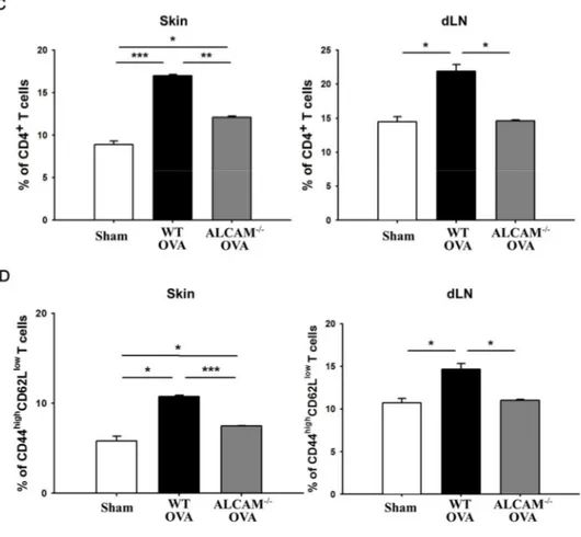
3. ALCAM deficiency suppresses the activation of T cell
To determine that ALCAM mediates the activation of T cell to develop an immune response in the skin and secondary lymphatic organ, flow cytometry was used to analyze the population of CD4+T cells and the activation of CD4+T cells in the skin lesion and skin draining lymph nodes (dLN) obtained from axillary and brachial sites.
CD4+ T cells were detected by CD3+CD4+ surface marker on the cells. The population of CD4+ T cells located in the skin and dLNs respectively was significantly increased in WT AD mice, comparing with a sham. Although mice were induced to AD, CD4+ T cells of ALCAM deficient mice were significantly lower than WT AD mice in both of the tissues (Fig. 3. A, C). Activated CD4+T cells were called to effector CD4+ T cells. They were detected by CD4+CD44highCD62Llow surface marker on the cells. Importantly, the dominant expression of CD4+CD44highCD62Llowwas observed in the skin and dLNs of WT AD mice, comparing ALCAM deficient AD mice (Fig. 3. B, D).
These findings suggest that ALCAM has an ability to activate CD4+T cells in the skin and dLNs.
Figure 3. Suppression of T cell activation in ALCAM deficient mice (A) The
population of CD4+T cells in the skin and dLNs. Cells were stained with anti-CD3, anti-CD4. (B) The activation of CD4+ T cells in the skin and dLNs. Cells were stained with anti-CD4, anti-CD44, anti-CD62L. The figure represented CD4+cells stained with anti-CD44 and anti-CD62L. (C) The percentage of CD4+ T cells. (D) The percentage of CD4+CD44highCD62Llow (effector) T cells. The cells were analyzed by flow cytometry. The experiments were representative of at least two independent experiments. The data represent means ± SD. * P < 0.05, ** P < 0.005, *** P < 0.001, n = 3 mice / group.
4. ALCAM deficiency diminishes the skin barrier dysfunction
To determine the effect of ALCAM in the skin barrier dysfunction, this study investigated that the changes of skin barrier genes and lamellar body in the skin lesion after mice were induced to AD by OVA. The mRNA levels of skin barrier related proteins (filaggrin, loricrin, involucrin) were quantified by real-time PCR. ALCAM deficient mice showed lower reduction of skin barrier genes expression than WT AD mice (Fig. 3. A). Moreover, ALCAM-/- AD mice showed lower decrease of the number (Fig. 3. B, C) of lamellar bodies located in SG than WT mice induced to AD. The aggregated shape was also decreased in the epidermis of ALCAM deficiency (Fig. 3. D). The number of lamellar bodies located in SG was expressed by the average of sum. The sum was gained from three sites chosen randomly of a mouse.
These findings suggest that ALCAM is involved in the regulation of skin barrier.
Figure 4. Inhibition of skin barrier dysfunction in ALCAM deficient mice (A)
The mRNA levels of genes associated with skin barrier. (B) The population of lamellar body in the stratum granulosum (× 50,000, Bar = 500 nm). (C) The number of lamellar bodies located in stratum granulosum. (D) The aggregation of lamellar body in epidermis (× 120,000). The data represent means ± SD. * P < 0.05, ** P < 0.005, *** P < 0.001, n = 2 - 6 mice / group.
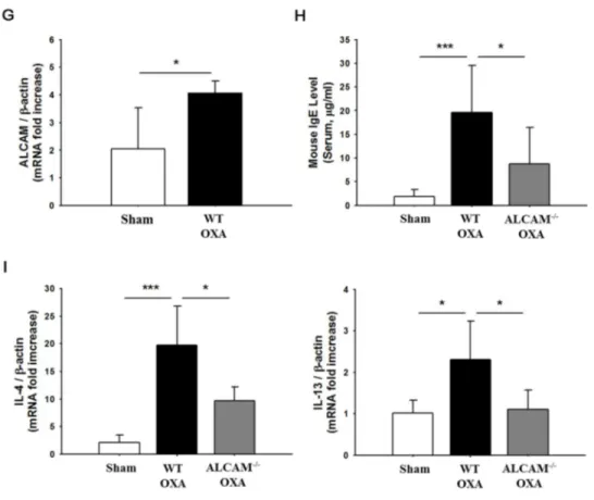
5. ALCAM deficiency attenuates Th2 inflammatory responses in OXA-induced AD mice
This study also performed in OXA-induced AD mice. WT and ALCAM -/-mice were induced to AD by OXA using the protocol (Fig. 4. A). OXA-induced AD ALCAM-/- mice showed same effects compared with OVA-induced AD murine models. ALCAM-/-mice lower increased clinical score (Fig. 4. B) and TEWL (Fig. 4. C) than WT AD mice. Ear thickness also was reduced in ALCAM-/-AD mice (Fig. 4. D). It could be observed by histological results (Fig. 4. E, F). Like OVA-induced AD, mRNA ALCAM expression was increased in skin lesion of WT AD mice induced by OXA (Fig. 4. G). Moreover, IgE level and IL-4, IL-13 mRNA level were quantified to investigate the effect of ALCAM in Th2 dominant condition. ALCAM -/-mice significantly decreased serum IgE level (Fig. 4. H) and Th2 type cytokines (Fig. 4. I).
These findings suggest that ALCAM affects to the development of Th2 dominant AD induced by OXA in mice.
Figure 5. Attenuation of oxazolone-induced atopic dermatitis in ALCAM deficient mice (A) Experimental protocol for oxazolone-induced atopic dermatitis
model. (B) Clinical score. (C) Transepidermal water loss (TEWL). (D) Ear thickness. (E) Histological score. (F) Hematoxylin and eosin (H&E) staining (× 100, Bar = 200 μm). (G) The mRNA expression of ALCAM in the skin lesions. (H) The level of IgE in the serum. (I) The level of Th2 type cytokines. The data represent means ± SD. * P < 0.05, *** P < 0.001, n = 2 - 8 mice / group.
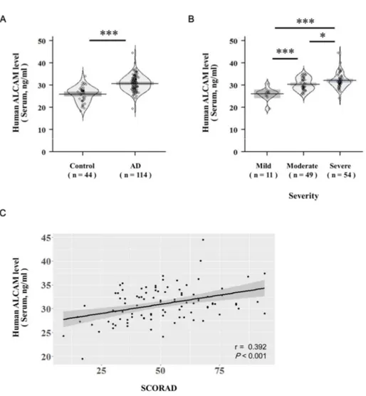
6. ALCAM is increased in the serum of pediatric AD patients
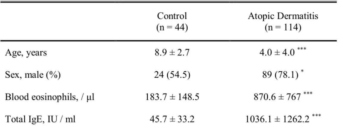
In addition to mice, identification of ALCAM level in human serum was performed as well. The clinical characteristics of the study subjects are summarized in Table 1. AD group was significantly younger in age and male dominant compared to control group. Serum total eosinophil count and IgE levels were significantly higher in AD group than healthy controls. Serum ALCAM level was also significantly elevated in AD group (26 ± 3.4 ng/ml vs. 30.8 ± 3.5 ng/ml, P < 0.001) (Fig. 5. A). After adjusting age and gender, serum ALCAM level was still statistically significantly increased in AD group (β = 3.68, P < 0.001). Moreover, ALCAM levels showed tendency of increase in children with AD as the severity increases, and revealed significantly positive correlations with SCORAD score (r = 0.392, P < 0.001) (Fig. 5. B, C).
These findings also suggest that ALCAM may be involved in the pathogenesis of human AD.
Figure 6. Increase of ALCAM in pediatric atopic dermatitis patients The level
of ALCAM was measured by ELISA in the human serum of healthy controls (n = 44) and atopic dermatitis patients (n = 114). Pirateplots represent the distribution of ALCAM levels of each sample, with the horizontal mean line and boxes representing the 95% confidence intervals. (A) The level of ALCAM in human serum. (B) Increment of serum ALCAM level as atopic dermatitis severity increases. (C) The correlation of serum ALCAM level with atopic dermatitis SCORAD index. The data represent means ± SD. * P < 0.05, *** P < 0.001.
Table 1. Characteristics of the study population Control (n = 44) Atopic Dermatitis (n = 114) Age, years 8.9 ± 2.7 4.0 ± 4.0 *** Sex, male (%) 24 (54.5) 89 (78.1)* Blood eosinophils, / μl 183.7 ± 148.5 870.6 ± 767*** Total IgE, IU / ml 45.7 ± 33.2 1036.1 ± 1262.2*** The study population is included control (n = 44) and atopic dermatitis (n = 114). Values are presented as mean ± standard deviations, or number (%).
Abbreviation; IgE, immunoglobulin E
IV. DISCUSSION
AD skin lesion contains a number of resident and infiltrated immune cells, such as dendritic cells, macrophages, mast cells, neutrophils, basophils, eosinophils, innate lymphoid cells (ILCs), natural killer cells, fibroblast and various T cell subsets. Functional interactions of these skin immune cells are important for the pathogenesis of AD.31Leukocytes coordinately use adhesion molecules in a highly regulated immune process to be trafficked from the blood into target peripheral tissues. Adhesion molecules are consisted of selectins, integrins and chemoattractant receptors. These molecules are up-regulated in tissue and vascular beds and provide directional cues for inflammatory T cells. The altered expression of trafficking receptors is involved in the states (activation, polarization and differentiation) of T cells.32
As an adhesion molecule, ALCAM is detectable in a wide range of vertebrate tissues of fetal, neonatal, adult stages in all three embryonic lineages. Although it is associated with T cell activation, hematopoiesis, growth of primary tumor, inflammation and migration of neutrophils, ALCAM does not appear abundant in the body. Its expression is also usually restricted to subsets of cells and selected time frames. ALCAM also mediates homophilic avidity as well as heterophilic avidity with CD6, forming the clustering at the cell surface. Meanwhile, CD6 is surface receptors expressed on T cells, thymocytes and a subset of B cells. ALCAM binds to the membrane proximal scavenger-receptor cysteine-rich (SRCR) domain of CD6. In several studies, CD6 has been reported as a co-stimulatory molecule to activate T-cell function. ALCAM-CD6 interaction recruits T cell and APC (antigen presenting cell) and contributes for the stabilization of the immune synapse. The interaction also mediates a long term engagement of CD6 and ALCAM to induce T-cell activation and proliferation. In addition, ALCAM-CD6 can result in activation of three signals, mitogen activated protein kinase (MAPK) cascades, extracellular signal-regulate kinases 1 and 2 (ERK1/2), p38 and c-Jun N-terminal kinase (JNK) to develop the adaptive immune response.16,33-35 Interestingly, ALCAM positive
transcription profile shows overexpression of protein kinase Cα (PKCα) and suppression of PKCε in metastatic melanoma cells. PKCθ, other PKC subfamily, is a kinase in mediating TCR signals and regulates the activation of transcriptional factors (NFκB, AP-1) in autoimmunity. ALCAM activation and PKC signaling can be a consecutive signaling event in immune response.36-38 On the other hand, the receptor for advanced glycation end products (RAGE) is a single transmembrane receptor of immunoglobulin superfamily mainly expressed on immune cells, neurons and a variety of cancer cells. RAGE binds with high mobility group box1 (HMGB1) or S100B and activates NFκB pathway to mediate inflammation in house dust mite induced AD NC/Nga transgenic mice. ALCAM and RAGE have a structural similarity and express complementarily. Other reports demonstrated that possible time- and spatial-dependent contributions of ALCAM and RAGE in the context of S100B-driven initiation, perpetuation and possibly termination of inflammation.39-43
The role of ALCAM in the pathogenesis of AD related with intra-cutaneous T-cell activation and impaired skin barrier function has not been well resolved.4,44-46 Classically AD is defined by Th2 type dominant condition. Th2 type cytokines are consisted of IL-4, IL-5, IL-13. IL-4 is a critical key for Th2 cell differentiation, IgE production and eosinophil recruitment. IL-5 is an essential cytokine for eosinophil development, survival and proliferation. It is related in epidermal thickening in AD. IL-13 is a mediator of allergic inflammation and it is expressed in both acute and chronic lesions of AD.5 In this study, WT AD-like mice induced by repeated exposure of OVA or OXA in the skin showed immune cell infiltration, altered the levels of IgE and IL-4, IL-5, IL-13, comparing with a sham group. However, ALCAM deficient AD-like mice showed attenuation of AD symptoms and lower levels of IgE and Th2 type cytokine, comparing with WT AD-like mice. The study also demonstrated that CD4+T cell activation and population were decreased in the skin and dLNs of ALCAM deficient AD-like mice. The results suggest that ALCAM affects to Th2 dominant immune response on the skin lesion by
co-stimulatory interaction of CD6-ALCAM in immune synapse.
This study also showed that ALCAM deficient AD-like mice were lower reduced skin barrier related gene expressions (Filaggrin, Loricrin, Involucrin) than WT AD-like mice. ALCAM deficiency also attenuates the damage of lamellar body located in the stratum granulosum after induction of AD. Cytokine signaling has a pivotal role in multiple consequences for the barrier function of the skin. The signaling affects to keratinocyte proliferation and differentiation. Thus, altered cytokine expression may contribute to dysregulation of the epidermal barrier and elicit to skin diseases, including AD and psoriasis.17,47 Although the correlation of Th2 type cytokine and skin barrier related gene expression still remain to be elucidated, IL-4 and IL-13 were down regulated to loricrin and involucrin using primary human keratinocyte differentiated with CaCl2or filaggrin deficient skin equivalent in vitro.3,48,49 An another study also demonstrated that the absence of IL-4 increased epidermal differentiation complex gene expression such as filaggrin in the presence of high Th2 environment, using OVA-induced mice only lacked to IL-4 but expressed active Stat6 on T cell specifically.50With recent reports, the findings may be suggested that ALCAM indirectly regulates to the function of skin barrier in Th2 dominant immune response.
However, there are some limitations in this study. First, ALCAM can be expressed in diverse type of cell. Further study is needed to determine the effect of ALCAM expressed on specific tissue or cell in the pathogenesis of AD. Second, T-cell can be divided into two major subsets, αβ and γδ T-T-cells which can differentiate to Th2 type. Human and mice have different populations of αβ and γδ T-cell on the skin.51 Other reports indicated that ALCAM is recruited for the activation of γδ T-cells but also αβ T-cell interacting with CD6 in the tumor T-cells sensitized with non-peptide antigen.52 Taken together, this study demonstrated that ALCAM regulates the expression of Th2 type cytokines and indirectly affects the skin barrier functions in OVA or OXA-induced AD mice. Therefore, ALCAM contributes to elicit an immune response in the pathogenesis of AD.
V. CONCLUSION
ALCAM is expressed on a variety cell types, including in neurons, fibroblasts, endothelial cells and dendritic cells. ALCAM also has been reported to have important roles in cell trafficking, tumor metastasis and a ligand for CD6 to stabilize the immunological synapse and activate T cell. Although ALCAM has been demonstrated to an effect to mediate immune response, the role of ALCAM has not been identified in the pathogenesis of AD.
In the study demonstrated that 1) ALCAM level was increased in the serum and skin lesion of OVA-induced WT AD mice. 2) ALCAM deficiency showed attenuation of Th2 dominant AD symptoms. 3) ALCAM deficiency also alleviated to activate T cell. 4) ALCAM was related to impaired function of skin barrier in AD.
5) ALCAM showed attenuation of AD symptoms in OXA-induced WT AD mice.
Moreover, 6) ALCAM level was increased in the serum of pediatric AD patients. Thus, the study has a hypothesis that ALCAM is involved in the expression of Th2 type cytokines in AD development. ALCAM also mediates to T cell activation and impaired skin barrier function in the AD skin lesion.
Taken together, ALCAM might play a potential role to develop inflammatory responses in the pathogenesis of Th2-dominant AD.
REFERENCES
1. Hennino A, Vocanson M, Toussaint Y, Rodet K, Benetiere J, Schmitt AM, et al. Skin-infiltrating CD8+ T cells initiate atopic dermatitis lesions. J Immunol 2007;178:5571-7.
2. Yanaba K, Kamata M, Asano Y, Tada Y, Sugaya M, Kadono T, et al. CD19 expression in B cells regulates atopic dermatitis in a mouse model. Am J Pathol 2013;182:2214-22.
3. Honzke S, Wallmeyer L, Ostrowski A, Radbruch M, Mundhenk L, Schafer-Korting M, et al. Influence of Th2 Cytokines on the Cornified Envelope, Tight Junction Proteins, and ss-Defensins in Filaggrin-Deficient Skin Equivalents. J Invest Dermatol 2016;136:631-9.
4. Nakajima S, Kitoh A, Egawa G, Natsuaki Y, Nakamizo S, Moniaga CS, et al. IL-17A as an inducer for Th2 immune responses in murine atopic dermatitis models. J Invest Dermatol 2014;134:2122-30.
5. Brandt EB, Sivaprasad U. Th2 Cytokines and Atopic Dermatitis. J Clin Cell Immunol 2011;2.
6. Di Cesare A, Di Meglio P, Nestle FO. A role for Th17 cells in the immunopathogenesis of atopic dermatitis? J Invest Dermatol 2008;128:2569-71.
7. Leung DY, Boguniewicz M, Howell MD, Nomura I, Hamid QA. New insights into atopic dermatitis. J Clin Invest 2004;113:651-7.
8. Fesenkova VI, Kurchenko AI, Castellani ML, Conti P, Anogeianaki A, Caraffa A, et al. Expression of Co-stimulatory molecules on langerhans cells in lesional epidermis of human atopic dermatitis. Immunopharmacol Immunotoxicol 2007;29:487-98.
9. Bromley SK, Burack WR, Johnson KG, Somersalo K, Sims TN, Sumen C, et al. The immunological synapse. Annu Rev Immunol 2001;19:375-96. 10. Gutcher I, Becher B. APC-derived cytokines and T cell polarization in
autoimmune inflammation. J Clin Invest 2007;117:1119-27.
11. Menon R, David BG. Itolizumab - a humanized anti-CD6 monoclonal antibody with a better side effects profile for the treatment of psoriasis. Clin Cosmet Investig Dermatol 2015;8:215-22.
12. Donizy P, Zietek M, Halon A, Leskiewicz M, Kozyra C, Matkowski R. Prognostic significance of ALCAM (CD166/MEMD) expression in cutaneous melanoma patients. Diagn Pathol 2015;10:86.
13. Swart GW. Activated leukocyte cell adhesion molecule (CD166/ALCAM): developmental and mechanistic aspects of cell clustering and cell migration. Eur J Cell Biol 2002;81:313-21.
14. Singer NG, Mitra R, Lialios F, Richardson BC, Marks RM, Pesando JM, et al. CD6 dependent interactions of T cells and keratinocytes: functional evidence for a second CD6 ligand on gamma-interferon activated keratinocytes. Immunol Lett 1997;58:9-14.
15. van Kempen LC, van den Oord JJ, van Muijen GN, Weidle UH, Bloemers HP, Swart GW. Activated leukocyte cell adhesion molecule/CD166, a marker of tumor progression in primary malignant melanoma of the skin. Am J Pathol 2000;156:769-74.
16. Zimmerman AW, Joosten B, Torensma R, Parnes JR, van Leeuwen FN, Figdor CG. Long-term engagement of CD6 and ALCAM is essential for T-cell proliferation induced by dendritic T-cells. Blood 2006;107:3212-20. 17. Hanel KH, Cornelissen C, Luscher B, Baron JM. Cytokines and the skin
barrier. Int J Mol Sci 2013;14:6720-45.
18. Kondo H, Ichikawa Y, Imokawa G. Percutaneous sensitization with allergens through barrier-disrupted skin elicits a Th2-dominant cytokine response. Eur J Immunol 1998;28:769-79.
19. Elias PM, Wakefield JS. Mechanisms of abnormal lamellar body secretion and the dysfunctional skin barrier in patients with atopic dermatitis. J Allergy Clin Immunol 2014;134:781-91.e1.
20. Galand C, Leyva-Castillo JM, Yoon J, Han A, Lee MS, McKenzie AN, et al. IL-33 promotes food anaphylaxis in epicutaneously sensitized mice by targeting mast cells. J Allergy Clin Immunol 2016;138:1356-66.
21. Venturelli N, Lexmond WS, Ohsaki A, Nurko S, Karasuyama H, Fiebiger E, et al. Allergic skin sensitization promotes eosinophilic esophagitis through the IL-33-basophil axis in mice. J Allergy Clin Immunol 2016;138:1367-80.e5.
22. Wang LF, Lin JY, Hsieh KH, Lin RH. Epicutaneous exposure of protein antigen induces a predominant Th2-like response with high IgE production in mice. J Immunol 1996;156:4077-82.
23. Guttman-Yassky E, Nograles KE, Krueger JG. Contrasting pathogenesis of atopic dermatitis and psoriasis--part I: clinical and pathologic concepts. J Allergy Clin Immunol 2011;127:1110-8.
24. Moniaga CS, Jeong SK, Egawa G, Nakajima S, Hara-Chikuma M, Jeon JE, et al. Protease activity enhances production of thymic stromal lymphopoietin and basophil accumulation in flaky tail mice. Am J Pathol 2013;182:841-51.
25. Na K, Yoo HS, Zhang YX, Choi MS, Lee K, Yi TG, et al. Bone marrow-derived clonal mesenchymal stem cells inhibit ovalbumin-induced atopic dermatitis. Cell Death Dis 2014;5:e1345.
26. Daszkiewicz L, Vazquez-Mateo C, Rackov G, Ballesteros-Tato A, Weber K, Madrigal-Aviles A, et al. Distinct p21 requirements for regulating normal and self-reactive T cells through IFN-gamma production. Sci Rep 2015;5:7691.
27. Almeida FF, Tenno M, Brzostek J, Li JL, Allies G, Hoeffel G, et al. Identification of a novel lymphoid population in the murine epidermis. Sci Rep 2015;5:12554.
28. Eichenfield LF, Hanifin JM, Luger TA, Stevens SR, Pride HB. Consensus conference on pediatric atopic dermatitis. J Am Acad Dermatol
2003;49:1088-95.
29. Severity scoring of atopic dermatitis: the SCORAD index. Consensus Report of the European Task Force on Atopic Dermatitis. Dermatology 1993;186:23-31.
30. Oranje AP, Glazenburg EJ, Wolkerstorfer A, de Waard-van der Spek FB. Practical issues on interpretation of scoring atopic dermatitis: the SCORAD index, objective SCORAD and the three-item severity score. Br J Dermatol 2007;157:645-8.
31. Peng W, Novak N. Pathogenesis of atopic dermatitis. Clin Exp Allergy 2015;45:566-74.
32. Islam SA, Luster AD. T cell homing to epithelial barriers in allergic disease. Nat Med 2012;18:705-15.
33. Weidle UH, Eggle D, Klostermann S, Swart GW. ALCAM/CD166: cancer-related issues. Cancer Genomics Proteomics 2010;7:231-43.
34. Hassan NJ, Simmonds SJ, Clarkson NG, Hanrahan S, Puklavec MJ, Bomb M, et al. CD6 regulates T-cell responses through activation-dependent recruitment of the positive regulator SLP-76. Mol Cell Biol 2006;26:6727-38.
35. Ibanez A, Sarrias MR, Farnos M, Gimferrer I, Serra-Pages C, Vives J, et al. Mitogen-activated protein kinase pathway activation by the CD6 lymphocyte surface receptor. J Immunol 2006;177:1152-9.
36. Swart GW, Lunter PC, Kilsdonk JW, Kempen LC. Activated leukocyte cell adhesion molecule (ALCAM/CD166): signaling at the divide of melanoma cell clustering and cell migration? Cancer Metastasis Rev 2005;24:223-36. 37. Kwon MJ, Wang R, Ma J, Sun Z. PKC-theta is a drug target for prevention
of T cell-mediated autoimmunity and allograft rejection. Endocr Metab Immune Disord Drug Targets 2010;10:367-72.
38. Manicassamy S, Gupta S, Huang Z, Sun Z. Protein kinase C-theta-mediated signals enhance CD4+ T cell survival by up-regulating Bcl-xL. J Immunol
2006;176:6709-16.
39. von Bauer R, Oikonomou D, Sulaj A, Mohammed S, Hotz-Wagenblatt A, Grone HJ, et al. CD166/ALCAM mediates proinflammatory effects of S100B in delayed type hypersensitivity. J Immunol 2013;191:369-77. 40. Karuppagounder V, Arumugam S, Thandavarayan RA, Pitchaimani V,
Sreedhar R, Afrin R, et al. Resveratrol attenuates HMGB1 signaling and inflammation in house dust mite-induced atopic dermatitis in mice. Int Immunopharmacol 2014;23:617-23.
41. Karuppagounder V, Arumugam S, Thandavarayan RA, Pitchaimani V, Sreedhar R, Afrin R, et al. Modulation of HMGB1 translocation and RAGE/NFkappaB cascade by quercetin treatment mitigates atopic dermatitis in NC/Nga transgenic mice. Exp Dermatol 2015;24:418-23. 42. Watanabe K, Karuppagounder V, Arumugam S, Thandavarayan RA,
Pitchaimani V, Sreedhar R, et al. Pruni cortex ameliorates skin inflammation possibly through HMGB1-NFkappaB pathway in house dust mite induced atopic dermatitis NC/Nga transgenic mice. J Clin Biochem Nutr 2015;56:186-94.
43. Riehl A, Nemeth J, Angel P, Hess J. The receptor RAGE: Bridging inflammation and cancer. Cell Commun Signal 2009;7:12.
44. Wang G, Savinko T, Wolff H, Dieu-Nosjean MC, Kemeny L, Homey B, et al. Repeated epicutaneous exposures to ovalbumin progressively induce atopic dermatitis-like skin lesions in mice. Clin Exp Allergy 2007;37:151-61.
45. Kaesler S, Volz T, Skabytska Y, Koberle M, Hein U, Chen KM, et al. Toll-like receptor 2 ligands promote chronic atopic dermatitis through IL-4-mediated suppression of IL-10. J Allergy Clin Immunol 2014;134:92-9. 46. Lee JH, Lee YS, Lee EJ, Lee JH, Kim TY. Capsiate Inhibits DNFB-Induced
Atopic Dermatitis in NC/Nga Mice through Mast Cell and CD4+ T-Cell Inactivation. J Invest Dermatol 2015;135:1977-85.
47. Strid J, McLean WH, Irvine AD. Too Much, Too Little or Just Enough: A Goldilocks Effect for IL-13 and Skin Barrier Regulation? J Invest Dermatol 2016;136:561-4.
48. Kim BE, Leung DY, Boguniewicz M, Howell MD. Loricrin and involucrin expression is down-regulated by Th2 cytokines through STAT-6. Clin Immunol 2008;126:332-7.
49. Di ZH, Ma L, Qi RQ, Sun XD, Huo W, Zhang L, et al. T Helper 1 and T Helper 2 Cytokines Differentially Modulate Expression of Filaggrin and its Processing Proteases in Human Keratinocytes. Chin Med J (Engl) 2016;129:295-303.
50. Sehra S, Yao Y, Howell MD, Nguyen ET, Kansas GS, Leung DY, et al. IL-4 regulates skin homeostasis and the predisposition toward allergic skin inflammation. J Immunol 2010;184:3186-90.
51. Dar AA, Patil RS, Chiplunkar SV. Insights into the Relationship between Toll Like Receptors and Gamma Delta T Cell Responses. Front Immunol 2014;5:366.
52. Kato Y, Tanaka Y, Hayashi M, Okawa K, Minato N. Involvement of CD166 in the activation of human gamma delta T cells by tumor cells sensitized with nonpeptide antigens. J Immunol 2006;177:877-84.
ABSTRACT (IN KOREAN)
아토피피부염 병인기전에서
Activated leukocyte cell adhesion molecule
(ALCAM/CD166)의 역할 규명
< 지도교수 손 명 현 >
연세대학교 대학원 의과학과
오 미 선
Activated leukocyte cell adhesion molecule의 약자인 ALCAM은
5개의 면역글로불린 도메인의 구조로 이루어진 막관통단백질이다. ALCAM은 체내의 신경세포, 내피세포, 섬유아세포 등 다양한 세포에서 발현한다고 알려져 있으며, 특히 항원제시세포인 수지상세포에서 가장 높게 발현된다. 또한, T 림프구 표면에서 발현하는 CD6의 리간드로 알려진 ALCAM은 T 림프구와 수지상세포의 접촉면에서 형성되는 면역시냅스를 안정적으로 유지하는데 기여하고, T 림프구의 활성화와 분화를 매개하여 면역반응을 조절하는 보조인자로써 보고된 바가 있다. 따라서 ALCAM이 면역시냅스에서 T 림프구의 활성화에 기여한다는 점과 아토피피부염이 T 림프구 매개의 만성적인 알레르기성 피부 질환이라는 점을 바탕으로, 본 연구에서는 아토피피부염 병인기전에서 ALCAM의 역할을 규명하고자 하였다. 아토피피부염 병인기전에서 ALCAM의 역할을 확인하기 위해, C57BL/6를 유전적 배경으로 갖는 7-10주 령의 야생형 마우스와 ALCAM
결핍마우스를 사용하였다. 마우스의 등 피부 털을 제거한 후, 피부에 테이프를 붙였다가 떼어냄을 5회 반복하는 것과 거즈에 분주한 100 μg의 난백 알부민을 반창고를 사용하여 등 피부에 노출시키는 것을 통해 아토피피부염을 유도하였다. 그 결과, 아토피피부염을 유도한 야생형 마우스의 피부와 혈청에서 ALCAM의 수치가 높게 측정됨을 확인했다. ALCAM 결핍마우스에서는 아토피피부염을 유도했음에도 불구하고, 야생형 마우스보다 T 림프구의 활성화가 적게 일어나며 혈청에서의 면역글로불린 E와 제 2형 T 도움세포에서 생성되는 사이토카인의 수치가 낮았다. 또한 ALCAM이 결핍된 마우스에서 난백알부민을 노출시켜도 피부장벽을 조절하는 단백질의 발현의 감소가 적었다. 합텐인 옥사졸론으로 마우스의 귀에 아토피피부염을 유도한 연구에서도 난백알부민으로 유도한 아토피피부염 마우스와 같은 결과를 보였다. 더 나아가, 아토피피부염이 없는 소아의 혈청과 비교하여 아토피 피부염이 있는 소아의 혈청에서 ALCAM의 수치가 증가되어 있음을 확인했다. 위의 결과를 통해서, 본 연구에서는 ALCAM이 T 림프구의 활성화와, 피부에서 발생하는 제 2형 T 도움 세포의 면역반응 및 피부장벽의 기능조절장애를 매개할 수 있음을 확인하였다.