저작자표시-비영리-변경금지 2.0 대한민국 이용자는 아래의 조건을 따르는 경우에 한하여 자유롭게 l 이 저작물을 복제, 배포, 전송, 전시, 공연 및 방송할 수 있습니다. 다음과 같은 조건을 따라야 합니다: l 귀하는, 이 저작물의 재이용이나 배포의 경우, 이 저작물에 적용된 이용허락조건 을 명확하게 나타내어야 합니다. l 저작권자로부터 별도의 허가를 받으면 이러한 조건들은 적용되지 않습니다. 저작권법에 따른 이용자의 권리는 위의 내용에 의하여 영향을 받지 않습니다. 이것은 이용허락규약(Legal Code)을 이해하기 쉽게 요약한 것입니다. Disclaimer 저작자표시. 귀하는 원저작자를 표시하여야 합니다. 비영리. 귀하는 이 저작물을 영리 목적으로 이용할 수 없습니다. 변경금지. 귀하는 이 저작물을 개작, 변형 또는 가공할 수 없습니다.
The Effect of Biological Aging of Implant
on Osseointegration in the Dog
Byung-Moon Hwang
The Graduate School
Yonsei University
Department of Dentistry
The Effect of Biological Aging of Implant
on Osseointegration in the Dog
A Dissertation Thesis
Submitted to the Department of Dentistry
and the Graduate School of Yonsei University
In partial fulfillment of the
Requirements for the degree of
Master of Dental Science
Byung-Moon Hwang
감사의 글
본 논문이 완성되기까지 부족한 저에게 아낌없는 조언을 해주시고 관심으로 이끌어 주신 이재훈 교수님께 깊은 감사를 드립니다. 그리고 이번 논문이 나 올 수 있도록 연구를 도와주신 박영범 교수님께 진심으로 감사드리며, 보다 더 좋은 논문이 나올 수 있도록 심사해 주신 김지환, 정의원 두 교수님께도 감사드립니다. 연구 내내 많은 도움을 준 장성호 선생님, 최현민 선생님과 영문 교정을 해 주신 강세영 선생님께 고마움을 전합니다. 그리고, 늘 아낌없는 사랑과 헌신적인 도움으로 든든하고 따뜻한 버팀목이 되어준 사랑하는 나의 아내 경희와 귀염둥이 연우에게 사랑과 고마움의 마음 을 전하며, 바쁘신 가운데에도 연우를 봐주시고, 물심양면으로 도와주신 장모 님께도 진심으로 감사드립니다. 마지막으로 지금 이 자리에 설 수 있도록 낳아주시고 길러주신 어머니께 감사드립니다. 2015년 12월 저자 씀Table of Contents
Abstract (English) ··· iii
Ⅰ. Introduction ··· 1
Ⅱ. Materials and Methods ··· 5
1. Implant samples and surface characterization ··· 5
2. Experimental design ··· 5 3. Surgical procedure ··· 6 4. Histological preparation ··· 7 5. Histomorphometry ··· 7 6. Statistical analysis ··· 8 Ⅲ. Results ··· 9 Ⅳ. Discussion ··· 11 Ⅴ. Conclusion ··· 14 References ··· 15 Legends ··· 19 Figures ··· 20 Abstract (Korean) ··· 24
List of Figures
Figure 1. Experimental timeline and schedule ··· 20 Figure 2. Segmentation of peri-implant tissue for bone histomorphometry
··· 21 Figure 3. 100x magnification microscopic images of peri-implant tissues
around implant at week 4 of healing ··· 22 Figure 4. 100x magnification microscopic images of peri-implant tissues
around implant at week 12 of healing ··· 23
List of Table
Table 1. Comparison of bone to implant contact (BIC%) between the groups ··· 10 Table 2. Comparison of bone volume (BV%) between the groups ··· 10
Abstract
The Effect of Biological Aging of Implant
on Osseointegration in the Dog
Byung-Moon Hwang, D.D.S.
Department of Dentistry Graduate School, Yonsei University
(Directed by Prof. Jae-Hoon Lee, D.D.S., M.S.D., PhD.)
A series of recent studies reported time-dependent biological degradation of implant, which is called as biological aging of implant. Although many studies have been performed on the implant aging and its resolution, there might not be as yet a study which measures bone to implant contact (BIC) and bone volume (BV) to examine the effect of implant aging in animals larger than the rat. The objective of this study is to investigate the effect of biological aging of implant on osseointergration in the dog.
Thirty six implants (3.5 mm in diameter and 8.5 mm in length); all with sandblasted/acid-etched surface were used in the experiment. The
implants were divided into 3 groups of 12 implants each; control
(6-month-old implants after manufacture), newly prepared implant with acid-etching (surface rejuvenation), and 2-week-old implant (stored for 2
weeks after surface rejuvenation). Six young adult mongrel male dogs were used. BIC and BV were evaluated by histometric measurements following a 4- and 12-week healing interval.
There were statistically significant differences between the groups in the lower zone of the implant at week 4 of healing (p<0.05). According to multiple comparisons, there was significant difference in BIC between control and 2-week-old implants (p=0.016), and between control and newly prepared implants with acid-etching (p=0.019). But there was no significant difference in BIC between newly prepared implants with acid-etching and 2-week-old implants. In all groups, BIC at week 12 was significantly higher than that of week 4 (p<0.05). In BV, there were no significant differences regardless of area and time.
In conclusion, biological aging of implant might affect osseointergration in bone marrow zone at week 4 of healing. Although implant aging did not greatly affect BIC and BV at week 12 of healing in this study, further study will be required to illustrate the standard period of biological aging of implant which would have significant clinical effects.
Key words: Biological aging of implant; surface rejuvenation; single etching; osseointegration; bone to implant contact; bone volume
The Effect of Biological Aging of Implant
on Osseointegration in the Dog
Byung-Moon Hwang, D.D.S.
Department of Dentistry Graduate School, Yonsei University
(Directed by Prof. Jae-Hoon Lee, D.D.S., M.S.D., PhD.)
Ⅰ. Introduction
Titanium implant has become indispensable to dental treatment since Brånemark discovered osseointegration in 1952 (Branemark, 1983). Long healing time was required for the early implant which had a smooth surface by machine milling, to have successful function. Clinicians were merely interested in replacing missing tooth with the implant in the past. Now they are reducing the healing time to enable immediate loading of dental implant.
One of the most important factors for immediate loading is implant stability which is also known as total stability. Total stability is the sum of primary stability during implant placement and secondary stability during healing period (Raghavendra et al., 2005), it is commonly known
that total stability of the implant reaches the lowest point at 4-6 weeks after implant placement. This phenomenon is called a “stability dip” (Raghavendra et al., 2005; Aparicio et al., 2006; Simunek et al., 2012) and influences success of immediate loading. Therefore, increasing primary stability and reducing stability dip are essential for immediate loading. To attain these requirements, implant design and surface treatment for enhanced early function and reduced healing time of implant have been developed ever since.
Meanwhile, time-dependent degradation of surface bioactivity of dental implant after manufacture was discovered (Att et al., 2009; Hori et al., 2009; Att et al., 2012). Deterioration of bioactivity of the implant surface occurred by absorbing organic materials such as hydrocarbons which come from the atmosphere, cleansing solution, and water during manufacture and storage (Kasemo et al., 1988; Kilpadi et al., 2000). Upon the investigation of the atomic percentage of carbon on the implant surface with X-ray photoelectron spectroscopy spectra, the value increased from 16 to 62% as time went by.
Hydrocarbon contamination changes the electric property of implant surface, which is naturally negatively charged. A divalent cation such as Ca is attracted to the negatively charged implant surface which is then followed by negatively charged proteins before cells adhere to the implant
surface. However, osseointegration is interrupted as proteins and
extracellular matrix cannot combine with oxide layer of implant surface when implant surfaces are contaminated by hydrocarbons (Aita et al.,
2009). As a consequence, hydrocarbon contamination results in undesirable effects, altering the characteristics of titanium surface from bioactive to bioinert.
Protein absorption, attachment and proliferation of osteogenic cells, and
mineralization on implant surface are very essential to accomplish
successful osseointegration. It was reported that aged-implant surface showed inferior performance compared to newly prepared acid-etched implant surface in protein absorption, attachment and proliferation of osteogenic cells, and mineralization on aged-implant surface (Att et al., 2009).
In vivo experiment using a rat model revealed that biomechanical strength of bone-titanium integration for 4-week-old acid-etched implants was less than half that for the newly prepared implants. It was also found
that the percentage of BIC was lower than 60% for 4-week-old
acid-etched implants whereas that of newly prepared acid-etched implants was more than 90% (Att et al., 2009).
Although many studies regarding implant aging and its resolution have been published (Aita et al., 2009; Att et al., 2009; Hori et al., 2010; Att et al., 2012; Lee et al., 2012; Ueno et al., 2012; Pyo et al., 2013), most of the studies were cellular experiments and a few studies were carried out with small animals such as rats to examine the effect of implant aging. There might not be a study which shows the effect of implant aging in the animals larger than the rat yet. Even though the application of the results from the experiment using the dog to human has limitations, the data from
the test using larger animals such as the dog might be more useful compared to cellular experiments or tests with small animals. The objective of this study is to investigate the effect of implant aging in the dog via histomorphometry.
Ⅱ. Materials & methods
1. Implant samples and surface characterization
Thirty six implants (3.5 mm in diameter and 8.5 mm in length*); all
with sandblasted/acid-etched surface were used in the experiment. All implants were made at the same time and placed in a sealed container. For surface rejuvenation, 24 implants were treated with 67% sulfuric acid (H2SO4) at 120℃ for 75 seconds (Att et al., 2009). Twelve out of 24
prepared implants were placed in a sealed container and stored in a dark room (temperature, 23℃; humidity, 60%) for 2 weeks (Att et al., 2009).
2. Experimental design
Group A (control): 6-month-old implants after manufacture.
Group B: implants which have fresh surfaces after the preparation that followed the protocol mentioned above.
Group C: implants which have 2-week-old surfaces after the preparation that followed the protocol mentioned above.
Half of each group and the other half were obtained from the animals 4 weeks and 12 weeks after implant installation, respectively.
3. Surgical procedure
Six young adult mongrel male dogs weighing approximately 30kg were used in this study. The animals had intact maxillae and mandibles and no periodontitis with normal dentition. The animals were in good general health. Animal care and treatment protocols were approved by the Animal Care and Use Committees, Yonsei Medical Center, Seoul, Korea (Approval no. 2013-0109).
All surgeries were performed by the same operator under general
anesthesia in a sterile operating room. The animals received a
subcutaneous injection of atropine (0.06㎎/㎏) and an intravenous injection of xylazine* (0.2㎎/㎏) and tiletamine/zolazepam† (5㎎/㎏). Inhalation
anesthesia was performed using 2% isoflurane. During the surgery, heating pad was applied for the animals. The P1, P2, P3, and P4 mandibular premolars on both sides were extracted. After 2 months, the implants were placed under the same general anesthesia condition as teeth extraction according to manufacturer’s recommendation. The same post-operative management was performed as the extraction of teeth. All sutures were removed after 7 days. The animals fed a liquid diet. The animals were sacrificed by anesthesia drug overdose 4 weeks and 12 weeks after implants placement (Figure 1).
* Rompun™, Bayer , KS, USA † Zoletil®, Virbac, TX. USA
4. Histological preparation
Specimens were fixed in 10% buffered formaldehyde solution (pH 7) and dehydrated in ascending concentrations of alcohol (up to 100%), and
embedded in methacrylate. Embedded specimens were sectioned
bucco-lingually and ground to a thickness of less than 35㎛. Sectioned specimens were stained with hematoxylin-eosin stain and observed with light microscopy.
5. Histomorphometry
Each implant section was analyzed using light microscopy* coupled to
a videocamera capture system. Magnification was 100x and 200x.
Measurements were made with computer-based histomorphometric
measurements†. The peri-implant tissue was divided into upper zone (blue
line) and lower zone (red line) of implant (Figure 2); Both zones were within a 500㎛ vicinity. BIC of bone tissue located within 50㎛ of the implant surface without intervention of soft tissue was calculated (Pyo et al., 2013).
BIC (%) = (sum of the length of bone to implant contact)/(circumference of the implant) x 100
BV (%) = (bone area in the area of interest)/(area of interest) x 100 * BX50, Olympus, Tokyo, Japan
6. Statistical analysis
Statistical analyses were performed using SPSS 12.0 for Windows*.
Kruskal-Wallis test was used to assess differences in BIC and BV; p<0.05 was considered significant. To avoid accumulation of errors from multiple comparisons, Mann-Whitney test with Bonferroni correction was performed.
Ⅲ. Results
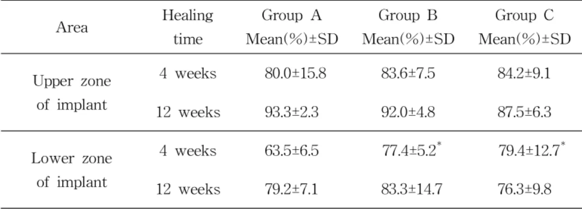
There were statistically significant differences in BIC between the groups in the lower zone of implant at week 4 of healing (p<0.05) (Figure 3). According to multiple comparisons, there was significant difference in BIC between group A and group C (p<0.017), and between group A and group B (p≒0.017). There was no significant difference in BIC between group B and group C. In all groups, BIC at week 12 of healing was significantly higher than that of week 4 (p<0.05).
The results showed that there were no significant differences in BIC in the upper zone of implant at week 4 and 12 of healing between the groups. At week 12 of healing, there were no significant differences in BIC between the groups in the lower zone of implant (Figure 4).
In the upper zone of implant, BV at week 4 was significantly higher than at week 12 (p<0.05). However there was no significant difference in BV in the lower zone of implant regardless of the healing time. Table 2 shows that there were no significant differences in BV between the groups at week 4 and 12 of healing regardless of the area.
Table 1. Comparison on bone to implant contact (BIC%) between groups Area Healing time Group A Mean(%)±SD Group B Mean(%)±SD Group C Mean(%)±SD Upper zone of implant 4 weeks 80.0±15.8 83.6±7.5 84.2±9.1 12 weeks 93.3±2.3 92.0±4.8 87.5±6.3 Lower zone of implant 4 weeks 63.5±6.5 77.4±5.2 79.4±12.7 12 weeks 79.2±7.1 83.3±14.7 76.3±9.8
: Statistically significant difference compared to group A(P<0.05)
Table 2. Comparison on bone volume (BV%) between groups
Area Healing time Group A Mean(%)±SD Group B Mean(%)±SD Group C Mean(%)±SD Upper zone of implant 4 weeks 80.5±8.3 72.1±17.2 81.5±14.7 12 weeks 66.5±12.6 72.1±15.9 69.8±8.4 Lower zone of implant 4 weeks 32.2±20.9 33.0±22.1 53.2±23.5 12 weeks 43.4±14.7 42.0±21.4 37.5±20.6
Ⅳ. Discussion
This study shows the effect of surface rejuvenation appeared in the bone marrow zone at week 4. Surface rejuvenation with acid-etching would help to increase success rate of immediate loading in patients by improving osseointegration between cancellous bone and implant before stability dip. The previous study reported that the implants treated for surface rejuvenation before implant placement showed no stability dip, regardless of the degree of primary stability (Suzuki et al., 2013).
BIC of group B was not generally higher than that of group C. This result suggests that even if the period of implant aging is shorter, BIC and BV can be lower depending on several factors such as implant thread design, surface treatment, condition of host, etc. It was reported that dental implant thread geometry was the factor that affects BIC in vivo study using the tibiae of rabbits (Steigenga et al., 2004). At celluar level, 2 weeks of implant aging might be enough time to influence osteoblast cell density, alkaline phosphatase activity, and calcium deposition whereas this time frame might not have profound impact on BIC and BV in large animals such as the dog.
All groups showed high percentage of BIC in upper zone of implant because the quality of cortical bone in the mandible of the dogs was good (Figure 3 and 4). Albrektsson and Johansson hypothesized that approximately 50% BIC is necessary for successful prosthetic result (Albrektsson et al.,
1991). All groups in this study satisfied this requirement. This result suggests that host bone quality (bone density and the amount of cortical bone) would play an important role in limiting the effect of implant aging.
Group A did not show any significant differences from the comparisons with group B and C except the data at week 4 of healing in lower zone of implant. This indicates that 6-month-old implants that are commercially used have no clinical problems although the implant surface undergoes changes such as loss of hydrophilicity by implant aging. It is widely known that most implants on the market in South Korea have 5 years of shelf life yet there is no vivid evidence for this period. There are few
studies that indicate the standardized period to actually reduce
osseointegration due to its biological aging. Further researches on shelf life of implant will be required. In recent progress of dental implant on the market, the implants are embedded in liquid such as calcium solution and stored in sterilized containers. The storage in liquid seems to prevent hydrocarbon contamination and surface deterioration, eventually promoting osteogenesis.
Surface rejuvenation with acid-etching was effective to sightly increase BIC. However surface rejuvenation with acid-etching seems to be less effective compared to the other methods used for surface rejuvenation in previous in vivo studies (Att et al., 2009; Pyo et al., 2013, Suzuki et al.,
2013). This might be ascribed to different mechanisms regarding
hydrocarbon removal, protein absorption, proliferation of osteogenic cells and osteoblast differentiation. The exact mechanism of surface rejuvenation
has not been elucidated and investigation in identifying the mechanism might be of further interest.
Ⅴ. Conclusion
Requiring a considerate interpretation of our data due to the limited number of samples, surface rejuvenation with acid-etching to offset the biological aging of implant enhanced BIC in the lower zone of implant at week 4. This result suggests that newly prepared implant might be more effective in successful loading of implant before stability dip than biologically aged implant by slightly improving osseointergration in bone marrow zone and reducing the period of stability dip.
In the case of enough healing period which is more than 12 weeks, implant aging did not affect BIC and BV in large animals such as the dog. However further study will be required to illustrate the standard period of biological aging of the implant which would have enough clinical effects. It will be also required to elucidate the mechanism of biological aging of implant and that of surface rejuvenation.
References
Aita, H., et al., The effect of ultraviolet functionalization of titanium on integration with bone. Biomaterials, 2009. 30(6): p. 1015-1025.
Albrektsson, T.a.C.J., Quantified bone tissue reactions to various metallic materials with reference to the so-called osseointegration concept. In: The Bone Biomaterial Interface, II volume Davies JE and Albrektsson T (Editors). University of Toronto Press: Toronto, 1991: p. 357-363.
Aparicio, C., N.P. Lang, and B. Rangert, Validity and clinical significance of biomechanical testing of implant/bone interface. Clin Oral Implants Res, 2006. 17 Suppl 2: p. 2-7.
Att, W., et al., The effect of UV-photofunctionalization on the time-related bioactivity of titanium and chromium-cobalt alloys. Biomaterials, 2009. 30(26): p. 4268-76.
Att, W., et al., Time-dependent degradation of titanium osteoconductivity: an implication of biological aging of implant materials. Biomaterials, 2009. 30(29): p. 5352-63.
Att, W. and T. Ogawa, Biological aging of implant surfaces and their restoration with ultraviolet light treatment: a novel understanding of osseointegration. Int J Oral Maxillofac Implants, 2012. 27(4): p. 753-61.
Brånemark, P.I., Osseointegration and its experimental background. J Prosthet Dent, 1983. 50(3): p. 399-410.
Hori, N., et al., Age-dependent degradation of the protein adsorption capacity of titanium. J Dent Res, 2009. 88(7): p. 663-667.
Hori, N., et al., Ultraviolet light treatment for the restoration of age-related degradation of titanium bioactivity. Int J Oral Maxillofac Implants, 2010. 25(1): p. 49-62.
Kasemo, B. and J. Lausmaa, Biomaterial and implant surfaces: on the role of cleanliness, contamination, and preparation procedures. J Biomed Mater Res, 1988. 22(A2 Suppl): p. 145-158.
Kilpadi, D.V., et al., Cleaning and heat-treatment effects on unalloyed titanium implant surfaces. Int J Oral Maxillofac Implants, 2000. 15(2): p. 219-230.
Lee, J.H. and T. Ogawa, The biological aging of titanium implants. Implant Dent, 2012. 21(5): p. 415-421.
Pyo, e.a., Photofunctionalization enhances bone-implant contact, dynamics of interfacial osteogenesis, marginal bone seal, and removal torque value of implants: a dog jawbone study. Implant Dent, 2013. 22(6): p. 666-675.
Raghavendra, S., M.C. Wood, and T.D. Taylor, Early wound healing
around endosseous implants: a review of the literature. Int J Oral
Maxillofac Implants, 2005. 20(3): p. 425-431.
Simunek, A., et al., Development of implant stability during early healing of immediately loaded implants. Int J Oral Maxillofac Implants, 2012. 27(3): p. 619-627.
Steigenga, J., et al., Effects of implant thread geometry on percentage of osseointegration and resistance to reverse torque in the tibia of rabbits. J Periodontol, 2004. 75(9): p. 1233-1241.
Suzuki, S., H. Kobayashi, and T. Ogawa, Implant stability change and osseointegration speed of immediately loaded photofunctionalized implants. Implant Dent, 2013. 22(5): p. 481-490.
Ueno, T., et al., Gamma ray treatment enhances bioactivity and osseointegration capability of titanium. J Biomed Mater Res B Appl Biomater, 2012. 100(8): p. 2279-2287.
Legends
Figure 1. Experimental time line and schedule.
Figure 2. Segmentation of peri-implant tissue for bone histomorphometry. BIC and BV were analyzed separately in upper zone and lower zone of implant.
Figure 3. 100x magnification microscopic images of peri-implant tissues around implant at week 4 of healing.
Figure 4. 100x magnification microscopic images of peri-implant tissues around implant at week 12 of healing.
Figure
국문요약
임플란트의 생물학적 노화가 성견의 임플란트
골유착에 미치는 영향
<지도교수 이 재 훈>
연세대학교 대학원 치의학과
황 병 문
최근 일련의 연구들은 표면 처리 후 시간이 지남에 따라 티타늄의 생물학 적 특징이 감소한다는 것을 보고하였다. 이러한 현상을 티타늄의 생물학적 노화 라 부른다. 많은 연구들이 임플란트의 생물학적 노화와 그 해결방법에 대해 발 표하고 있지만 대부분이 세포 수준의 실험이거나 쥐와 같은 소형 동물을 이용 한 실험이었고, 아직까지 대형 동물을 대상으로 조직학적 계측방법을 통해 임플 란트의 생물학적 노화의 효과에 대하여 연구한 논문은 찾아보기 어렵다. 비록 성견에서의 실험 결과를 인간에게 적용하는 것은 한계가 있지만, 세포 실험이나 소형 동물을 이용한 실험과 비교하였을 때 성견과 같은 대형 동물을 대상으로 얻은 결과가 더 유용할 것이라 생각된다. 이번 연구의 목적은 시중에 판매되는 임플란트 표면을 산부식 처리하고 노화 기간을 달리하여 성견에게 식립한 후, 노화의 정도가 임플란트 골유착에 어떠한 영향을 미치는지 알아보고자 한다. 직경 3.5mm, 길이 8.5mm의 산부식과 블라스팅의 조합 표면을 가진 제작 된 지 6개월 된 티타늄 임플란트 36개를 각각 12개씩 제조된 후 아무 처리도하지 않은 군, 산부식 후 바로 식립한 군, 산부식 처리 후 2주 노화시킨 후 식 립한 군으로 나누어 성견 6마리의 하악골에 식립하였다. 술 후 4 주와 12 주 에 희생시켜 치유 결과를 조직형태계측을 통해 비교 관찰하였다. 조직형태계측학적 분석 결과, 술후 4주의 치유기간을 가진 임플란트는 임 플란트의 하부에서 군들 간 골-임플란트 접촉에서 유의한 차이가 있었다 (p<0.05). 다중 비교 결과, 제작된 지 6개월 된 군과 산부식 처리 후 2주 노화 시킨 후 식립한 군 (p=0.016), 제작된 지 6개월 된 군과 산부식 처리 후 바로 식립한 군 (p=0.019)은 통계적으로 유의한 결과를 보였다. 그러나 산부식 처리 후 바로 식립한 군과 2주 노화시킨 후 식립한 군 간의 통계적으로 유의한 차 이는 없었다. 모든 군에서, 술 후 12주의 골-임플란트 접촉은 4주의 골-임플 란트 접촉보다 유의하게 높았다 (p<0.05). 임플란트의 상부에서는 각 군 간의 유의차는 관찰하기 어려웠다. 골량은 임플란트 상부에서 술후 4주의 골량이 술후 12주의 골량보다 유의하게 높은 것을 제외하고, 노화 정도나 술 후 치유 시기, 측정 부위에 관계없이 모든 군에서 통계적으로 유의한 차이가 없었다. 결론적으로, 임플란트의 생물학적 노화는 임플란트 식립 후 4주 경 임플 란트 하부의 골유착에 유의한 영향을 미치는 것으로 나타났다. 비록 이번 실 험에서 치유기간이 12주인 경우 임플란트의 생물학적 노화로 인한 영향이 골-임플란트 접촉과 골량에 큰 영향을 주지는 않는 것으로 나타났으나 정확하게 어느 정도의 노화 기간이 임상적으로 영향을 줄 수 있는지에 대해서는 추가적 인 연구가 필요하다. 핵심되는 말: 임플란트의 생물학적 노화, 산부식 처리, 골유착, 골-임플란트 접촉, 골량