Copyright © 2004, American Society for Microbiology. All Rights Reserved.
Biochemical Characterization of the Drosophila Wingless Signaling
Pathway Based on RNA Interference
Hiroko Matsubayashi,
1† Sonoka Sese,
1† Jong-Seo Lee,
1‡ Tadaoki Shirakawa,
1Takeshi Iwatsubo,
2Taisuke Tomita,
2and Shin-ichi Yanagawa
1*
Department of Viral Oncology, Institute for Virus Research, Kyoto University, Sakyo-Ku, Kyoto 606-85071,1and
Department of Neuropathology and Neuroscience, Graduate School of Pharmaceutical Sciences, University of Tokyo, Bunkyo-Ku, Tokyo 113-00332,2Japan
Received 2 July 2003/Returned for modification 12 August 2003/Accepted 25 November 2003
Regulation of Armadillo (Arm) protein levels through ubiquitin-mediated degradation plays a central role in the Wingless (Wg) signaling. Although zeste-white3 (Zw3)-mediated Arm phosphorylation has been impli-cated in its degradation, we have recently shown that casein kinase I␣ (CKI␣) also phosphorylates Arm and
induces its degradation. However, it remains unclear how CKI␣ and Zw3, as well as other components of the
Arm degradation complex, regulate Arm phosphorylation in response to Wg. In particular, whether Wg signaling suppresses CKI␣- or Zw3-mediated Arm phosphorylaytion in vivo is unknown. To clarify these
issues, we performed a series of RNA interference (RNAi)-based analyses in Drosophila S2Rⴙ cells by using
antibodies that specifically recognize Arm phosphorylated at different serine residues. These analyses revealed that Arm phosphorylation at serine-56 and at threonine-52, serine-48, and serine-44, is mediated by CKI␣ and
Zw3, respectively, and that Zw3-directed Arm phosphorylation requires CKI␣-mediated priming
phosphory-lation. Daxin stimulates Zw3- but not CKI␣-mediated Arm phosphorylation. Wg suppresses Zw3- but not
CKI␣-mediated Arm phosphorylation, indicating that a vital regulatory step in Wg signaling is Zw3-mediated
Arm phosphorylation. In addition, further RNAi-based analyses of the other aspects of the Wg pathway clarified that Wg-induced Dishevelled phosphoylation is due to CKI␣ and that presenilin and protein kinase
A play little part in the regulation of Arm protein levels in Drosophila tissue culture cells.
The Wnt/Wingless (Wg) signaling pathway is essential for many aspects of animal development, and mutations in com-ponents of the Wnt pathway are oncogenic (reviewed in ref-erences 3, 30, and 45). Wnts are secreted glycoproteins that exert their effects on neighboring cells by binding to a receptor protein complex consisting of the Frizzled (Fz) transmembrane receptor family and the single-pass transmembrane proteins of the low density lipoprotein receptor-related protein family. A variety of studies have set a general framework for the Wnt/Wg pathway and revealed that components of this pathway are structurally and functionally conserved in various species. However, it remains unclear how the Wnt signal is transduced from receptors to downstream components such as Dvl/Di-shevelled (Dsh). In this pathway, the stabilization of cytoplas-mic pools of -catenin/Armadillo (Drosophila homolog of -catenin, Arm) is a key regulatory step.
Several components of this pathway, including Dvl/Dsh, gly-cogen synthase kinase-3 (GSK-3)/zeste-white3 (Zw3), -catenin/Arm, adenomatous polyposis coli (APC) protein/ Dapc, and protein phosphatase 2A (PP2A), have been shown to form a large multimeric protein complex on the scaffold protein Axin/Daxin, and Wnt/Wg-regulated phosphorylation of-catenin/Arm known to occur in this complex (6, 11, 20, 31,
32, 33, 47). In the absence of Wnt/Wg signaling,-catenin/Arm is phosphorylayted at four conserved serine (Ser) and threo-nine (Thr) residues at the N terminus of the protein (1, 2, 22, 50), and phosphorylated-catenin/Arm is targeted to the ubi-quitin-proteasome pathway for degradation via-Trcp/Slimb, a subunit of the E3 ubiquitin ligase (1). Moreover, the impor-tance of-catenin phosphorylation in controlling degradation has been inferred from mutations at four conserved Ser and Thr, residues of-catenin in tumor cells (reviewed in reference 30). Upon Wnt/Wg stimulation, Dvl/Dsh, by an unknown mechanism, inhibits -catenin/Arm phosphorylation, thereby allowing it to accumulate in the cytoplasm (41). -Catenin/ Arm then forms a complex with the Tcf-Lef/D-Tcf family of transcription factors and activates the transcription of specific target genes.
A group of GSK-3 substrates are formed by prior phos-phorylation from other kinases, an event known as “priming,” to generate the sequence S/T-X-X-X-S/T-PO4where S/T
cor-responds to Ser or Thr and X refers to any other residues (reviewed in reference 10). Indeed, recent crystallographic studies of GSK-3 have revealed the existence of a phosphate-binding site, which explains the unique specificity for primed substrates and inactivation by phosphorylation (7, 13). Because suppression of GSK-3/Zw3 led to an elevation in -catenin/ Arm levels, and the four conserved Ser and Thr residues at the N terminus of-catenin/Arm match the consensus target se-quences for GSK-3/Zw3 phosphorylation, GSK-3/Zw3 has been assumed to phosphorylate these sites (28, 36, 50). On the other hand, by using double-stranded RNA-mediated interfer-ence (RNAi), we have demonstrated that casein kinase I␣
* Corresponding author. Mailing address: Department of Viral On-cology, Institute for Virus Research, Kyoto University, Shyo-goin-Kawahara-Cho, Sakyo-Ku, Kyoto 606-8507, Japan. Phone: 81-75-751-3996. Fax: 81-75-751-3995. E-mail: syanagaw@virus.kyoto-u.ac.jp.
† H.M. and S.S. contributed equally to this study.
‡ Present address: Lab Frontier Co., Ewha University, 120-750 Seoul, Korea.
2012
on March 19, 2017 by Ewha Womans Univ
http://mcb.asm.org/
(CKI␣) stimulates Arm degradation, thus functioning as a neg-ative regulator of Wg signaling, and that CKI␣ phosphorylates Arm at Ser56, one of the four conserved Ser and Thr residues in vitro (48).
It has long been believed that-catenin/Arm did not require a priming phosphate and may rely on high-affinity interactions in a multiprotein complex with GSK-3/Zw3 (13). Indeed, GSK-3-mediated phosphorylation of -catenin is stimulated 20,000-fold in the presence of Axin (6). Nonetheless, it has recently been shown in Xenopus embryos and mammalian cells that the GSK-3-mediated sequential phosphorylation of -catenin at Thr41, Ser37, and Ser33 requires CKI-mediated priming phosphorylation at Ser45 (2, 22; reviewed in reference 9). Our finding that CKI␣-mediated phosphorylation of Arm at Ser56 is essential for its degradation is consistent with the notion that this phosphorylated Ser56 could function as a prim-ing phosphate for the Zw3-dependent phosphorylation of Arm at Thr52, Ser48, and Ser44 (48).
The presenilins (PS) are structurally and functionally well-conserved polytopic proteins with six to eight transmembrane domains that are required for the regulated intramembrane proteolysis of the amyloid precursor protein and the Notch receptors and thereby associated with familial Alzheimer’s dis-ease and Notch signaling (16, 27, 38). On the other hand, recent reports implicate PS as a negative regulator of -cate-nin/Arm independent of Wnt/Wg signaling. A PS1 (one of the two PS genes in mice) deficiency in primary fibroblasts stabi-lizes levels of free-catenin (16). In a genetic screen,
Drosoph-ila presenilin (DPS) was identified as a negative modifier of Wg
signaling and a DPS deficiency resulted in an increase in cyto-plasmic Arm at the expense of its adherens junction-associated pools (27). Moreover, using mammalian tissue culture cells, Kang et al. have found that PS1 functions as a scaffold that couples-catenin phosphorylation through two sequential ki-nase activities, a protein kiki-nase A (PKA)-mediated priming phosphorylation at Ser45 and subsequent GSK-3-dependent phosphorylation at Thr41, Ser37, and Ser33 and that this PS1-dependent control of-catenin phosphorylation and degrada-tion occurs outside of the Wnt-regulated Axin complex (16).
Although the Wnt/Wg pathway has been intensively investi-gated, it is not yet verified in Drosophila whether CKI␣ and Zw3 phosphorylate Arm cooperatively in the suspected man-ner. In addition, it remains unclear and controversial whether CKI-mediated priming phosphorylaytion of-catenin/Arm is affected by Wnt/Wg signaling (2, 16, 22), how exactly compo-nents of the Arm destruction complex (Daxin, Dsh, and PP2A) regulate distinct Arm phosphorylations in response to Wg (20, 21, 31, 32, 41, 44), and whether two isoforms of CKI (CKI␣ and CKIε) exert similar or different functions in Wnt/Wg signaling (2, 15, 17, 18, 23, 26, 29, 33, 34, 35, 48).
In the present study, we performed a series of RNAi-based loss-of-function analyses in Drosophila tissue culture cells to address these issues. These analyses revealed the following: (i) Zw3-mediated progressive phosphorylation of Arm at Thr52, Ser48, and Ser44 absolutely requires CKI␣-mediated priming phosphorylation at Ser56; (ii) Daxin markedly stimulates Zw3-mediated but not CKI␣-Zw3-mediated Arm phosphorylation; (iii) Wg signaling suppresses Zw3-mediated Arm phosphorylation, whereas CKI␣-mediated priming phosphorylation of Arm is not affected, indicating that CKI␣-mediated Arm
phosphory-lation is constitutive and not modulated by Wg signaling; (iv) Wg-induced phosphorylation of Dsh is abolished by CKI␣-RNAi, suggesting that CKI␣ is responsible for the Dsh phos-phorylation evoked by Wg treatment; (v) and finally, in con-trast to earlier reports, DPS- and PKA-RNAi have little effect on Arm protein levels in Drosophila cultured cells, suggesting that the proposed role of DPS and PKA in Arm degradation must be revaluated.
MATERIALS AND METHODS
Cell cultures and transfections.Drosophila S2R⫹ cells, a subline of Schneider S2 cells which respond to Wingless signaling, and mouse L cells were cultured as described previously (19, 40, 46). The Kc cell line and ML-DmBG2cl6 (abbre-viated as BG2), a cell line established from the central nervous system of Dro-sophila third-instar larvae (39) were maintained in Schneider insect medium supplemented with 10% heat-inactivated fetal bovine serum. Expression plas-mids were introduced into S2R⫹ cells by using Effectine reagent (Qiagen). Stable S2R⫹ transfectants that expressed the Zw3, CKI␣, CKIε, or PKA cata-lytic subunit, tagged with the hemagglutinin (HA) sequences, Flag-tagged-Daxin, and myc-tagged-Dsh were generated with pMK33-based-vectors as described previously (19, 47, 48, 49). Expression of the transfected genes was induced by adding 0.5 mM CuSO4.
Immunoblot analyses and antibodies.The cell lysate was subjected to Western blot analysis as described previously (49). For phosphorylation analysis of -cate-nin and Arm, two anti--cate-cate-nin phosphopeptide antibodies (Cell Signaling Technology) were used; anti-phospho-The41/Ser45- and anti-phospho-Ser33/37/ Thr41--catenin. To analyze the phosphorylation status of Zw3 at Ser9, anti-phospho-Ser21-GSK-3␣ and anti-phospho-Ser9-GSK-3 (both from Cell Signal-ing Technology) were used. The rabbit polyclonal anti-Par-1 antibody is a gift from D. St Johnston (Wellcome/CRC Institute and University of Cambridge). The other antibodies used were as follows, and their origins were described previously (19, 38, 46, 47, 48, 49): the mouse monoclonal anti-Arm antibody N2-7A1; the rat polyclonal anti-Dsh region I antibody; the rat monoclonal anti-Drosophila-␣-catenin antibody DCAT-1; the rat antibody against the C-terminal region of Daxin; the mouse monoclonal anti-Zw3 antibody; the rabbit polyclonal anti-Drosophila protein phosphatase 2A catalytic subunit antibody; the rabbit polyclonal anti-human CKIεantibody; the rabbit polyclonal anti-DPS antibody; the mouse monoclonal anti--catenin antibody; the mouse monoclonal anti-GSK-3 antibody; the rabbit polyclonal anti-glutathione S-transferase (GST) antibody Z-5 (Santa Cruz Biotechnology); the rat monoclonal anti-␣-catenin antibody␣18; the rabbit polyclonal anti-Axin antibody H-98 (Santa Cruz Biotechnology); the mouse monoclonal anti-Flag antibody M2 (Sigma); the mouse monoclonal anti-HA antibody 12CA5 (Roche); the mouse monoclonal anti-human c-myc antibody 9E10 (Calbiochem); and peroxidase-conjugated sec-ondary antibodies against mouse immunoglobulin G (IgG; Bio-Rad), rat IgG (Santa Cruz Biotechnology), and rabbit IgG (New England Biolabs). The blots were visualized with enhanced chemiluminescence reagent (Amersham Pharma-cia Biotech).
Wg and Wnt3A treatment of cells.Details of Wg treatment of S2R⫹ cells were described previously (40, 47). Wnt3A treatment of mouse L cells and the method used for the preparation of conditioned medium (CM) from Wnt-3A-producing L cells or control L cells were described previously (19). Briefly, confluent S2R⫹ cell cultures and subconfluent cultures of L cells, both preincubated or not with lactacystin (20M) for 4 h, were cocultivated with S2-HS-wg (S2 cells expressing Wg under the control of a heat shock promoter) or control S2 cells and incubated in Wnt-3A-CM or Control-CM, respectively, for a further 1 or 3 h in the presence or absence of lactacystin. Then the cell extracts for sodium dodecyl sulfate-polyacrylamide gel electrophoresis were prepared. Since Kc cells are not adher-ent, Wg treatment of Kc cells was performed as follows. Kc cell suspensions preincubated or not with lactacystin were cocultivated with confluent cultures of S2-HS-Wg or S2 cells in each well of a six-well plate. After Wg treatment, Kc cells were recovered and subjected to Western blot analysis.
His6-tagged Arm proteins and GST-Arm fusion proteins.GST-Arm fusion proteins containing the N-terminal 39 amino acids (from codons 37 to 75) of the wild-type and mutant forms of Arm and various myc-tagged Arm cDNA in pBluescript II were described previously (48). Using the pQE 30 (Qiagen) vector, His6tag was added to the N terminus of wild-type and mutant forms of the
myc-tagged Arm, and the Arm proteins doubly tagged with His6 and myc
epitopes thus generated were purified as described previously (48).
on March 19, 2017 by Ewha Womans Univ
http://mcb.asm.org/
dsRNA production and RNAi procedures.The RNAi experiments in Drosoph-ila S2R⫹, BG2, and Kc cells were performed as described previously (106cells
were incubated with 15g of double-stranded RNA [dsRNA] in each well of a six-well plate [4]). The S2R⫹ cell cultures preincubated with various dsRNAs for 30 h were further incubated for 6 h in the presence or absence of 20M lactacystin and then harvested. Primer sequences used to generate dsRNAs were as follows: Daxin, DDBJ/EMBL/GenBank accession no. AF086811, sense-primer (S-P) 221-241, antisense sense-primer (AS-P) 882-901; Par-1, accession no. AF258462, S-P 974-992, AS-P 1619-1637; Drosophila PKA catalytic subunit, accession no. M18655, S-P 931-948, AS-P 1613-1630; Drosophila PP2A-C, acces-sion no. X55199, S-P 189-209, AS-P 882-902; Drosophila presenilin (DPS), ac-cession no. U78084, S-P 83-97, AS-P 769-783; and CKIε(new version), accession no. AF055583, S-P 950-975, AS-P 1361-1387. Primers to generate LacZ-, CKI␣-, D␣-catenin-, Zw3-, and casein kinase II-dsRNA, as well as the old version of CKIε-dsRNA, were described previously (48).
Expression constructs. pMK33, CKI␣HA, CKIεHA, pMK-Zw3HA, pMK-DaxinFlag, and pMK-DshMyc plasmids were described previ-ously (47, 48, 49). To express wild-type and mutant forms of myc-tagged Arm under the control of the Drosophila actin 5C gene promoter, various myc-tagged Arm cDNAs in pBluescript II were digested with BamHI and blunted, and the resulting 2.7-kb fragments were inserted into the EcoRV site of pAC5.1/V5-His-C vector (Invitrogen). To add the HA epitope to the carboxyl terminus of full-length PKA-C, the entire coding sequence of PKA-C was amplified by reverse transcription-PCR with the single-stranded cDNA synthesized from Dro-sophila embryonic poly(A)⫹RNA and the following set of primers: S-P with a
XhoI site (5-TAGCTCGAGGAGCAGCTAGCCAGGATGGACAAG-3⬘) and AS-P with a SpeI site (5⬘-CAGACTAGTGTCCGCGATCAGGGGCTTGCCG TT-3⬘). The PKA-C–reverse transcription-PCR product was double-digested with XhoI and SpeI before being cloned into the XhoI-SpeI-cleaved pMK33-HA. The resulting plasmid was named pMK-PKA-HA.
In vitro kinase assay.The stable pMK-CKI␣HA or pMK-Zw3HA transfec-tants were treated with CuSO4for 14 h. From the lysates of these cells,
HA-tagged C⌲I␣ or HA-HA-tagged Zw3 was immunoprecipitated with the rabbit poly-clonal anti-HA antibody (Y-11; Santa Cruz Biotechnology) and protein A-Sepharose (Amersham Pharmacia Biotech). The HA-immunoprecipitate from naive S2R⫹ cells was used as a negative control. The immune complexes were washed with lysis buffer and with kinase buffer (10 mM HEPES [pH 7.5], 75 mM KCl, 5 mM MgCl2, 20M ATP, and 1 mM dithiothreitol) before being
sus-pended in 60l of kinase buffer containing either 10 g of various GST-Arm proteins or 10g of various His6-tagged Arm proteins. Ten minutes after
incu-bation at 30°C, 5l of the reaction mixture was taken and subjected to Western blot analysis.
The sequential phosphorylation of Arm with CKI and GSK-3 was performed as follows: 3g of the wild-type or S56A-mutant form of Arm, which was doubly tagged with His6and myc epitopes, was incubated in 40l of kinase buffer
containing 125 U of CKI for 10 min at 30°C. Then, 360l of immunoprecipita-tion buffer (10 mM Tris-HCl [pH 7.5], 1% NP-40, 150 mM NaCl, 20 mM -glycerophosphate, 0.5 mM Na3VO4), 5g of goat anti-myc antibody (A-14;
Santa Cruz Biotechnology), and 50l of protein G-Sepharose (Amersham Phar-macia Biotech) were added, and the mixture was incubated for 1 h at 4°C. Next, the immune complexes were washed twice with immunoprecipitation buffer and once with kinase buffer before being suspended in 40l of kinase buffer con-taining 150 U of GSK-3. Ten minutes after incubation at 30°C, 5 l of the reaction mixture was taken and subjected to Western blot analysis.
RESULTS
Characterization of anti--catenin phosphopeptide
anti-bodies in Drosophila system.Recent studies showed that anti-bodies specifically recognizing either Ser45-phosphorylated -catenin or -catenin phosphorylated at Ser33, Ser37, and Thr41 are quite useful for analyzing how Wnt signaling regu-lates -catenin phosphorylation in vivo (2, 16, 22). On the other hand, except our previous study suggesting cooperation of CKI␣ with Zw3 in Arm phosphorylation (48), there has been no study on how Arm phosphorylation is regulated in vivo. Alignment of the N-terminal amino acid sequences of Arm and-catenin (Fig. 1A) suggests that antibodies for phos-phorylated -catenin could cross-react with corresponding phosphorylated Arm and would become valuable tools for the
analysis of Arm phosphorylation in Drosophila. Thus, we first clarified the specificity of the commercial phospho-41/45--catenin and phospho-33/37/41--phospho-41/45--catenin antibodies (data not shown) and confirmed the results described by Kang et al. (16): the phospho-41/45 antibody only recognized phospho-45; thus, it should be designated phospho-45–-catenin antibody. More-over, neither phospho-41/45 nor phospho-33/37/41 antibody reacted with the -catenin peptide singly phosphorylated at Thr41 or the unphosphorylated recombinant GST–-catenin fusion protein, indicating that there is no overlap in antigen recognition between the two phospho-specific antibodies.
Wnt3A treatment for 3 h elevated steady-state levels of total, as well as the Ser45- and Ser33/37/Thr41-phosphorylated -catenin in mouse L cells. Similarly, Wg treatment for 3 h elevated the amount of total Arm, as well as putative phorylated Arm species, which were recognized by the phos-pho-41/45- and phospho-33/37/41--catenin antibodies in S2R⫹ cells (Fig. 1C). Thus, the phospho-specific -catenin antibodies appeared to cross-react with the corresponding phosphorylated Arm.
To confirm the phosphorylated amino acid sequences in Arm, which are recognized by the phospho-41/45- and phos-pho-33/37/41--catenin antibodies, various GST-Arm fusion proteins (Fig. 1D) or His6-tagged full-length Arm proteins
(Fig. 1E) with wild-type or mutant forms of Arm sequences were phosphorylated by Zw3 or CKI␣, and their reactivities with the phospho-specific-catenin antibodies were examined by Western blotting. As expected, GST-Arm fusion protein with the wild-type Arm sequence was recognized by both phos-pho-specific -catenin antibodies upon phosphorylation with CKI␣ or Zw3. The reactivity of these antibodies with GST-Arm fusion protein was not affected by alanine (Ala) and asparagine substitutions at Ser58 and Asp43, respectively, but was completely abolished by Ala substitutions at Ser44, Ser48, Thr52, and Ser56. Moreover, the reactivity of the phospho-41/ 45- but not phospho-33/37/41–-catenin-antibody was abol-ished by an Ala substitution at Ser56. Conversely, the reactivity of the phospho-33/37/41– but not phospho-41/45–-catenin an-tibody was abolished by Ala substitutions at Ser48 and Thr52. These results are consistent with the notion that phosphory-lated Ser44 and phosphoryphosphory-lated Ser48 of Arm, together with neighboring residues, constitute the recognition motif for the phospho-33/37/41–-catenin antibody, while phosphorylated Ser56 of Arm, together with neighboring residues, constitutes that of the phospho-41/45–-catenin-antibody (phosphory-lated Ser residues recognized by these two antibodies are sum-marized in Fig. 1B). Thus, based on the Arm codon, the phos-pho-33/37/41– and phospho-41/45–-catenin antibodies were renamed the anti (␣)-phospho-Ser44/48- and ␣-phospho-Ser56-Arm antibodies, respectively.
Analyses with His6-tagged Arm proteins confirmed the
re-sults with the GST-Arm fusion proteins described above, but the following points should be noted. (i) In contrast to GST-Arm fusion protein, Ser56 in His6-tagged Arm was efficiently
phosphorylated by CKI␣ but not by Zw3. (ii) Although both CKI␣ and Zw3 phosphorylated Ser44 and Ser48 in His6-tagged
wild-type-Arm, the Ala substitution at Ser56 severely sup-pressed Zw3-mediated but not CKI␣-mediated phosphoryla-tion at Ser44 and Ser48. (iii) Consistent with our previous report (48), replacement of a stretch of acidic amino acids (E
on March 19, 2017 by Ewha Womans Univ
http://mcb.asm.org/
and D, which are conserved in-catenin and plakoglobin) with Q and N (E at 61, 63, 64, and 66 to Q and D at 62 to N) suppressed CKI␣-mediated phosphorylation at Ser56 (Fig. 1E), suggesting that CKI␣-mediated phosphorylation of Ser56 requires a neighboring cluster of acidic amino acids (12). In this regard, Marin et al. (24) have recently reported that the sequence downstream from Ser45 of-catenin (Ser56 of Arm as well) is very similar to a sequence recognized by CKI in nuclear factor for activated T cells 4; the common features include an SLS (Ser-Leu-Ser) motif, followed two to five res-idues downstream by a cluster of acidic resres-idues. These authors also reported that the substitution of all five acidic residues in -catenin (E53A, D54A, E55A, D56A, and D58A) eliminated Ser45 phosphorylation by CKI. Interestingly, a search for teins with similar motifs yielded many important signaling pro-teins, including APC, Ras GTPase-activating protein 2, tumor suppressor P53 binding protein, and tyrosine-protein kinase receptor Ror2.
CKI functions as a priming kinase for Arm phosphorylation by Zw3.Using the wild-type and Ser56-mutated forms of full-length Arm doubly tagged with His6and myc epitope at the N
terminus and in the C-terminal region, respectively, we sought to determine which of the two kinases, CKI␣ and Zw3, pref-erentially phosphorylates Ser56 and two neighboring Ser resi-dues, Ser44 and Ser48, and evaluated whether CKI␣ really functions as a priming kinase for Zw3 in vitro, where a scaffold protein, Daxin, is not present (Fig. 2A). In this experiment, substrates had to be quantitatively and sequentially phosphor-ylated by two kinases. For this reason, purified recombinant CKI (rat CKI-␦ with a C-terminal truncation and thus struc-turally similar to CKI␣; New England Biolabs) and purified recombinant rabbit GSK-3 (New England Biolabs) were used instead of Drosophila CKI␣⌯〈 and Zw3HA immunoprecipi-tates. Western blotting with phospho-Ser44/48 and anti-phospho-Ser56-Arm antibodies demonstrated the following. (i) In these experimental conditions, Ser56 was phosphorylated solely by CKI but not GSK-3 and, conversely, Ser44 and Ser48 were phosphorylated by GSK-3 but not CKI. (ii) Al-though GSK-3 by itself weakly phosphorylated Ser44 and Ser48 in both wild-type and Ser56-mutated forms of Arm, prior phosphorylation of Ser56 by CKI in wild-type Arm markedly enhanced GSK-3-mediated phosphorylation of Ser44 and Ser48, whereas in the Ser56-mutated form prior treatment with CKI failed to stimulate GSK-3-mediated phosphorylation (Fig. 2A). These in vitro experiments with recombinant CKI and GSK-3 strongly suggested that CKI␣-mediated priming phosphorylation of Arm at Ser56 enhances Zw3-mediated phosphorylation of Arm at Ser44 and Ser48 in Drosophila even in the absence of Daxin.
Next, to clarify whether the priming phosphorylation at Ser56 is required for subsequent phosphorylation of Arm in vivo, we analyzed the effect of the Ala substitution at Ser56 on the phosphorylation at Ser44 and Ser48 in S2R⫹ cells (Fig.
with the His6and myc epitopes were phosphorylated by CKI␣ or Zw3 and subjected to Western blot analysis with the antibodies indicated. In the ED-to-QN mutant, a stretch of acidic amino acids (E and D) was replaced with Q and N (E at 61, 63, 64, and 66 to Q and D at 62 to N). Wild-type Arm not treated with kinase was used as a negative control. FIG. 1. Anti-phospho-Thr41/Ser45- and anti-phospho-Ser33/37/
Thr41–-catenin antibody recognizes phospho-Ser56 Arm and phos-pho-Ser44/48 Arm, respectively. (A) Alignment of conserved N-termi-nal amino acid sequences of Arm and-catenin. The Arm sequence from codons 37 to 60 and the-catenin sequence from codons 27 to 49 are shown. Ser, Thr, and Asp residues mutated in some constructs are shown with boldface. (B) Antibodies for phosphorylated -catenin cross-react with corresponding phosphorylated Arm counterparts. T41P in parentheses indicates that, in contrast to the manufacturer’s descriptions, the batches of the antibodies used in the present study did not recognize T41P. (C) Phosphorylated-catenin in L cells and pu-tative phosphorylated Arm in S2R⫹ cells accumulates in response to Wnt3A and Wg treatment, respectively. (D and E) Immunoblot anal-yses to confirm cross-reactivities of antibodies for phosphorylated -catenin with phosphorylated Arm proteins and to determine phos-phorylated amino acid sequences in the N-terminal region of Arm which were recognized by anti-phospho--catenin antibodies. (D) Var-ious GST-Arm fusion proteins treated with CKI␣ or ZW3 were sub-jected to Western blot analysis with the antibodies indicated. In the S/T-to-A mutant, the Sers at codons 44, 48, and 56 and the Thr at codon 52 were changed to Ala. A GST fusion protein with the wild-type Arm sequence not treated with kinase was used as a negative control. (E) The wild-type and mutated Arm proteins doubly tagged
on March 19, 2017 by Ewha Womans Univ
http://mcb.asm.org/
2B). Because an actin 5C promoter with very strong transcrip-tional activity was used for the protein expression, all myc-tagged Arm species showed similar expression levels, although the stability of each protein appeared to differ significantly. Thus, among various Arm proteins, almost the same amounts were immunoprecipitated with anti-myc antibody. Although
the Ala substitution at Ser58 affected neither Ser56 phosphor-ylation nor Ser44 plus Ser48 phosphorphosphor-ylation, the Ala substi-tution occurring at Ser56 not only abolished Ser56 phosphor-ylation but also Ser44 plus Ser48 phosphorphosphor-ylation in S2R⫹ cells, indicating that prior phosphorylation at Ser56 is essential for the subsequent phosphorylation of Arm at Ser44 plus Ser48 in vivo.
Priming phosphorylation of Arm at Ser56 and phosphory-lation at Ser44 plus Ser48 is mediated by CKI␣ and Zw3,
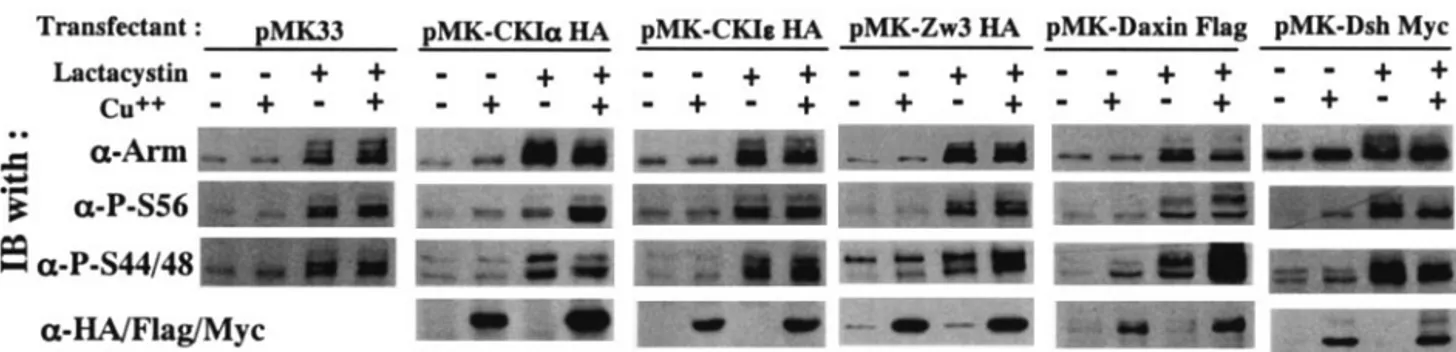
respectively, in vivo.The notion that the priming phosphory-lation of Arm at Ser56 and subsequent phosphoryphosphory-lations at Ser44 plus Ser48 are due to CKI␣ and ZW3, respectively, in vivo remains unproven in Drosophila. To address this issue, we upregulated and downregulated (by overexpression and RNAi, respectively) CKI␣, Zw3, and Daxin expression in S2R⫹ cells and examined its effect on the amount of Arm phosphorylated at Ser56 (P-S56-Arm) and at Ser44 plus Ser48 (P-S44/48-Arm). Since Arm is a protein with a rapid turnover, these analyses were performed in S2R⫹ cells treated or not with lactacystin, a specific inhibitor of proteasome. Actually, only in the pres-ence of lactacystin were the effects of overexpression of CKI␣, ZW3, and Daxin prominent (Fig. 3). Overexpression of CKI␣ elevated P-S56-Arm levels but not P-S44/48-Arm protein lev-els. Conversely, overexpression of Zw3 elevated P-S44/48-Arm but not P-S56-Arm levels. At least in this system, overexpres-sion of CKIεaffected neither Ser56 nor Ser44 plus Ser48 phos-phorylation. These results suggested that phosphorylation of Arm at Ser56 and at Ser44 plus Ser48 is mediated by CKI␣ and Zw3, respectively.
On the other hand, Daxin overexpression resulted in a marked and slight elevation of P-S44/48-Arm and P-S56-Arm, respectively, in the presence of lactacystin, indicating that Daxin mainly facilitates Zw3-mediated Arm phosphorylation in Drosophila. This result is in striking contrast to earlier re-ports for Xenopus and the mammalian system that expression of Axin dramatically stimulated-catenin priming-phosphory-lation at Ser45 (2, 22). In the presence of lactacystin, overex-pression of Dsh diminished P-S56-Arm, as well as P-S44/48-Arm levels, whereas in the absence of lactacystin, total P-S44/48-Arm levels, as well as P-S56- and P-S44/48-Arm levels, increased upon overexpression. These findings are in line with the notion that Dvl/Dsh is a positive regulator of the Wnt/Wg pathway
FIG. 2. CKI functions as a priming kinase for Zw3 in Arm phos-phorylation. (A) in vitro kinase experiment to demonstrate that prim-ing phosphorylation by CKI facilitates subsequent GSK-3-mediated Arm phosphorylation. The wild-type or S56A-mutant form of Arm proteins doubly tagged with the His6and myc epitopes was pretreated or not with CKI, immunoprecipitated with anti-myc antibody, again treated or not with GSK-3, and then subjected to Western blot analysis. The top and second panels show long and short exposures, respectively of the same immunoblot. (B) Arm phosphorylation at Ser44 and Ser48 requires phosphorylation at Ser56 in vivo. First, 1g of pAc5.1/V5-His C vector or various pAcArm-Myc constructs were transfected into S2R⫹ cells and, after 48 h, the myc-tagged Arm was immunoprecipitated with the goat polyclonal anti-myc antibody in combination with protein G-Sepharose. The cell lysates and the im-munoprecipitates were subjected to Western blot analysis with the antibodies indicated.
FIG. 3. Effect of overexpression of CKI␣, CKIε, Zw3, Daxin, and Dsh on Arm phosphorylation status. Stable S2R⫹ transfectants which could overexpress CKI␣, CKIε, or Zw3 (all of which are HA tagged), as well as Daxin (Flag tagged) and Dsh (myc tagged), were cultured in the presence or absence of CuSO4for 8 h. The cultures were then incubated for a further 6 h in the presence or absence of lactacystin (20M). Cell lysates were subjected to Western blot analysis with the antibodies indicated. Total cell lysates were used for this analysis because the amount of membrane-bound Arm is very limited (⬍5%) in lactacystin-treated S2R⫹ cells.
on March 19, 2017 by Ewha Womans Univ
http://mcb.asm.org/
and consistent with the earlier report that in the presence of MG132, a proteasome inhibitor, overexpression of Dvl1 sup-pressed both priming phosphorylation of -catenin at Ser45 and GSK-3-mediated -catenin phosphorylation at Ser33 plus Ser37 (2).
By using RNAi, we further characterized the roles of CKI␣, Zw3, and Daxin in the phosphorylation of Arm in a reciprocal way. The S2R⫹ cells and BG2 cells were used for this analysis and both of these cell lines gave similar results (Fig. 4). Since CKI␣, Zw3, and Daxin are all negative regulators of Wg sig-naling, RNAi of these proteins all resulted in a marked eleva-tion of Arm in the absence of lactacystin, confirming RNAi is working. In the presence of lactacystin, however, CKI␣- but not Zw3- or Daxin-RNAi decreased P-S56-Arm, which is con-sistent with the observation that overexpression of CKI␣ but not Zw3 or Daxin induced a large increase in P-S56-Arm levels (Fig. 3). On the other hand, CKI␣-, Zw3- or Daxin-RNAi all decreased P-S44/48-Arm. We interpreted this as follows. Zw3 appears to be directly responsible for the phosphorylation of Arm at Ser44 plus Ser48, and Daxin brings Arm and Zw3 into close proximity, thereby enhancing the Zw3-mediated phos-phorylation of Arm. Thus, it is convincing that Zw3- and Daxin-RNAi suppressed the phosphorylation at Ser44 plus Ser48. Moreover, the decrease in the P-S44/48-Arm caused by CKI␣-RNAi appears to indicate that CKI␣-RNAi diminished the phosphorylation at Ser56 that was required for subsequent Zw3-mediated phosphorylation at Ser44 plus Ser48. Taken together, a series of gain-of-function analyses in S2R⫹ cells and loss-of-function analyses in S2R⫹ and BG2 cells showed that the phosphorylation of Arm at Ser56 and at Ser44 plus Ser48 is mediated by CKI␣ and Zw3, respectively, in vivo. In addition, Daxin markedly enhances the Zw3- but not CKI␣-mediated phosphorylation, indicating that, in Drosophila scaf-folding protein, Daxin is not essential for the initial priming phosphorylation but is necessary for bridging the two phos-phorylation events.
CKI␣ but not CKI destabilizes Arm. Using S2R⫹ trans-fectants overexpressing HA-tagged CKIε or CKI␣ (because good antibody against Drosophila CKI␣ is not available), we have previously shown that dsRNA corresponding to the cat-alytic region of CKIεselectively suppressed expression of CKIε
but not of CKI␣ (48). In addition, although the induction level was much lower than that of CKI␣-dsRNA, CKIε-dsRNA evoked a significant accumulation of Arm protein (Fig. 5B). Thus, in a previous study, we concluded that CKIεby itself destabilizes Arm (48). However, in view of the nucleotide sequence similarity between CKIεand CKI␣ in their kinase-encoding domains, we could not rule out the possibility that in naive S2R⫹ cells, CKIε-dsRNA slightly diminished endoge-nous CKI␣ protein levels, thereby moderately inducing the accumulation of Arm. In addition, using a Drosophila cultured cell-based Tcf-dependent reporter assay, Lum et al. recently found that CKIε-RNAi has no effect on the Wg pathway, whereas overexpression of CKIεelevates the basal activity of the pathway (23).
To determine the effect of CKIεspecific-RNAi on Wg sig-naling, we generated a new CKIε-dsRNA corresponding to the C-terminal extracatalytic region (which is unique for this iso-form) of CKIε and evaluated its influence on Arm protein levels. Northern analysis revealed that whereas the old version of CKIε-dsRNA reduced steady-state levels of CKI␣ mRNA by 60%, the new one had no effect on CKI␣ mRNA levels (Fig. 5A). Moreover, in contrast to the old version, this new CKIε -dsRNA did not elevate Arm, although both of the CKIε
-FIG. 4. Effect of RNAi-mediated downregulation of CKI␣, Zw3, and Daxin expression on Arm phosphorylation. To the S2R⫹ or BG2 cell cultures preincubated with dsRNA for 30 h, 20M lactacystin was added or not, and then the cultures were further incubated for 6 h. Western blots of the cell lysates are shown. The bottom panel shows the effect of various RNAi on steady-state levels of Arm protein in the absence of lactacystin.
FIG. 5. Arm protein levels are not affected by CKIε-RNAi. (A) Northern blot analysis showing the effect of various dsRNAs on CKI␣ and Arm mRNA levels. Northern blots were produced as de-scribed previously (48). The bottom panel is an ethidium bromide staining of the gels showing that the same amount (10 g) of total RNA was loaded in each lane. (B) Effect of various dsRNAs on Arm, CKIε, and ZW3 protein levels. In the middle blot, lysates from the pMK-CKIεHA transfectant treated with or without CuSO4were used to demonstrate that the antibody against human CKIεrecognizes
Dro-sophila CKIe.
on March 19, 2017 by Ewha Womans Univ
http://mcb.asm.org/
dsRNA reduced CKIεprotein levels to the same extent (Fig. 5B). Therefore, we corrected the previous report (48) and concluded that only CKI␣ and not CKIε destabilizes Arm. These results are in line with the observation that overexpres-sion of CKIεdid not stimulate Arm phosphorylation (Fig. 3). The finding that overexpression of CKIεhad no effect on Arm protein levels (Fig. 3) appears to be inconsistent with the result from the reporter assay that overexpression of CKIεelevates basal activity of the Wg pathway (23). However, the report that phosphorylation of Tcf3 by CKIε stimulates its binding to -catenin might explain this discrepancy (18).
Effect of Wg/Wnt3A signaling on CKI␣- and
Zw3/GSK-3-mediated phosphorylation of Arm/-catenin in S2Rⴙ, Kc, and
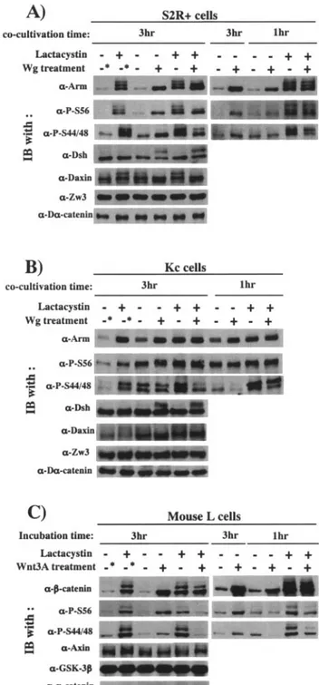
L cells. There is disagreement as to whether Wnt signaling inhibits GSK-3- or CKI-mediated -catenin phosphorylation in mammalian cells (2, 16, 22). Moreover, the effect of Wg signaling on Zw3- and CKI␣-mediated Arm phosphorylation has not been documented. Therefore, S2R⫹, Kc, and L cells were each treated with Wg and Wnt3A, and their effects on CKI␣- and Zw3/GSK-3-mediated-phosphorylation of Arm/-catenin were analyzed in the presence or absence of lactacystin (Fig. 6). In addition, to compare the effects of short and long-term Wg/Wnt treatment, cells treated with Wg/Wnt3A for 1 and 3 h were examined.
Consistent with the results shown in Fig. 1C, treatment of S2R⫹, Kc, and L cells with Wg or Wnt3A for 3 h in the absence of lactacystin elevated steady-state levels of total Arm/ -catenin as well as Arm/-catenin species undergoing CKI␣-or Zw3/GSK-3-mediated phosphCKI␣-orylation. Thus, in cells treated with Wg/Wnt3A for a long time, the steady-state levels of Arm/-catenin phosphorylated by either CKI␣ or Zw3/ GSK-3 appear to increase dependent on total Arm/-catenin levels. Consistent with previous reports, Wg signaling induced and suppressed phosphorylation of Dsh (19, 49) and Daxin (32, 44), respectively. Further analysis with cells treated with Wg/ Wnt3A for 1 h in the absence of lactacystin, however, revealed the following. Total Arm or-catenin levels in cells subjected to a 1-h treatment were lower than those in cells subjected to a 3-h treatment; Wnt3A treatment rapidly decreased steady-state levels of Ser33/37-phosphorylated-catenin (detected by ␣-P-S44/48 antibody), but those of Ser45-phosphorylated -catenin were not affected. Similarly, a 1-h Wg treatment markedly decreased P-S44/48-Arm levels in Kc cells, but this was not obvious in S2R⫹ cells. In S2R⫹ cells, a 1-h Wg treatment slightly elevated P-S56-Arm protein levels.
Avoiding the effect of Arm/-catenin turnover, we also per-formed similar analyses in the presence of lactacystin. As ex-pected, either in S2R⫹, Kc, and L cells, the total amount of Arm or-catenin was little affected by Wg or Wnt3A treat-ment under these conditions. Irrespective of the length of treatment, Wg markedly decreased P-S44/48-Arm protein lev-els, whereas P-S56-Arm protein levels remained constant in both S2R⫹ and Kc cells (Fig. 6A and B). In S2R⫹ cells treated with Wg for 3 h, the Arm with the highest electrophoretic mobility increased at the expense of Arm proteins showing less electrophoretic mobility, which may correspond to Arm spe-cies fully phosphorylated by both CKI␣ and Zw3 and ubiqui-tinated to various extents, but we noted that the total amount of P-S56-Arm protein was not affected by Wg treatment (Fig. 6A). In accordance with the Wg result, 1 or 3 h of Wnt3A
FIG. 6. Wg/Wnt3A signaling-induced changes in Arm/-catenin phosphorylation in the presence or absence of lactacystin. Effects of Wg/ Wnt3A treatment for 1 and 3 h were compared. Effects of Wg signaling on phosphorylation of Arm, Dsh, and Daxin were analyzed in S2R⫹ (A) and Kc (B) cells by Western blotting. Asterisks indicate that these cultures were treated in the same way as the others, except that the cocultivation with plain S2 cells for 3 h was replaced with incubation in plain medium (in the presence or absence of lactacystin) for 3 h. Zw3- and D␣-catenin blots were used as loading controls. (C) Effect of Wnt3A signaling on phosphorylation status of-catenin in L cells. Asterisks indicate that these cultures were treated in the same way as the others, except that they were incubated for a further 3 h in plain medium (in the presence or absence of lactacystin) but not in the control conditioned medium. Total cell lysates were used for this analysis, because L cells do not express cadherin; thus, the amount of membrane-bound-catenin is very limited. Amounts of P-Ser45- and P-Ser44/48–-catenin were monitored by using ␣-P-S56 and␣-P-S44/48 antibodies, respectively.
on March 19, 2017 by Ewha Womans Univ
http://mcb.asm.org/
treatment in the presence of lactacystin markedly decreased Ser33/37-phosphorylated -catenin levels. However, in con-trast to the Wg result, but in line with a previous report (2), Wnt3A diminished Ser45-phosphorylated-catenin levels, and this effect was prominent in the 3-h Wnt3A treatment. This appeared to be related to the earlier reports in mammalian cells that CKI␣-mediated -catenin phosphorylation is highly dependent on Axin (2, 22) and that prolonged Wnt3A treat-ment decreased total Axin protein levels (44). Against expec-tation, however, either in the presence or in the absence of lactacystin, 3-h Wnt3A treatment little affected total Axin pro-tein levels (Fig. 6C).
Taken together, Wg signaling suppresses ZW3-mediated Arm phosphorylation at Ser44 and Ser48 but not CKI␣-medi-ated phosphorylation at Ser56. Wnt3A signaling mainly inhib-its GSK-3-mediated -catenin phosphorylation at Ser33/37, but prolonged Wnt3A treatment also decreases CKI␣-medi-ated-catenin phosphorylation at Ser45.
Nevertheless, the primary target of Wnt3A signaling is clearly GSK-3-mediated phosphorylation of -catenin, be-cause short Wnt3A treatment rapidly decreased Ser33/37- but not Ser45-phosphorylated -catenin levels in the absence of lactacystin. Kang et al. (16) and Liu et al. (22) have reported basically the same results with Wnt3A-treated L cells and Wnt1-treated Rat2 cells, respectively. On the other hand, Amit et al. have concluded that Wnt regulates the pathway primarily by inhibiting CKI-mediated-catenin phosphorlation at Ser45, because in the presence of MG132, Wnt3A treatment for 5 h (in L, HeLa, and Jurkat cells) and Dvl overexpression (in 293T cells) both resulted in inhibition of Ser45 and Ser33⫹Ser37 phosphorylation (2).
Our finding that Wg/Wnt3A signaling suppresses ZW3/ GSK-3-mediated Arm/-catenin phosphorylation appears to be inconsistent with the observation that treatment of S2R⫹, Kc, and L cells with Wg or Wnt3A for 3 h (but not 1 h), in the absence of lactacystin, elevated steady-state-levels of P-S44/48-Arm and Ser33/37-phosphorylated-catenin (Fig. 1C and 6). To address these questions, we analyzed in detail (0, 30, 60, 90, 120, 150, and 180 min after Wg/Wnt3A treatment) the kinetics of Wg/Wnt3A-induced changes in levels of total Arm/-cate-nin, as well as Arm/-catenin species undergoing CKI␣- or Zw3/GSK-3-mediated phosphorylation in S2R⫹ and L cells (data not shown). The results can be summarized as follows. (i) Consistent with the notion that CKI␣-mediated phosphoryla-tion of Arm/-catenin is constitutive, the amounts of both P-S56-Arm and Ser45-phosphorylated-catenin were elevated progressively in proportion to the total amount of Arm/-catenin, which was markedly elevated depending on the length of Wg/Wnt3A treatment. (ii) In contrast, in the lag phase (up to 30 and 60 min after the Wg and Wnt3A treatments, respec-tively), where increase in total Arm/-catenin protein levels was little or very limited, the amounts of P-S44/48-Arm and Ser33/37-phosphorylated -catenin decreased slightly and sharply, respectively (as shown in Fig. 6C). (iii) However, at 60 min after Wg treatment, the amount of P-S44/48-Arm recov-ered to the same level as that at 0 min (as shown in Fig. 6A). After 90 min, amount of P-S44/48-Arm exceeded that at 0 min, and it elevated gradually as the total amount of Arm protein elevated progressively. (iv) Similarly, at 90 min after Wnt3A treatment, the amount of Ser33/37-phosphorylated-catenin
recovered to the same level as that at 0 min in L cells. After 120 min, the amount of Ser33/37-phosphorylated -catenin be-came greater than that at 0 min and elevated gradually in a time-dependent fashion.
Based on these observations, the following explanation is possible for the elevation of Zw3/GSK-3-phosphorylated Arm/-catenin in cells treated with Wg/Wnt3A for 3 h: even though Wg/Wnt3A suppresses Zw3/GSK-3-mediated phos-phorylation of Arm/-catenin, it also induces a marked in-crease in total amount of Arm/-catenin protein, a substrate for Zw3/GSK-3 at the later phase of treatment. Because of this secondary effect, long-term Wg/Wnt3A treatment causes a significant increase in the amount of Arm/-catenin phosphor-ylated by Zw3/GSK-3. However, we could not rule out the possibility that Wg/Wnt3A signaling not only regulates Arm/ -catenin phosphorylation but also blocks Arm/-catenin deg-radation downstream of the phosphorylation events (at the step of ubiquitination of the phosphorylated Arm/-catenin, for example), thus causing the elevation of Zw3/GSK-3-phos-phorylated Arm/-catenin.
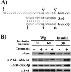
Wg signaling does not affect Zw3 phosphorylation at Ser9.
Thus far, we have shown that Wg/Wnt diminishes the amount of Arm/-catenin undergoing Zw3/GSK-3-mediated phos-phorylation. However, the molecular mechanisms underlining this process are poorly understood. Using GSK-3 substrate peptide, it was shown that the kinase activity of GSK-3/Zw3 was partially inhibited by Wnt-1 in 293, CHO, and C57MG cells (8) and by Wg in mouse 10T1/2 fibroblasts (5) and
Dro-sophila clone-8 cells (32). In spite of these reports, the effect of
Wg/Wnt signaling on the kinase activity of Zw3/GSK-3 still remains controversial. In insulin signaling, on the other hand, protein kinase B/Akt-mediated inhibitory phosphorylation at either Ser9 (GSK-3) or Ser21 (GSK-3␣) is well established as a molecular mechanism by which insulin inhibits GSK-3 activ-ity. Surprisingly, the fundamental question to date of whether or not Wg increases the inhibitory N-terminal phosphorylation of Zw3 remains unanswered. Therefore, we analyzed this by using anti-phospho-GSK-3 (Ser9) and anti-phospho-GSK-3␣ (Ser21) antibodies, both of which are found to cross-react with Zw3 only when its Ser9 is phosphorylated (Fig. 7A). Insulin but not Wg treatment promoted the inhibitory phosphorylation of Zw3 at Ser9 in S2R⫹ cells, indicating that stimulation of in-hibitory phosphorylation of Zw3 is not the mechanism by which Wg suppresses Zw3-mediated-phosphorylation of Arm (Fig. 7B). Similarly, insulin but not Wnt3A induced inhibitory phosphorylation of GSK-3 in L, Rat2, and NIH 3T3 cells (data not shown). These results appear to substantiate reports ex-cluding the involvement of the components of the Akt pathway in Wg signaling: (i) Wg signaling was not affected by inhibitors of phosphatidylinositol 3-kinase (5); (ii) the kinase activity of
Drosophila and mammalian Akt was not stimulated by Wg and
Wnt-1, respectively (8, 32); and (iii) Wg signaling was not affected by RNAi of Drosophila Akt (23).
PP2A catalytic subunit counteracts Arm and Daxin phos-phorylation but does not appears to play an indispensable role in Wg signaling.PP2A is a multisubunit Ser/Thr phosphatase that has a wide range of substrates and is involved in many cellular processes. It comprises a regulatory A subunit, a cat-alytic C subunit, and variable regulatory B subunits, which may target the location and/or action of the holoenzyme (reviewed
on March 19, 2017 by Ewha Womans Univ
http://mcb.asm.org/
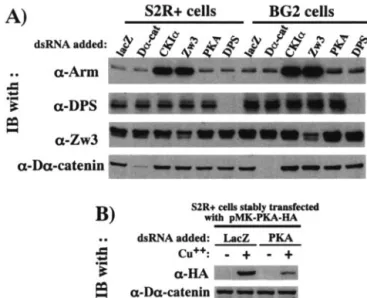
in reference 42). The catalytic subunit of PP2A (PP2A-C) is known to bind to Axin (31, 47), whereas regulatory subunits of the PP2A-B⬘ family can interact with APC (21). Thus, PP2A could serve to modulate the effect of CKI␣ and Zw3/GSK-3 on Arm/-catenin or other proteins in the Arm/-catenin deg-radation complex. An earlier report with Xenopus and mam-malian cells has indicated a positive regulatory role for PP2A-C at multiple levels in the Wnt pathway (31). However, the specific role of PP2A-C in the Wg pathway remains un-clear. Therefore, using RNAi, we determined whether PP2A-C antagonizes CKI␣- and Zw3-mediated-Arm phosphorylation, as well as Daxin phosphorylation (Fig. 8A), and whether PP2A-C promotes Arm dephosphorylation in a Wg-dependent manner, thereby playing a pivotal role in Wg-induced Arm accumulation (Fig. 8B).
In the absence of lactacystin, PP2A-C-RNAi slightly de-creased the total Arm protein level, which is consistent with the report that PP2A-C functions as a positive regulator of Wnt signaling (31). Either in the presence or in the absence of lactacystin, PP2A-C–RNAi slightly elevated both P-S56-Arm and P-S44/48-Arm, as well as hyperphosphorylated Daxin, sug-gesting that, in vivo, PP2A-C antagonizes CKI- and Zw3-me-diated Arm phosphorylation and Daxin phosphorylation, which appears to be mediated by Zw3 (32, 44).
In Fig. 6A, we have shown that Wg reduces Arm phosphor-ylation at Ser44 and Ser48. Although the N-terminal inhibitory phosphorylation of Zw3 is not associated with this process
(Fig. 7B), it might also involve alterations in phosphatase ac-tivity: Wg/Wnt signaling in some way enhances the enzymatic activity of PP2A, thereby abolishing Zw3/GSK-3-mediated Arm/-catenin phosphorylation that eventually leads to its ac-cumulation. If so, PP2A-C-RNAi may severely block the Wg-induced increase in Arm in the absence of lactacystin and strongly inhibit the Wg-induced decrease in P-S44/48-Arm in the presence of lactacystin. However, PP2A-C RNAi induced neither of these molecular events. Upon stimulation with Wg, S2R⫹ cells treated with PP2A-C-dsRNA showed levels of Arm similar to those of S2R⫹ cells treated with Laz-dsRNA. In the presence of lactacystin, PP2A-C-RNAi had no effect on the Wg-induced decrease in P-S44/48-Arm (Fig. 8B). Hence, as far as this RNAi-based analysis is concerned, PP2A-C counteracts Arm and Daxin phosphorylation, but this PP2A-C function does not appear to play a crucial role in normal Wg signaling.
Characterization of kinases involved in Wg-induced Dsh phosphorylation.We have previously shown that Dsh/Dvl pro-tein becomes phosphorylated in response to Wg/Wnt3A treat-ment in S2R⫹ and mouse L cells (19, 49), indicating that Dsh/Dvl phosphorylation occurs concomitantly with
Wg/Wnt-FIG. 7. Zw3 phosphorylation at Ser9 is affected by insulin but not Wg in S2R⫹ cells. (A) Alignment of conserved N-terminal amino acid sequences of GSK-3␣, GSK-3, and Zw3. The GSK-3␣ (human) se-quence from codons 1 to 27 and Zw3 and GSK-3 (human) sese-quences from codons 1 to 15 are shown. Ser residues phosphorylated upon insulin treatment are shown in boldface. Anti-phospho-GSK-3␣ and anti-phospho-GSK-3 antibody detects endogenous levels of GSK-3␣ and GSK-3, respectively, only when Ser21 of GSK-3␣ and Ser9 of GSK-3 are phosphorylated. Both of these antibodies recognize the Ser9-phosphorylated form of Zw3. (B) Wg signaling does not induce Zw3 phosphorylation at Ser9. Confluent S2R⫹ cell cultures were ei-ther cocultivated with S2-HS-wg or plain S2 cells for the time indicated or treated with 200 nM insulin for 30 min, and the cell lysates were subjected to Western blot analysis with the antibodies indicated.
FIG. 8. RNAi experiments to evaluate the function of PP2A-C in Wg signaling. (A) PP2A-C counteracts Arm phosphorylation at Ser56 and at Ser44 plus Ser48, as well as Daxin phosphorylation. To the S2R⫹ cell cultures preincubated with dsRNA for 30 h, 20 M lacta-cystin was added or not, and then the cultures were further incubated for 6 h. The cell lysates were subjected to Western blot analysis with the antibody indicated. (B) Wg-induced decrease in Arm phosphory-lation at Ser44 plus Ser48 is not caused by phosphatase activity of PP2A-C. The S2R⫹ cell cultures were preincubated with dsRNA for 30 h, further treated or not with lactacystin for 6 h, cocultivated with S2 or S2-HS-wg cells for a further 3 h, and then harvested for Western blot analysis.
on March 19, 2017 by Ewha Womans Univ
http://mcb.asm.org/
induced Arm/-catenin elevation. However, whether this Dsh/ Dvl phosphorylation is an essential integral process in Wg/Wnt signal transduction or just a Wg/Wnt-induced molecular event which by itself has little function in the Wg/Wnt pathway re-mains unclear. In addition, it is still controversial which of the two Dsh-associated kinases, CKI or Par-1, is responsible for Wg/Wnt-induced Dsh/Dvl phosphorylation (15, 18, 26, 29, 37). In some reports, CKI-7, a CKI inhibitor was used at higher concentrations (100 to 600M) to show that Wnt-3A-induced Dvl phosphorylation in L cells was due to CKI (15, 26). How-ever, we have noticed that at higher concentrations, CKI-7 does not necessarily work as a specific inhibitor of CKI in tissue culture systems (see Discussion).
To identify the kinase responsible for the Wg-induced Dsh phosphorylation in a more convincing way, the expression of four Dsh-associated kinases—CKI␣, CKIε, casein kinase II (CKII [43]), and Par-1—was downregulated by RNAi, and their effects on Dsh phosphorylation were examined. The Wg-induced Dsh phosphorylation was almost completely abolished by CKI␣-RNAi but not by RNAi of the other Dsh-associated kinases (Fig. 9). Strictly speaking, this result only indicates CKI␣ is required for the Wg-induced Dsh phosphorylation, but we conclude that CKI␣ is the kinase directly responsible for the Wg-induced Dsh phosphorylation because CKI␣ is a Dsh-associated kinase and its overexpression in vivo induces hyperphosphorylation of Dsh (48). On the other hand, Par-1-, CKIε-, or CKII-RNAi had little effect on the Wg-induced accumulation of Arm, suggesting that these three kinases do not play an essential role in Wg signaling. The present results are inconsistent with the report that Par-1 potentiates Wg/Wnt signaling and is responsible for the Wg/Wnt-induced phos-phorylation of Dsh/Dvl in Drosophila and mammalian cells (37). The reason for the discrepancy is not clear.
PKA and PS play little part in Arm degradation in
Drosoph-ila tissue culture cells.A recent report documented that PS functions as a scaffold linking PKA-mediated priming phos-phorylation of-catenin at Ser45 to subsequent
GSK-3-me-diated phosphorylation at Ser33, Ser37, and Thr41, thereby downmodulating -catenin expression in mammalian cells (16). To elucidate whether this PS-controlled regulation of Arm family protein is conserved in Drosophila and to biochem-ically evaluate the report in Drosophila embryos that a DPS deficiency led to a cytoplasmic Arm elevation (27), expression of the PKA catalytic subunit and DPS was disrupted by RNAi, and their effects on Arm levels were examined in S2R⫹ and BG2 cells. PKA-RNAi suppressed the expression of HA-tagged PKA in pMK-PKA-⌯〈 transfectant, indicating that PKA-RNAi is working (Fig. 10B). Furthermore, DPS protein levels expressed in both S2R⫹ and BG2 cells were diminished by DPS-RNAi. Although Zw3- and CKI␣-RNAi markedly el-evated Arm protein levels in both S2R⫹ and BG2 cells, DPS-and PKA-RNAi had little effect, suggesting that the assumed Arm degradation complex consisting of PKA, DPS, and Zw3 is not present or plays a marginal role in Arm protein metabo-lism at least in these Drosophila tissue culture cells (Fig. 10A). Although Drosophila genetic studies support the negative reg-ulation of Arm by DPS, our RNAi-based findings in tissue culture cells demonstrated that the model recently proposed in mammalian cells is not applicable to Drosophila. Further study is required to clarify the biochemical relationship between DPS and Arm.
DISCUSSION
In this RNAi-based study, we have provided compelling evidence that phosphorylation of Arm at Ser56 by CKI␣ primes it for subsequent phosphorylation at Thr52, Ser48, and
FIG. 9. Wg-induced phosphorylation of Dsh is due to CKI␣. Ex-pression of each of four Dsh-associated kinases was suppressed by preincubating S2R⫹ cells with dsRNA for 30 h, and these S2R⫹ cell cultures were treated or not with Wg for 3 h and then harvested. The amounts of phosphorylated Dsh (shown with an arrow), Arm, CKIε, Par-1, CKII␣ subunit, and D␣-catenin among these cells were com-pared by Western blottings. LacZ dsRNA was used as a negative control.
FIG. 10. Arm protein levels are little affected by DPS and PKA in
Drosophila tissue culture cells. (A) Expression of CKI␣, ZW3, PKA, and DPS was downregulated by RNAi, and their effects on Arm pro-tein levels were analyzed in S2R⫹ and BG2 cells. S2R⫹ or BG2 cells were incubated with dsRNA for 60 h. The cell lysates were then subjected to Western blot analysis with the antibody indicated. LacZ-and D␣-catenin-RNAi were used as negative controls. Total cell ly-sates were used for this analysis, because the amount of membrane-bound Arm is very limited in S2R⫹ and BG2 cells. (B) The PKA-RNAi experiment works. The stable pMK-PKA-HA transfectant was incubated with dsRNA for 60 h, treated or not with CuSO4 for a further 12 h, and subjected to Western blotting.
on March 19, 2017 by Ewha Womans Univ
http://mcb.asm.org/
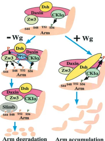
Ser44 by Zw3 and that Daxin enhances the Zw3- but not CKI␣-mediated Arm phosphorylation. Moreover, we have clarified that Wg signaling inhibits the Zw3- but not CKI␣-mediated phosphorylation of Arm and that PP2A-C does not play a primary role in this Wg-induced decrease in Arm phos-phorylation. In addition, we have confirmed that Wg-induced Dsh phosphoylation is due to CKI␣. Taking these findings into account, we present a model of how Wg signaling modulates the phosphorylation of Arm, as well as other components of the destruction complex, thereby regulating the stability of Arm (Fig. 11).
As shown in Fig. 9, CKI␣ plus CKIε-RNA elevates Arm levels, suggesting that the overall effect of inhibition of the CKI family is to induce the accumulation of Arm in S2R⫹ cells. In addition, the Wg-induced accumulation of Arm was not blocked by any of the CKI-RNAi (data not shown). Given that the 50% inhibitory concentration of CKI-7 for CKI is 9.5M, we treated S2R⫹ cells with various concentrations (10 to 1,000 M) of CKI-7 for 5 h and analyzed their effect on Arm levels. CKI-7 did not induce Arm accumulation, whereas at higher concentrations (ⱖ300 M), it inhibited Wg-induced Arm ele-vation and Dsh phosphorylation. Similarly, CKI-7 did not in-duce -catenin elevation but inhibited both Wnt3A-induced -catenin accumulation and Dvl phosphorylation in L cells (data not shown). These results indicate that CKI-7-induced changes in Wnt/Wg signaling components are not necessarily due to its function as a CKI inhibitor.
In contrast to the finding in mammalian fibroblasts that PS1 and PKA functions as a scaffold and a priming kinase, respec-tively, for-catenin degradation (16), RNAi in S2R⫹ and BG2 cells showed that DPS and PKA play little part in Arm degra-dation (Fig. 10). The reason for this discrepancy is not clear, but it should be noted that even in a mammalian system, Marambaud et al. have reported PS1 as a positive regulator of -catenin: PS1 cleaves E-cadherin at the membrane-cytoplas-mic interface, thereby releasing its intracellular domain, which eventually leads to disassembly of the E-cadherin-catenin com-plex and an increase in the cytoplasmic pools of-catenin (25). However, this mechanism of Arm regulation may not exist in S2R⫹ and BG2 cells because these cells express little
Drosoph-ila E-cadherin (46).
Using RNAi in combination with a Drosophila Kc cell-based Tcf-dependent reporter assay, Lum et al. have systematically screened the functional roles of all kinases and phosphatases encoded by the Drosophila genome in Wg signaling (23). This screen identified Zw3 and CKI␣ but not CKIε, PKA, and PP2A as components of the Wg/Arm pathway, a finding which is in line with our results that CKIε-, PKA-, and PP2A-C-RNAi have little effect on Arm protein levels.
There is disagreement as to whether different CKI isoforms play the same or different roles (both positive or negative) in Wnt/Wg signaling. The different experimental systems and ap-proaches used in different studies make the situation more complicated. Mainly based on observations that overexpres-sion of CKI induced dorsal axis duplication in Xenopus em-bryos and stimulated the Tcf/lef reporter in mammalian cells, Graff’s group have reported that all CKI isoforms except CKI␥ positively regulate Wnt signaling in vertebrates (26, 29), whereas Sakanaka et al. have identified CKIεbut not CKI␣ as a positive regulator of the Wnt pathway (34). Lee et al. have
FIG. 11. Model depicting how the Wg signal controls phosphory-lation-dependent degradation of Arm. Daxin assembles Zw3, CKI␣, Dsh, PP2A-C, and Arm in close proximity. Drosophila APC is not shown in this figure because the present study does not deal with this protein. CKI␣ constitutively primes Arm phosphorylation at Ser56. In the absence of Wg, Zw3 recognizes this priming phosphate then se-quentially phosphorylates Thr52, Ser48, and Ser44. The latter two phosphorylations are required for the binding of an F-box protein, Slimb, that targets Arm for ubiquitination and degradation. PP2A-C appears to counteract the CKI␣-mediated-phosphorylation of Arm, as well as the Zw3-mediated phosphorylation of Arm and Daxin. Al-though its molecular mechanism is unknown, the binding of Wg to the Dfrizzled2-Arrow receptor complex is speculated to induce conforma-tional changes in Dsh that prevent Zw3 from phosphorylating Arm at Thr52, Ser48, and Ser44 on the one hand and elicit CKI␣-mediated phosphorylation of Dsh itself on the other. However, CKI␣-mediated Arm phosphorylation at Ser56 is not affected by Wg signaling. The mechanism by which Wg signaling counteracts Zw3-mediated phos-phorylation of Arm is poorly understood, but the present study ruled out both Ser9 phosphorylation-induced inactivation of Zw3 kinase (Fig. 7B) and the involvement of PP2A (Fig. 8B). In this process, Frat/GBP has been proposed to evoke the dissociation of GSK-3 from Axin in vertebrates (6, 11, 20). However, Drosophila has no apparent Frat/GBP counterpart. Wnt/Wg signaling also diminishes GSK-3/Zw3-mediated phosphorylation of Axin/Daxin (Fig. 6A), which appears to lower Axin/Daxin’s affinity to-catenin (44) and Zw3 (32), which were shown to stimulate the release of-catenin from the degradation complex and speculated to reduce Arm phosphorylation, respectively. In Drosophila, it still remains unclear whether Wg-in-duced phosphorylation of Dsh by CKI␣ is essential for normal Wg signal transduction. However, in vertebrates Wnt-induced Dvl/Xdsh phosphorylation by CKIεhas been shown to enhance the binding of Frat/GBP to Dvl/Xdsh that promotes the disintegration of the -cate-nin destruction complex, which eventually leads to activation of the Wnt pathway (15, 18).
on March 19, 2017 by Ewha Womans Univ
http://mcb.asm.org/
reported that CKIε strengthened Tcf3–-catenin and GBP/ Frat-Xdsh interactions, which led to -catenin stabilization (18), whereas Rubinfeld et al. have demonstrated that CKIε
mediates Axin-dependent phosphorylation of APC, thereby stimulating -catenin degradation (33). However, these pro-posals for mechanisms of CKI action have not been evaluated by others.
In contrast, in three recent studies (2, 22, 48) and in the present study, the function of CKI␣ as a priming kinase for GSK-3/Zw3 in -catenin/Arm phosphorylation, and thus its negative role in Wnt/Wg signaling, has been established in both vertebrates and Drosophila. Liu et al. (22) and the present study have ruled out a role for CKIε as a priming kinase, whereas Ben-Neriah’s group have demonstrated that in some cell types CKIεalso functions as a priming kinase (2, 14). In addition, Schwarz-Romond et al. have reported that the ankyrin repeat protein Diversin recruits CKIεbut not CKI␣ to the -catenin degradation complex and allows efficient phos-phorylation of-catenin (35). These reports support negative roles for CKIε(as a priming kinase) in Wnt signaling. How-ever, in two consecutive papers (15, 17), a positive role for CKIεbut not CKI␣ in Wnt signaling has been reported: CKIε
phosphorylates Dvl and thereby enhances the binding of Frat to Dvl, which leads to inhibition of Dvl-Axin interaction and a malfunction of the -catenin degradation complex. Clearly, further studies are necessary to reconcile discrepancies about CKI’s role in Wnt/Wg signaling and to figure out the real functions of CKI in the Wnt/Wg pathway.
ACKNOWLEDGMENTS
We are grateful to D. St Johnston (University of Cambridge) and K. Tei (University of Tokyo) for providing anti-PAR-1 antibody and BG2 cells, respectively.
This study was supported by a grant-in-aid from the Ministry of Education, Science, Sports, and Culture of Japan to S.Y.
REFERENCES
1. Aberle, H., A. Bauer, J. Stappert, A. Kispert, and R. Kemler. 1997.-Catenin is a target for the ubiquitin-proteasome pathway. EMBO J. 16:3797–3804. 2. Amit, S., A. Hatzuba, Y. Birman, J. S. Andersen, E. Ben-Shushan, M. Mann,
Y. Ben-Neriah, and I. Alkalay.2002. Axin-mediated CKI phosphorylation of -catenin at Ser 45: a molecular switch for the Wnt pathway. Genes Dev.
16:1066–1076.
3. Cadigan, K., and R. Nusse. 1997. Wnt signaling: a common theme in animal development. Genes Dev. 11:3286–3305.
4. Clements, J. C., C. A. Worby, N. Simonson-Leff, M. Muda, T. Maehara, B. A.
Hemmings, and J. E. Dixon.2000. Use of double-stranded RNA interference in Drosophila cell lines to dissect signal transduction pathways. Proc. Natl. Acad. Sci. USA 97:6499–6503.
5. Cook, D., M. J. Fry, K. Hughes, R. Sumathipala, J. R. Woodgett, and T. C.
Dale.1996. Wingless inactivates glycogen synthase kinase-3 via an intracel-lular signaling pathway which involves a protein kinase C. EMBO J. 15:4526– 4536.
6. Dajani, R., E. Fraser, S. M. Roe, M. Yeo, V. M. Good, V. Thompson, T. C.
Dale, and L. H. Pearl.2003. Structural basis for recuruitment of glycogen synthase kinase 3 to the axin-APC scaffold complex. EMBO J. 22:494–501. 7. Dajani, R., E. Fraser, S. M. Roe, N. Young, V. Good, T. C. Dale, and L. H.
Pearl.2001. Crystal structure of glycogen synthase kinase 3: structural basis for phosphate-primed substrate specificity and auto-inhibition. Cell 105:721– 732.
8. Ding, V. W., R.-H. Chen, and F. McCormick. 2000. Differential regulation of glycogen synthase kinase 3 by insulin and Wnt signaling. J. Biol. Chem.
275:32475–32481.
9. Ding, Y., and T. Dale. 2002. Wnt signal ttransduction: kinase cogs in a nano-machine? Trends Biochem. Sci. 7:327–329.
10. Doble, B. W., and J. R. Woodgett. 2003. GSK-3: tricks of the trade for a multi-tasking kinase. J. Cell Sci. 116:1175–1186.
11. Ferkey, D. M., and D. Kimelman. 2002. Glycogen synthase kinase3 mu-tagenesis identifies a common binding site for GBP and Axin. J. Biol. Chem.
277:16147–16152.
12. Flotow, H., and P. J. Roach. 1991. Role of acidic residues as substrate determinants for casein kinase I. J. Biol. Chem. 266:3724–3727.
13. Frame, S., P. Cohen, and M. Biondi. 2001. A common phosphate binding site explains the unique substrate specificity of GSK3 and its inactivation by phosphorylation. Mol. Cell 7:1321–1327.
14. He, X. 2003. A Wnt-Wnt situation. Dev. Cell 4:791–797.
15. Hino, S., T. Michiue, M. Asashima, and A. Kikuchi. 2003. Casein kinase Iε enhances the binding of Dvl-1 to Frat-1 and is essential for Wnt-3a-induced accumulation of-catenin. J. Biol. Chem. 278:14066–14073.
16. Kang, D. E., S. Soriano, X. Xia, C. G. Eberhart, B. De Strooper, H. Zheng,
and E. H. Koo.2002. Presenilin couples the paird phosphorylation of -cate-nin independent of axin: implications for-catenin activation in tumorigen-esis. Cell 110:751–762.
17. Kishida, M., S.-i. Hino, T. Michiue, H. Yamamoto, S. Kishida, A. Fukui, M.
Asashima, and A. Kikuchi.2001. Synergistic activation of the Wnt signaling pathway by DVl and casein kinase 1ε. J. Biol. Chem. 276:33147–33155. 18. Lee, E., A. Salic, and M. W. Kirschner. 2001. Physiological regulation of
-catenin stability by Tcf3 and CK1ε. J. Cell Biol. 154:983–994.
19. Lee, J.-S., A. Ishimoto, and S. Yanagawa. 1999. Characterization of mouse Dishevelled (Dvl) proteins in Wnt/Wingless signaling pathway. J. Biol. Chem. 274:21464–21470.
20. Li, L., H. Yuan, C. D. Weaver, J. Mao, G. H. Farr III, D. J. Sussman, J.
Jonker, D. Kimelman, and D. Wu.1999. Axin and Frat1 interact with Dvl and GSK, bridging Dvl to GSK in Wnt-mediated regulation of LEF-1. EMBO J. 18:4233–4240.
21. Li, X., H. J. Yost, D. M. Virshup, and J. M. Seeling. 2001. Protein phospha-tase 2A and its B56 regulatory subunit inhibit Wnt signaling in Xenopus. EMBO J. 20:4122–4131.
22. Liu, C., Y. Li, M. Semenov, C. Han, G.-H. Bang, Y. Tan, Z. Zhang, X. Lin,
and X. He.2002. Control of-catenin phosphorylation/degradation by a dual-kinase mechanism. Cell 108:837–847.
23. Lum, L., S. Yao, B. Mozer, A. Rovescalli, D. Von Kessler, M. Nirenberg, and
P. A. Beachy. 2003. Identification of hedgehog pathway components by RNAi in Drosophila cultured cells. Science 299:2039–2045.
24. Marin, O., V. H. Bustos, L. Cesaro, F. Meggio, M. A. Pagano, M. Antonelli,
C. C. Allende, L. A. Pinna, and J. E. Allende.2003. A noncanonical sequence phosphorylated by casein kinase 1 in-catenin may play a role in casein kinase 1 targeting of important signaling proteins. Proc. Natl. Acad. Sci. USA 100:10193–10200.
25. Marmbaud, P., J. Shioi, G. Serban, A. Georgakopoulos, S. Sarner, V. Nagy,
L. Baki, P. Wen, S. Efthimiopoulos, Z. Shao, T. Wisniewski, and N. K. Robakis.2002. A presenilin-1/␥-secretase claevage release the E-cadherin intracellular domain and regulates disassembly of adherens junctions. EMBO J. 21:11948–11956.
26. Mckay, R. M., J. M. Peters, and J. M. Graff. 2001. The casein kinase I family in Wnt signaling. Dev. Biol. 235:388–396.
27. Noll, E., M. Medina, J. D. Hartley, Zhou, N. Perrimon, and K. S. Kosik. 2000. Presenilin affects Arm/-catenin localization and function in Drosoph-ila. Dev. Biol. 227:450–464.
28. Pai, L.-M. S., S. Orsulic, A. Bejsovec, and M. Peifer. 1997. Negative regu-lation of armadillo, a wingless effector in Drosophila. Development 124: 2255–2266.
29. Peters, J. M., R. M. Mckay, J. P. McKay, and J. M. Graff. 1999. Casein kinase I transduces Wnt signals. Nature 401:345–350.
30. Polakis, P. 2000. Wnt signaling and cancer. Genes Dev. 14:1837–1851. 31. Ratcliffe, M. J., K. Itoh, and S. Sokol. 2000. A positive role for the PP2A
catalytic subunit in the Wnt signal transduction. J. Biol. Chem. 275:35680– 35683.
32. Ruel, L., V. Stambolic, A. Ali, A. S. Manoukian, and J. R. Woodgett. 1999. Regulation of the protein kinase activity of Shaggy by components of the Wingless pathway in Drosophila cells. J. Biol. Chem. 274:21790–21796. 33. Rubinfeld, B., D. A. Tice, and P. Polakis. 2001. Axin-dependent
phosphor-ylation of the adenomatous polyposis coli protein mediated by casein kinase 1 epsilon. J. Biol. Chem. 276:39037–39045.
34. Sakanaka, C., P. Leong, L. Xu, S. D. Harrison, and L. T. Williams. 1999. Casein kinase Iεin the Wnt pathway: regulation of-catenin function. Proc. Natl. Acad. Sci. USA 96:12548–12552.
35. Schwarz-Romond, T., C. Asbrand, J. Bakkers, M. Kuhl, H.-J. Schaeffer, J.
Huelsken, J. Berhrens, M. Hammerscmit, and W. Birchmeier.2002. The ankyrin repeat protein Diversin recuruit casein kinase Iεto the-catenin degradation complex and acts in both canonical Wnt and Wnt/JNK signaling. Genes Dev. 16:2073–2084.
36. Siegfried, E., T.-B. Chou, and N. Perrimon. 1992. wingless signaling acts through zeste-white 3, the Drosophila homolog of glycogen synthase kinase-3 to regulate engrailed and establish cell fate. Cell 71:1167–1179.
37. Sun, T.-Q., B. Lu, J.-J. Feng, C. Reinhard, Y. N. Jan, W. J. Fanti, and L.
Williams.2001. PAR-1 is a dishevelled-associated kinase and a positive regulator of Wnt signalling. Nat. Cell Biol. 3:628–636.
38. Takasugi, N., Y. Takahashi, Y. Morohashi, T. Tomita, and T. Iwatsubo. 2002. The mechanism of␥-secretase activities through high molecular weight complex formation of presenilin is conserved in Drosophila melanogaster and mammals. J. Biol. Chem. 277:50198–50205.