저작자표시-비영리-동일조건변경허락 2.0 대한민국 이용자는 아래의 조건을 따르는 경우에 한하여 자유롭게 l 이 저작물을 복제, 배포, 전송, 전시, 공연 및 방송할 수 있습니다. l 이차적 저작물을 작성할 수 있습니다. 다음과 같은 조건을 따라야 합니다: l 귀하는, 이 저작물의 재이용이나 배포의 경우, 이 저작물에 적용된 이용허락조건 저작자표시. 귀하는 원저작자를 표시하여야 합니다. 비영리. 귀하는 이 저작물을 영리 목적으로 이용할 수 없습니다. 동일조건변경허락. 귀하가 이 저작물을 개작, 변형 또는 가공했을 경우 에는, 이 저작물과 동일한 이용허락조건하에서만 배포할 수 있습니다.
TheExpr
essi
on ofSyndecan-1i
sRel
at
edt
ot
he
Ri
sk ofEndomet
r
i
alHyper
pl
asi
aPr
ogr
essi
ng t
o
Endomet
r
i
alCar
ci
noma
by
Hyunj
i
n Ki
m
Maj
ori
n Medi
ci
ne
Depar
t
mentofMedi
calSci
ences
TheGr
aduat
eSchool
,Aj
ou Uni
ver
si
t
y
TheExpr
essi
on ofSyndecan-1i
sRel
at
edt
ot
he
Ri
sk ofEndomet
r
i
alHyper
pl
asi
aPr
ogr
essi
ng t
o
Endomet
r
i
alCar
ci
noma
by
Hyunj
i
n Ki
m
A Di
s
s
e
r
t
at
i
onSubmi
t
t
e
dt
oTheGr
a
dua
t
eSc
hoo
lofAj
ou
Uni
ve
r
s
i
t
y
i
nPar
t
i
alFul
f
i
l
l
me
ntoft
heRe
qui
r
e
me
nt
sf
ort
heDe
gr
e
eof
Ph.D.i
nMe
di
c
i
ne
Super
vi
sedby
Thi
scer
t
i
f
i
est
hatt
hedi
sser
t
at
i
on
ofHyunj
i
n Ki
m i
sappr
oved.
SUPERVI
SORY COMMI
TTEE
Hee-Sug Ryu
Ki
-Hong Chang
Haeng-SooKi
m
Young Don Lee
Yong-Man Ki
m
TheGr
aduat
eSchool
,Aj
ou Uni
ver
si
t
y
J
une,25t
h,2010
ABSTRACT
-TheExpr
essi
on ofSyndecan-1i
sRel
at
edt
ot
heRi
sk of
Endomet
r
i
alHyper
pl
asi
aPr
ogr
essi
ng t
oEndomet
r
i
al
Car
ci
noma
Objective: Aberrant expression of the cell surface proteoglycan, syndecan-1,isfound in many malignancies.Thecurrentstudy describesthe immunohistochemicalstudy ofsyndecan-1 expression in normal,hyperplastic, andmalignantendometrialtissuesforevaluationofapplicationasaparameter ofcancerprogressioninpatientswithendometrialhyperplasia.
Methods:Immunohistochemicalstaining ofsyndecan-1 was performed in 101 formalin fixed,paraffin embedded sections ofnormal,hyperplastic,and malignant endometrialtissues.We analyzed specimens from patients with normalendometrium (NE,n=10)ascontrols,and thoseofsimplehyperplasia (SH, n=20), complex hyperplasia without atypia (CH, n=20), atypical hyperplasia(AH,n=20),andendometrialcancer(EC,n=31).
Conclusion:Syndecan-1 expression appears to be usefulas a predictive indicator of endometrial carcinogenesis from normal endometrium or endometrialhyperplasiatoendometrialcancer.
TABLE OF CONTENTS
ABSTRACT ···ⅰ TABLE OF CONTENTS ···ⅲ LIST OF FIGURES···ⅳ LIST OF TABLES···ⅴ
Ⅰ.INTRODUCTION···1
Ⅱ.PATIENTS AND METHODS···3
A.Patientsandtissuesamples ……… 3
B.Immunohistochemicalstudy……… 5
C.Stainingevaluation……… 5
D.Statisticalanalysis……… 6
Ⅲ.RESULTS ···7
A.Patientcharacteristics……… 7
B.Expressionofsyndecan-1………8
Ⅳ.DISCUSSION ···12
REFERENCE ···15
Fig.1.Sectionsofformalin-fixed,paraffin-embedded samplesofendometrial tissues were stained with eosin and hematoxylin for hyperplasia classification (original magnification,×400).(a) Simple hyperplasia without atypia. (b) Simple hyperplasia with atypia. (c) Complex hyperplasiawithoutatypia.(d)Complexhyperplasiawithatypia……4
Fig.2.Immunohistochemicalstaining forsyndecan-1(originalmagnification, ×100).(a)Normalendometrium showingnegativesyndecan-1staining (frequency score:0).(b)Simplehyperplasia(frequency score:1).(c) Complex hyperplasiawithoutatypia(frequencyscore:2).(d)Complex hyperplasia with atypia (frequency score:3).(e)Almosttumorcells show positivesyndecan-1expression(frequencyscore:3) …………9
Fig.3.Immunostaining mean scores of syndecan-1 expression in normal control and in specimens of simple hyperplasia (SH), complex hyperplasia without atypia (CH),complex hyperplasia with atypia (AH),andendometrialcancer(EC)……… 10
Table1.Demographicdataofthepatientsbyhistologicdating……… 7
Ⅰ.I
NTRODUCTI
ON
Endometrialcancer is the mostcommon disease among gynecological cancersand isoneofthemain health concernsworldwide(Leeetal,2005; Jemal et al, 2006). In the United States alone, 41,200 women develop endometrialcancer,and about7,350 women die from this disease annually (Jemaletal,2006).According to the AnnualReportofGynecologic Cancer Registry Program in Korea,the incidence ofendometrialcancer in Korean womenisrecentlyincreasing (Leeetal,2005).Endometrialcancermayoccur denovo,butcan alsoarisewithin preexisting premalignantlesionsexhibiting advanceddegreesofhyperplasia(Silverbergetal,2003).In1994,accordingto theWHO classification,simpleand complex typesofhyperplasia arefurther subdivided into those with typicalor atypicalarchitecture (Kurman et al, 1985).Thisisthemostimportantdistinction from thestand pointofclinical management,because the risk ofmalignanttransformation ofhyperplasia to invasive carcinoma increases according to the classification of hyperplasia from 2 to 23% in premalignant lesions exhibiting without atypia to with atypia,respectively (Kendalletal,1998).Theprogression ofalesion from a premalignantstateto endometrialcancerisbelieved to betheoutcomeofa seriesofmetagenesisdriven by variousrisk factors(Silverberg etal,2003). The identification of specific intracellular events in carcinogenesis is a necessary prerequisite to the identification of therapeutics that target and interruptspecificstepsintheprogressionofcancer(DrukerandLydon,2000).
Previousstudieshavesuggestedacorrelationbetweentheexpressionof syndecan-1 and the development of cancer characteristics in the human endometrium (Choi et al, 2007). However, it is not yet clear whether syndecan-1iscloselycorrelatedwiththeonsetoftumorigenesisorifitplays
a role as a positive predictive marker in cancer development.For these reasons, we sought to determine if quantitative analysis of syndecan-1 correlated with the histologicalclassification of endometrialhyperplasia by evaluating syndecan-1 expression in endometrial hyperplasia and stage I, grade Icancer tissues by determining the percentage ofpositively stained cells to yield a quantitative value for syndecan-1 expression.Our results suggestthe role ofsyndecan-1 as a predictive factor ofprogression from hyperplasiatoendometrialcancer.
Ⅱ.PATI
ENTS AND METHODS
A.Pat
i
ent
sandt
i
ssuesampl
es
Tissuesampleswereobtained from theparaffin-embedded endometrium of study 101 patients who underwent endometrial curettage and/or hysterectomy and were followed up at the Department of Obstetrics and Gynecology ofAjou University Hospital,Suwon,Republic ofKorea between 1995 and 2007. The patient population consisted of individuals including endometrialhyperplasia(n=60).Endometrialdataobtained wereclassified into five groups:normalendometrium (NE,n=10) as the controlgroup,simple hyperplasia (SH, n=20), complex hyperplasia without atypia (CH, n=20), complex hyperplasia with atypia or atypical hyperplasia (AH,n=20),and endometrialcancer(EC,n=31)with grade1differentiation.NE wasobtained from patients with intramuralor subserosalmyomas showing grossly and microscopically intact endometrium. Specimen was obtained from representativeendometrialpathologies:hysterectomizedendometrium (n=62)or endometrialcurettage(n=38).
We performed a quantitative analysis of syndecan-1 expression employing a scoring method ofthe immunohistochemicalstaining frequency. Histopathologic classification of endometrialhyperplasia was based on the “InternationalSociety ofGynecologicalPathologists”criteria.The histological gradesaccording totheInternationalFederationofGynecology andObstetrics (FIGO)staging classification wereasfollows:31patientsin thisstudy were grade 1.Surgicalstaging was reviewed based on the FIGO staging system and allofthe endometrialcancerpatientswere stageIdisease.Endometrial hyperplasiaspecimenswerefurthersubdivided by thepresenceofatypiaand
degree ofdifferentiation.In allcases,atypicalcells were identified on the original H&E sections (Fig. 1). Follow-up for all patients who had endometrialhyperplasia in the endometrialcurettage specimen,as evidenced by staining with syndecan-1,wasobtainedwhen possible.Weretrospectively reviewedpatientchartstoidentifypatient’sdemographicfactors.
B.I
mmunohi
st
ochemi
calst
udy
Serial paraffin sections were cut to 4μm in thickness for immunohistochemical study of syndecan-1 expression in endometrial hyperplasia and endometrialcancer cells.Paraffin-embedded sections from clinical endometrial tissue samples were subjected to immunostaining for syndecan-1 using antihuman syndecan-1 mouse B-B4 monoclonalantibody and DAB visualization,as described in the manufacturer’s manual.In brief, afterdeparaffinationandtreatmentof3% H2O2for5min,thetissuesamples were blocked with a 10% serum in the blocking solution of the Histostain®-Plus Bulk Kit(Biocompare,San Francisco,CA) for 1 hr and incubated overnightat4°C with antisyndecan-1 mouse monoclonal(B-B4 clone)antibodies at1/400 dilution in the antibody diluent.Foreithertissue samples,DAB colorreaction was performed with the Histostain®-Plus Bulk Kitbeforebeing examinedunderanOlympusmicroscopeD50(Tokyo,Japan) and photographed using an AxioCam MRc5 camera (Carl Zeiss AG, Oberkochen,Germany).Then each stained section was counterstained with Mayer’shematoxylin.Also,thesectionsfrom thesame tissueswerestained withMayer’shematoxylinandeosinforhistologicalclassification.
C.St
ai
ni
ng eval
uat
i
on
The level of syndecan-1 immunoreactivity in endometrial cells was expressed by scoring the percentage ofsyndecan-1 positive cells into four groups:0,negativeofcellsstained;1,<33% ofcellsstained;2,33- 66% of cellsstained;and3,> 66% ofthecellsstained(Choietal,2007).Microscopic analyseswereevaluatedindependentlybyonepathologist(HJH)withnoprior knowledgeoftheclinicaldata.
D.St
at
i
st
i
calanal
ysi
s
Distribution ofthepatients’characteristicswaspresentedasmean (SD) forcontinuousvariables,andfrequency (%)forcategoricalvariablesincluding immunohistochemicalstaining.Fisher’sexacttestwasused to determinethe correlation between thetwocategoricalvariables.TheMann-Whitney U test or Kruskall-Wallis testwas used to compare the mean or median values between the two or more groups. P-values < 0.05 were considered statisticallysignificant.
Ⅲ.RESULTS
A.Pat
i
entchar
act
er
i
st
i
cs
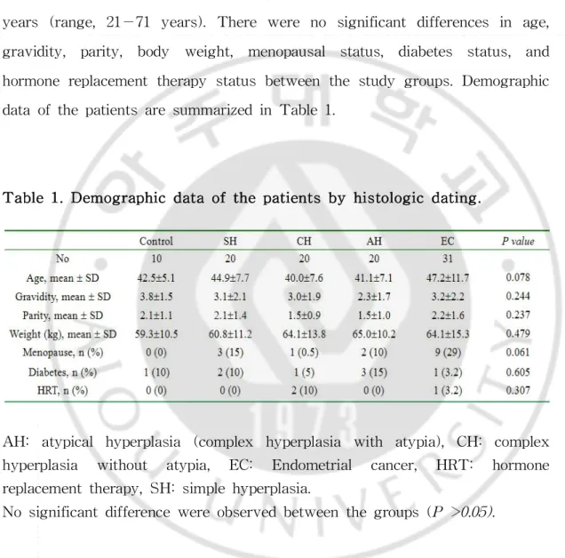
The mean age ofincluded subjects atthe time ofdiagnosis was 44 years (range,21−71 years).There were no significantdifferences in age, gravidity, parity, body weight, menopausal status, diabetes status, and hormonereplacementtherapy statusbetween thestudy groups.Demographic dataofthepatientsaresummarizedinTable1.
Table1.Demographicdataofthepatientsby histologicdating.
AH: atypical hyperplasia (complex hyperplasia with atypia),CH: complex hyperplasia without atypia, EC: Endometrial cancer, HRT: hormone replacementtherapy,SH:simplehyperplasia.
B.Expr
essi
on ofsyndecan-1
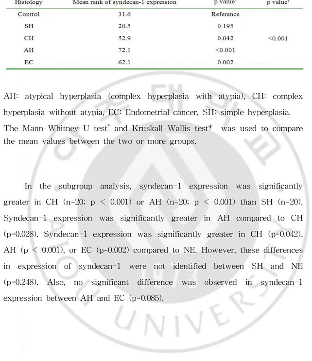
The immunostaining of syndecan-1 in the control, endometrial hyperplasias and endometrialcancertissues areshown in Fig.2.The mean scoreofsyndecan-1immunoreactivitywas1,0.6,1.9,2.6,and2.2inNE,SH, CH,AH,and EC,respectively (Fig.3).The mean rank score ofstaining according toclassification ofendometrialhyperplasiaisshown in Table2.In otherwords,themean ranksofexpression scoresbasedon thefrequency of syndecan-1 staining were 31.6,20.5,52.9,72.1,and 62.1 with NE,SH,CH, AH, and EC, respectively (Table 2). The differences were statistically significant(p< 0.001).
Fig. 2. Immunohistochemical staining for syndecan-1 (original magnification,×100).(a)Normalendometrium showing negativesyndecan-1 staining (frequency score:0).(b)Simplehyperplasia(frequency score:1).(c) Complex hyperplasia without atypia (frequency score: 2). (d) Complex hyperplasia with atypia (frequency score:3).(e)Almosttumor cells show positivesyndecan-1expression(frequencyscore:3).
Table2.Mean rankofsyndecan-1expression according tohistology.
AH: atypical hyperplasia (complex hyperplasia with atypia),CH: complex hyperplasiawithoutatypia,EC:Endometrialcancer,SH:simplehyperplasia. TheMann-Whitney U test*and Kruskall-Wallistest† wasusedtocompare themeanvaluesbetweenthetwoormoregroups.
In the subgroup analysis, syndecan-1 expression was significantly greaterin CH (n=20;p < 0.001)orAH (n=20;p < 0.001)than SH (n=20). Syndecan-1 expression was significantly greater in AH compared to CH (p=0.028).Syndecan-1 expression was significantly greaterin CH (p=0.042), AH (p< 0.001),orEC (p=0.002)comparedtoNE.However,thesedifferences in expression of syndecan-1 were not identified between SH and NE (p=0.248). Also, no significant difference was observed in syndecan-1 expressionbetweenAH andEC (p=0.085).
Ⅳ.DI
SCUSSI
ON
A number of immunohistochemical studies have demonstrated the aberrantexpression orimmunoreactivity ofsyndecan-1in many malignancies, aswellasacorrelationofsyndecan-1expressionwithneoplasticprogression, andinversesignificancehasbeen foundin differenttumors.Forexample,the expression of syndecan-1 in carcinomas of the head and neck regions (Anttonen etal,1999),esophagus(Mikamietal,2001),larynx (Pulkkinen et al,1997),liver (Matsumoto etal,1997),lung (Anttonen etal,2001),colon (Fujiyaetal,2001),anduterinecervix (Numaetal,2002)wasrelatedtolow clinicalstage,favorable outcome,and betterdifferentiation,whereas inverse results are noted in malignancies ofthe nasopharynx (Chen and Ou,2006), breast(Barbareschietal,2003),prostate(Zellwegeretal,2003),and thyroid (Itoetal,2003).
In endometrialcancer,there have been recent histologicalstudies on endometrialtissues suggesting thatthe expression ofsyndecan-1 is closely correlatedwiththegainofcarcinogenesisinthehumanendometrium (Choiet al,2007).
syndecan-1 in endometrial hyperplasia specimens by immunohistochemical stainwhichquantitavelyanalyzedsyndecan-1.
Also,syndecan-1expressionwasevaluatedforsignificanceaccording to the histologic types of endometrial hyperplasia, because the risk of endometrialhyperplasia progressing to carcinoma is related to the presence and severity of cytologic atypia (Hunter et al,1994;Amant et al,2005). Kurmanetal.foundthatprogressiontocarcinomaoccurredin1% ofpatients withSH,3% ofpatientswithCH,8% ofpatientswithatypicalSH,and29% ofpatients with AH (Kurman etal,1985).As the above results indicate, there was significantdifference in syndecan-1 expression according to the histologicclassificationofendometrialhyperplasia.Syndecan-1expressionwas significantly greater in AH than SH or CH.Our results therefore further suggestthatsyndecan-1 expression is positively correlated with the risk of progression of endometrial hyperplasia to endometrial cancer. Thus, syndecan-1 immunoreactivity in endometrial hyperplasia appears to be a usefulindicatorofhighriskfordevelopmentofendometrialcancer.
Smallsamplesizewasastudy limitation.Additionalpossiblelimitations of this study were sample selection and other confounders because of retrospective sample selection.Also,there is the possibility of decreased reproducibility in terms of reading and interpretation of the immunohistochemicalstaining for syndecan-1 because a single pathologist read and interpretated all slides. We made an effort to minimize these limitationsasmuchaspossible.
Future studies will determine whether the extent of syndecan-1 expression in endometrialcancercorrelates with prognosis.Also,there is a need for a prospective study for syndecan-1 as a reliable biomarker of aggressiveness in endometrialhyperplasia.Our findings lend support to a
suggestion thattherisk ofendometrialhyperplasia progressing to carcinoma is related to the presence and severity ofcytologic atypia.In addition,the current findings,which revealed that aberrant syndecan-1 expression is a property ofboth complex atypicalhyperplasia and invasivecarcinoma ofthe endometrium,raisethepossibility thatsomeorallfocidiagnosedascomplex atypicalhyperplasiainfactmay beendometrialadenocarcinomainsitu.Thus, syndecan-1 may be used as a diagnostic indicator of progression to endometrialcancer.
REFERENCES
1. Amant F,Moerman P,Neven P, Timmerman D,Van Limbergen E, VergoteI:Endometrialcancer.Lancet366:491-505,2005
2. Anttonen A, Heikkila P, Kajanti M, Jalkanen M, Joensuu H: High syndecan-1expression isassociatedwith favourableoutcomein squamous celllungcarcinomatreatedwithradicalsurgery.Lung Cancer32:297-305,
2001
3.Anttonen A,KajantiM,Heikkila P,Jalkanen M,Joensuu H:Syndecan-1 expression hasprognosticsignificancein head and neck carcinoma.BrJ
Cancer79:558-564,1999
4.BarbareschiM,Maisonneuve P,AldoviniD,CangiM G,PecciariniL, AngeloMauriF,VeroneseS,CaffoO,LucentiA,PalmaP D,GalligioniE, DoglioniC:High syndecan-1expression in breastcarcinoma isrelated to anaggressivephenotypeandtopoorerprognosis.Cancer98:474-483,2003
5.Bernfield M,KokenyesiR,Kato M,Hinkes M T,Spring J,Gallo R L, LoseE J:Biology ofthe syndecans:a family oftransmembrane heparan sulfateproteoglycans.AnnuRevCellBiol8:365-393,1992
6.Chen C L,Ou D L:Expression ofsyndecan-1(CD138)in nasopharyngeal carcinoma is correlated with advanced stage and poor prognosis.Hum
Pathol37:1279-1285,2006
7.ChoiD S,Kim JH,Ryu H S,Kim H C,Han JH,LeeJS,Min C K: Syndecan-1,a key regulatorofcellviability in endometrialcancer.IntJ Cancer121:741-750,2007
8.DrukerB J,Lydon N B:Lessonslearned from thedevelopmentofan abl tyrosinekinaseinhibitorforchronic myelogenous leukemia.J Clin Invest
105:3-7,2000
9.Fujiya M,WatariJ,Ashida T,Honda M,TanabeH,FujikiT,Saitoh Y, Kohgo Y:Reduced expression ofsyndecan-1 affects metastatic potential and clinicaloutcomein patientswith colorectalcancer.JpnJ CancerRes
92:1074-1081,2001
10.HunterJE,TritzD E,HowellM G,DepriestP D,GallionH H,Andrews S J,Buckley S B,KryscioR J,Van NagellJR,Jr.:Theprognosticand therapeutic implications of cytologic atypia in patients with endometrial hyperplasia.GynecolOncol55:66-71,1994
11.Ito Y,Yoshida H,Nakano K,Takamura Y,Miya A,KobayashiK, Yokozawa T, Matsuzuka F, Matsuura N, Kuma K, Miyauchi A: Syndecan-1 expression in thyroid carcinoma:stromalexpression followed by epithelialexpression is significantly correlated with dedifferentiation.
Histopathology43:157-164,2003
12.JemalA,SiegelR,Ward E,Murray T,Xu J,SmigalC,Thun M J: Cancerstatistics,2006.CA CancerJClin56:106-130,2006
13.KendallB S,RonnettB M,IsacsonC,ChoK R,HedrickL,Diener-West M, Kurman R J: Reproducibility of the diagnosis of endometrial hyperplasia,atypicalhyperplasia,and well-differentiated carcinoma.Am J
513-521,2003
16.Lee S E,Kim J W,Park N H,Song Y S,Kang S B,Lee H P: Contemporary trends ofendometrialcancerin Korean women.Korean J GynecolOncol.16:315-220,2005
17.Matsumoto A,Ono M,Fujimoto Y,Gallo R L,Bernfield M,Kohgo Y: Reducedexpression ofsyndecan-1in human hepatocellularcarcinomawith highmetastaticpotential.IntJCancer74:482-491,1997
18.MikamiS,OhashiK,UsuiY,Nemoto T,Katsube K,Yanagishita M, Nakajima M,Nakamura K,Koike M:Loss ofsyndecan-1 and increased expressionofheparanaseininvasiveesophagealcarcinomas.JpnJ Cancer Res92:1062-1073,2001
19.Mukunyadzi P,Liu K,Hanna E Y,Suen J Y,Fan C Y: Induced expression ofsyndecan-1 in thestroma ofhead and neck squamous cell carcinoma.ModPathol16:796-801,2003
20.NumaF,HirabayashiK,KawasakiK,SakaguchiY,SuginoN,SuehiroY, SuminamiY,Hirakawa H,Umayahara K,Nawata S,Ogata H,Kato H: Syndecan-1 expression in cancer ofthe uterine cervix:association with lymphnodemetastasis.IntJOncol20:39-43,2002
21. Pulkkinen J O, Penttinen M, Jalkanen M, Klemi P, Grenman R: Syndecan-1: a new prognostic marker in laryngeal cancer. Acta Otolaryngol117:312-315,1997
22.Soukka T,Pohjola J,InkiP,Happonen R P:Reduction ofsyndecan-1 expression is associated with dysplastic oralepithelium.J OralPathol Med29:308-313,2000
23.ZellwegerT,NinckC,MirlacherM,AnnefeldM,GlassaG,GasserT C, Mihatsch M J,Gelmann E P,BubendorfL:Tissue microarray analysis
reveals prognostic significance of syndecan-1 expression in prostate cancer.Prostate55:20-29,2003
국문 요약
-자궁내막증식증 및 자궁내막암과 syndecan-1의 발현과의
관련성
아주대학교 대학원의학과 김 현 진 (지도교수 :유 희 석) 목적:세포 표면의 프로테오글리칸인 syndecan-1의 비정형 발현은 여러 암종 에서 발견된다.본 연구는 정상,자궁내막증식증 및 자궁내막암에서 syndecan-1 의 면역조직화학 발현을 확인하여 자궁내막암성 변화의 표지자로써의 유용성을 확인하고자 한다. 방법:정상,자궁내막증식증 및 자궁내막암을 포함하는 101개의 포르말린에 고정된 파라핀조직을 이용하여 syndecan-1의 면역조직화학 발현 결과를 확인한 다. 비교군으로 정상 자궁내막 (n=10)을 사용하였고, 단순 자궁내막증식증 (n=20),이형성이 없는 복합 자궁내막증식증 (n=20),이형성이 동반된 복합 자궁 내막증식증 (n=20),자궁내막암 (n=31)이 포함되었다.결과:Syndecan-1발현의 순위 평균 (mean rank)은 정상 자궁내막,단순 자 궁내막증식증,이형성이 없는 복합 자궁내막증식증,이형성이 동반된 복합 자궁 내막증식증,자궁내막암이 각각 31.6,20.5,52.9,72.1,62.1였으며,이는 통계적인 차이를 보였다 (p < 0.001).Syndecan-1의 발현은 단순 자궁내막증식증과 비교 하여,이형성이 없는 복합 자궁내막증식증 (p < 0.001)과 이형성을 동반한 복합 자궁내막증식증 (p< 0.001)모두에서 유의하게 증가하였다.또한,이형성이 없는
복합 자궁내막증식증에 비해 이형성을 동반한 복합 자궁내막증식증에서 syndecan-1의 발현이 증가했다 (p=0.028).정상 자궁내막과 비교해서는 이형성이 없는 복합 자궁내막증식증 (p=0.042),이형성이 동반된 복합 자궁내막증식증 (p < 0.001),그리고 자궁내막암 (p=0.002)모두에서 syndecan-1의 발현이 유의하게 증가했다.그러나,정상 자궁내막과 단순 자궁내막증식증에서 syndecan-1의 발현 의 차이는 없었다 (p=0.248). 결론:자궁내막증식증에서 syndecan-1은 자궁내막암으로 진행을 예측하는 유 용한 인자이다. 핵심어 :Syndecan-1,자궁내막증식증,자궁내막암