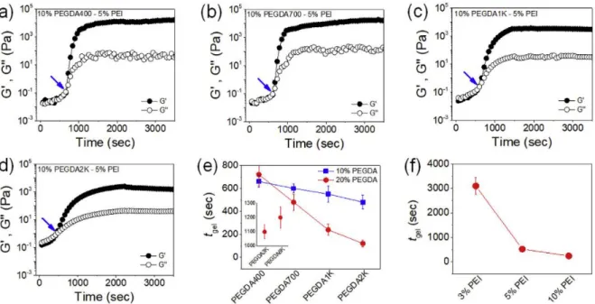
The arrows indicate the gel point. d) The ratio of the number densities of acrylate in PEGDA and amine in PEI (ФAc/Am) for varying MW of PEGDA at different PEI concentrations. 3-2 and (c) the kinetic rate constants (k1) were obtained. d) The latter drug release profiles were fitted with Eq.
Introduction
Research Background
A Schiff base is usually formed by the condensation of an aldehyde or ketone with a primary amine such as HA, chitosan, and dextran under physiological conditions. The dynamic balance between Schiff base bonds and aldehyde and amine reactants provides advantages for the formation of Schiff bases and ensures the self-healing capabilities of the hydrogel network through reversible reactions such as uncoupling and recoupling of imine bonds.
Research Purpose and Motivation
In general, their gelation time and physical properties can be varied depending on the ratio of amine and aldehyde groups. It can be easily used to construct an injectable hydrogel because aldehyde groups can react with other amine groups in tissues or organs.
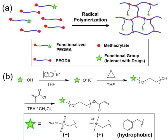
Modulation of functional pendant chains within poly(ethylene glycol) hydrogels
Introduction
Modulation of functional pendant chains within poly(ethylene glycol) hydrogels for enhanced protein control. Their release rates from PEG hydrogels with different numbers of charged pendant chains were measured.
Materials and Methods
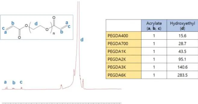
- Synthesis of Poly(ethylene glycol) dimethacrylate (PEGDA)
- Synthesis of heterobifunctional poly(ethylene glycol) monomethacrylate (PEGMA)
- Fabrication of hydrogels
- Evaluation of mechanical properties of hydrogels
- Evaluation of protein release from hydrogels
At a given PEGMA concentration, the fraction of heterobifunctional PEGMA was varied to control the physical properties of the pendant PEG chains. This relationship generally fits well for the first 60% of the release due to the fixed time dependence (t1/2).
Results and Discussion
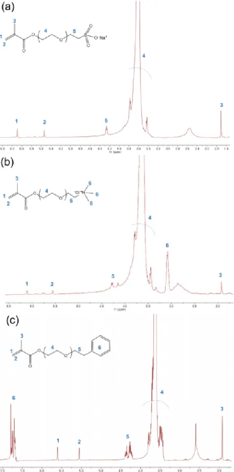
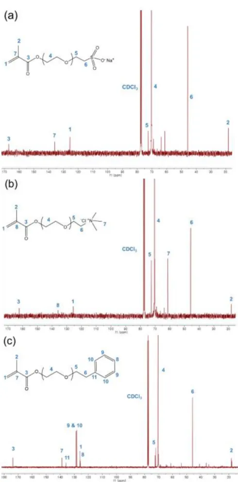
- Synthesis of heterobifunctional PEGMA
- Mechanical properties of PEG hydrogel with functional pendant chains
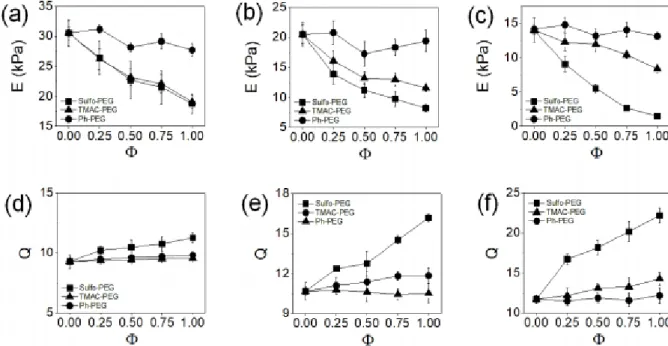
- Protein release from PEG hydrogel with functional pendant chains
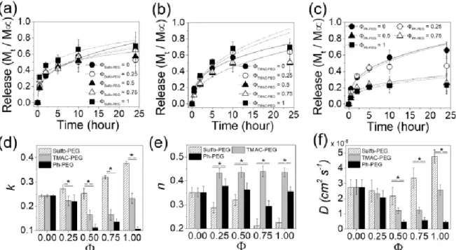
The release profiles of albumin from (b) Sulfo-PEG hydrogels, (c) TMAC-PEG hydrogels, and (d) Ph-PEG hydrogels. e). The release profiles of insulin from (a) Sulfo-PEG hydrogels, (b) TMAC-PEG hydrogels, and (c) Ph-PEG hydrogels.
Conclusion
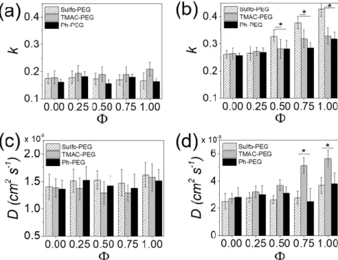
Similarly, for hydrogels at other PEGMA concentrations, the k and D values of trypsin in Sulfo-PEG hydrogels were significantly reduced compared to albumin and insulin, while those in TMAC-PEG hydrogels did not change significantly with Φ (Figure 2-16). ). Regardless of the crosslinking density of the hydrogels, k values in Ph-PEG hydrogels were not significantly affected by Φ. It is possible that the range of physical properties accommodated by the TMAC-PEG hydrogels and Ph-PEG hydrogels used in this study was not sufficient to significantly affect the rate of trypsin release. a,b) Kinetic rate constants (k) and (c,d) diffusion coefficients (D) of trypsin release from Sulfo-PEG, TMAC-PEG and Ph-PEG hydrogels obtained by fitting the release profiles with Eq. The release of albumin and insulin, whose pI values are lower than physiological pH, negatively charged, is more facilitated in Sulfo-PEG hydrogels due to electrostatic repulsion compared to TMAC-PEG hydrogels, which can provide an attractive force.
On the other hand, the release of trypsin, which has a positive charge due to a higher pI value than physiological pH, was significantly reduced in Sulfo-PEG hydrogels due to the repulsive force. These results suggest that PEG hydrogels with pendant functional groups could be successfully used as carriers for refined control of protein release for biomedical applications.
Integrative control of mechanical and degradation properties of in situ
- Introduction
- Materials and Methods
- Synthesis of poly(ethylene glycol) diacrylate (PEGDA)
- PEGDA-PEI hydrogel fabrication
- Mechanical properties and degradation of PEGDA-PEI hydrogels
- Drug release profiles from PEGDA-PEI hydrogels
- Results and Discussion
- Mechanical properties of PEGDA-PEI hydrogels
- Gelation kinetics
- Rigidity
- Degradation of PEGDA-PEI hydrogels
- Degradation of PEGDA-PEI hydrogels
- Conclusion
The reaction parameters include the concentrations of PEGDA and PEI and the MW of PEGDA. Therefore, the stiffness of the PEGDA-PEI hydrogels was also evaluated by measuring the elastic moduli from uniaxial compression experiments. Change in normalized elastic moduli (E/E0) of 20% PEGDA-5% PEI hydrogels with different MW PEGDA shown over time.
Similarly, at 20% PEGDA, the increase in kd with MW PEGDA became more pronounced with PEI concentration (Figure 3-11e). The mechanical properties of PEGDA-PEI hydrogels can be controlled over a wide range by varying the concentrations of PEGDA and PEI and the MW of PEGDA.
In situ facile-forming chitosan hydrogels with tunable physicomechanical and
Introduction
93-96] Due to this method of hydrogel fabrication, in situ forming hydrogels have been considered particularly attractive as an injectable and tissue-implantable form for biomedical applications, allowing for spontaneous hydrogel formation upon delivery. One of the main challenges of in situ hydrogel formation is to control the mechanical properties over a wide range while adequately maintaining the rate of the cross-linking reaction, at least in a manageable range for practical handling. In this study, the physical properties of the gel-forming polymer were modified to control the mechanical properties of the in situ-forming hydrogels while effectively maintaining the gelation kinetics.
More significantly, an extensive number of amine groups on chitosan, one on each saccharide monomer, provides ample opportunity for chemical modifications, including various nucleophilic reactions for the formation of hydrogels in situ. To further illustrate their potential application as a tissue sealant, the adhesive properties of the in situ forming Cs-PEG hydrogels on biological tissues were investigated both ex vivo and in vivo. a) PEG grafting of chitosan (PEG-g-Cs).
Materials and Methods
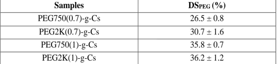
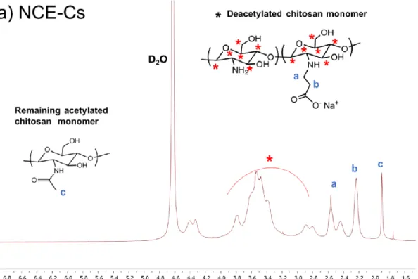
- Synthesis of PEG-grafted chitosan (‘PEG-g-Cs’) and PEG-dialdehyde
- Fabrication of Cs-PEG hydrogels
- Mechanical and rheological properties of Cs-PEG hydrogels
- Diffusional properties of Cs-PEG hydrogels
- Tissue adhesive properties of Cs-PEG hydrogels
- Anti-microbial properties of PEG-g-Cs
- Swept-Source Optical Coherence Tomography (SS-OCT)
The mechanical properties of Cs-PEG hydrogel were measured by uniaxial compression tests (Model 3343, Instron). The diffusion properties of Cs-PEG hydrogels were evaluated by measuring their swelling ratios and drug release profiles. BSA (5 mg mL-1) was encapsulated into the Cs-PEG hydrogel by dissolving in the PEG-g-Cs stock solution prior to hydrogel fabrication.
The adhesive properties of the Cs-PEG hydrogel were measured based on a standard method for lap shear strength properties of tissue adhesives under tension loading (ASTM F2255-05). The sample was first prepared by applying the Cs-PEG mixture (150 u L) to a hole (5 mm diameter) in a porcine skin specimen.
Results and Discussion
- Synthesis and physical properties of PEG-g-Cs
- Mechanical properties of Cs-PEG hydrogels
- Diffusional properties of Cs-PEG hydrogels
- Anti-microbial properties of Cs-PEG hydrogels
- Gelation kinetics of Cs-PEG hydrogels
- Tissue adhesive properties of Cs-PEG hydrogels
- In vivo evaluation of Cs-PEG hydrogel as tissue sealant
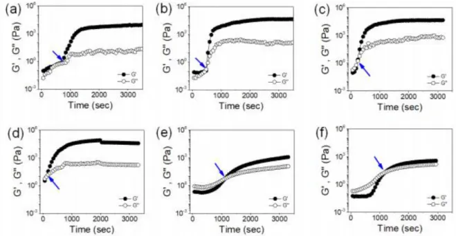
Elastic moduli (E) of Cs-PEG hydrogels. a) Elastic moduli (E) of Cs-PEG hydrogels made by cross-linking NCE-C with different MW of PEGDAld. This result indicated that the Cs-PEG hydrogels are highly elastic and their diffusive properties can be controlled by both the MW of PEGDAld and the PEG grafting of PEG-g-Cs. The effects of MW and PEGDAld concentration and PEG graft length and density on the gelation kinetics to form Cs-PEG hydrogels were evaluated by measuring the rheological properties of the precursor gel solution of PEG-g-Cs and PEGDAld during the cross-linking reaction.
This highlighted that the increased mechanical stiffness of Cs-PEG hydrogels with a larger extent of cross-linking generally increased their adhesion to biological tissues. Taken together, the adhesive strength of Cs-PEG hydrogels was strongly correlated with their mechanical properties, and there was a significant decrease at the highest PEG grafting density and length of PEG-g-Cs.
Conclusion
In situ facile-forming chitosan hydrogels with tunable physicomechanical and
Introduction
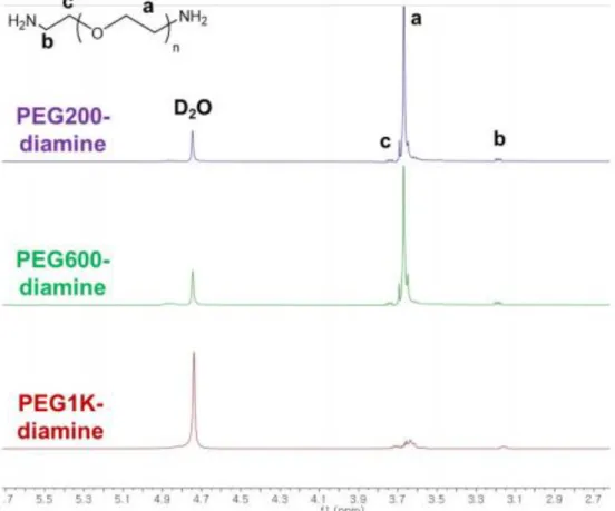
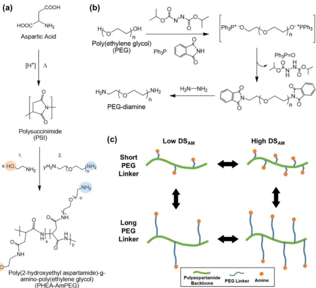
Therefore, here the grafting parameters, grafting density and length of reactive functional groups of polyaspartamide crosslinkers were modulated to control the mechanical properties of the resulting hydrogels. We synthesized PEG-diamine with different molecular weights and used it to represent amine groups on the polyaspartamide framework. The length of the PEG linker and the degree of amine substitution were varied to control the mechanical properties of the hydrogels.
Moreover, the unreacted amine groups on PHEA-PEGAm facilitated the degradation and final dissolution of hydrogels via hydrolysis under physiological conditions, and the rate of degradation was also controlled by the grafting parameters. To further control the degradation and mechanics, the alginate component of hydrogels was additionally cross-linked with divalent calcium ions.
Materials and Methods
- Synthesis of Polysuccinimide (PSI)
- Synthesis of PEG-Diamine
- Synthesis of Poly(2-hydroxyethyl aspartamide)-g-Amino-Poly(ethylene glycol) (PHEA–
- Trinitrobenzene sulfonic acid (TNBS) assay
- Synthesis of oxidized alginate
- Fabrication of PHEA–PEGAm Hydrogels
- Mechanical and Degradation Properties of Hydrogels
- Drug Release Kinetics
- Rheological Properties of Hydrogels
- In Vitro Cytotoxicity
- Ex Vivo Evaluation of Degradation Behavior of Hydrogel
The resulting poly(2-hydroxyethyl-aspartamide)-g-amino-poly(ethylene glycol) (PHEA-PEGAm) was then reacted with aldehyde-presenting alginate via Schiff base to develop hydrogels. PHEA–PEGAm with different degrees of amine substitution (DSAm), defined as the fraction of succinimidyl groups conjugated with amine groups, was obtained by simply changing the PEG-diamine reactant. As a control, PHEA-presenting short aminoethyl grafts, PHEA–PEGam, were also synthesized using ethylenediamine instead of PEGdiamine.
Alg-PHEA hydrogel was fabricated by reacting PHEA-PEGAm with alginate-presenting aldehyde groups (Alg-ALD) via Schiff base formation. Before hydrogel fabrication, BSA (5 mg mL-1) was dissolved in the PHEA-PEGAm precursor solution for encapsulation.
Results and Discussion
- Synthesis and Characterization of Amine-Presenting Polyaspartamide with
- Fabrication and Mechanical Properties of PHEA-Linked Hydrogels
- Degradation of PHEA-Linked Hydrogels
- Drug Release Kinetics of PHEA-Linked Hydrogels
- Ex Vivo Alg-PHEA Hydrogel Injection and Degradation
- Dual Ionic Cross-Linked Alg-PHEA Hydrogels
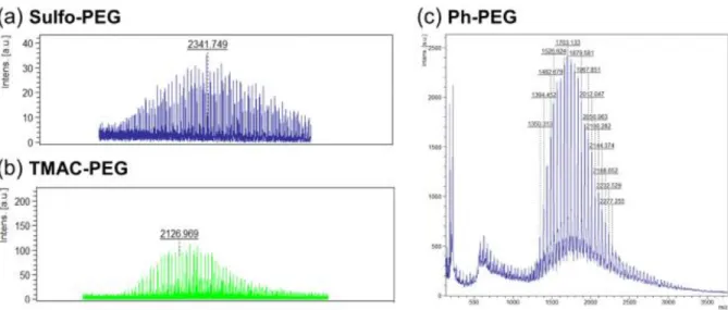
The chemical structures of PHEA-PEGAM with varying DSAm and length were analyzed with 1H NMR and FT-IR spectroscopy (Figure 5-4 and Figure 5-5). These results all confirmed the convenient control of length and DSAm of PHEA–PEGAm by reaction conditions. Further increase in amine graft length of PHEA-PEGam resulted in continued decrease in hydrogel rigidity, at all concentrations and DSAm (Figure 5-7c and d).
In contrast, for PHEA-PEGAm1K, the degradation rate decreased with increasing DSAm and concentration. On the other hand, by varying the DSAm of Alg-PHEA hydrogels at the same concentration (15%) of PHEA-PEGAm600, kR indeed increased with kD and did not correlate with the mechanics.
Conclusion
Drug release profiles using BSA as the model protein drug were also performed for Ca-Alg-PHEA hydrogels and the release rate (kR) was obtained from Eq. It is interesting to note that kR increased with Ca2+ concentration, from 1 to 5 M, despite having greater mechanical properties and structural stability. It is speculated that the excess of local Ca2+ within the hydrogels at higher concentrations that is mainly released at an earlier time frame (before 2 h) is likely to accelerate the release of BSA, since BSA is negatively charged under neutral buffered conditions that have an isoelectric point of 5.3 and, thus. , can interact electrostatically with Ca2+.
95] Taken together, in situ formation and mechanically tunable Alg-PHEA hydrogels that undergo facile dissolution can be further modulated by additional ionic crosslinking to slow and control the degradation and drug release rates.
Summary and Future Perspective
Fefelova, N.A., et al., Mucoadhesive interactions of amphiphilic cationic copolymers based on [2-(methacryloyloxy)ethyl]trimethylammonium chloride. Alexander, A., et al., Thermosensitive injectable hydrogels based on poly(ethylene glycol)–poly(lactic-co-glycolic acid) for biomedical applications. Li, Y., et al., In situ hydrogel constructed from starch-based nanoparticles via a Schiff base reaction.
Choi, S.M., et al., Synthesis and characterization of in situ gelable poly(glycerol sebacate)-co-poly(ethylene glycol) polymers. Zhang, H., et al., Mussel-inspired hyperbranched poly(aminoester) polymer as strong wet tissue adhesive.