The values of cell viability (%), obtained by the MTT assay, were calculated in comparison with cells treated only with DMSO (0–1%, v/v). The values of cell viability (%), measured by the MTT assay after 24 h incubation, were calculated in comparison with cells treated only with DMSO (0-1%, v/v). The values of cell viability (%), obtained by the MTT assay, were calculated in comparison with cells treated only with DMSO (0–1%, v/v).
Cell viability values (%), obtained by the MTT assay, were calculated relative to cells.
Introduction
Amyloid-b, Metals, and Oxidative Stress in Alzheimer’s Disease
Amyloid-b (Ab)
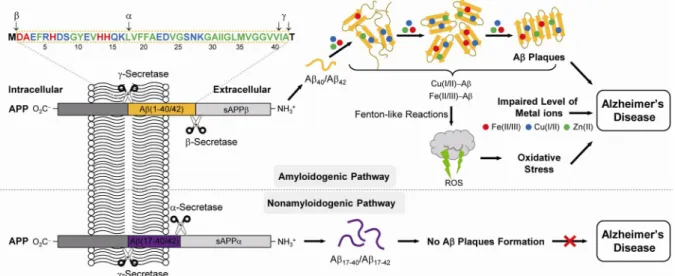
In the healthy brain, Ab production and clearance is well balanced by the activity of secretases and proteases; however, this balance is not maintained in the AD-affected brain, thus. Senile plaques in the brain affected by AD are observed to contain high levels of transition metals, such as Cu, Zn and Fe (Cu, ca.
Metals
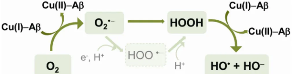
Redox active metal ions [i.e. Cu(I/II), Fe(II/III)] are involved in Fenton-like reactions that produce ROS such as superoxide anion (O2•-), hydrogen peroxide.
Oxidative Stress Induced by Metal-Ab
Human Islet Amyloid Polypeptide and Metals in Type II Diabetes Mellitus
Human Islet Amyloid Polypeptide
Several mechanisms of hIAPP fibril formation in T2DM have been proposed.38 Among these, the most widely accepted mechanism is that hIAPP accumulation and aggregation may result from its increased production and secretion with increased insulin demand as a result of insulin resistance. 38 However, the role and mechanism of hIAPP in the pathogenesis of T2DM is still not clear.34.
Metal Ions and hIAPP
Conclusions
Biological Studies of Small Molecules Rationally Designed to Target and Modulate
Results and Discussion
- Design Principle of the L2-b and ML Derivatives
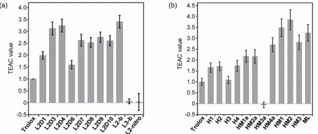
- Antioxidant Capacity of the L2-b and ML Derivatives
- Cytotoxicity of the L2-b and ML Derivatives
- Influence of the L2-b and ML Derivatives on Cytotoxicity Induced by Metal-free
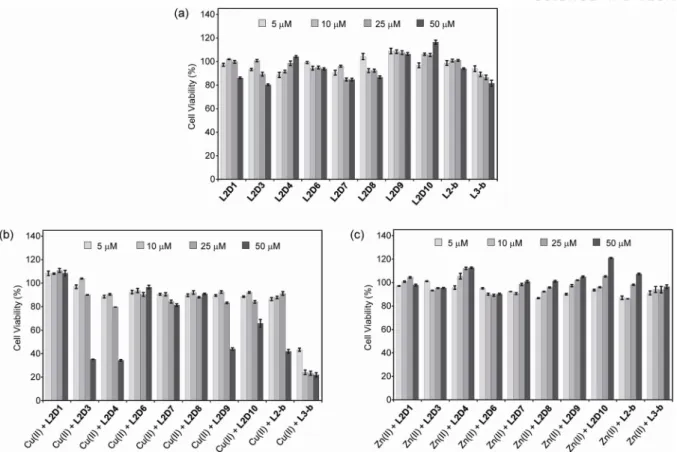
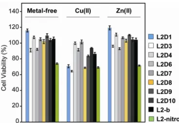
In the case of L2D3 and L2D8, cell viability was reduced when their concentrations were increased; However, L2D9 was indicated to be less toxic than L2-b, L2D3 and L2D8 (Figure 2.3a). The overall cell viability with the compounds was reduced in the presence of Cu(II) (Figure 2.3b). L2D1 showed higher cell viability in the absence and presence of Zn(II) than with Cu(II) (Figure 2.4).
65-70% cell viability was observed (Figure 2.4), indicating that the nitro group may cause more cytotoxicity than the dimethylamino functionality in L2-b. Cell viability upon treatment with compounds under conditions (a) without and with metal ions [(b) Cu(II) or (c) Zn(II)]. Cytotoxicity studies with the L2-b derivatives in human neuroblastoma SK-N-BE(2)-M17 (M17) cells. a) Cell viability upon incubation with compounds in the absence and presence of CuCl2 or ZnCl2.
In the case of the ML derivatives, cell viability of ML and its derivatives was also determined. Taken together, in both N2a and M17 cell lines, similar cell viability with compounds was indicated despite structural variation. The values of cell viability (%) obtained by the MTT assay after 24 h of incubation at 37 °C were calculated.
Conclusion
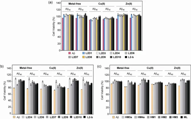
In the case of ML derivatives, the effect of small molecules to regulate metal-free Ab/metal-Ab induced toxicity was investigated in N2a cells. All compounds except HM3a were shown to attenuate cytotoxicity upon Ab/metal-Ab induction (Figure 2.7c). Overall, Ab-/metal-Ab-induced cytotoxicity was attenuated by L2-b, ML, and their derivatives to different degrees, which may be influenced by the structural variables of the ligands.
Effect of compounds on survival of cells incubated with metal-free Ab and metal Ab. c) Effect of ML derivatives on the survival of cells incubated with metal-free Ab and metal Ab. This chapter describes our studies on elucidating the relationship between chemical structures (L2-b and ML derivatives), antioxidant activity and cytotoxicity. L2-b/ML derivatives have been shown to exhibit antioxidant activity and cytotoxicity, although the structural modifications of the parent molecules are to varying degrees.
Our results and observations regarding a relationship between structure, antioxidant activity, and cytotoxicity may provide guidance in the design of small molecules applicable for biological applications.
Experimental Sections
- Ab Preparation
- Trolox Equivalent Antioxidant Capacity (TEAC) Assay
- Cell Viability Studies
The TEAC value of ligands was calculated as a ratio of the slope of the compound's standard curve to that of Trolox. The N2a and M17 cells were treated with or without Ab and CuCl2 or ZnCl2, followed by the addition of compounds (1% v/v final concentration of DMSO) and incubated for 24 h. Formazan produced by the cells was solubilized overnight at room temperature in the dark using an acidic solution of N,N-dimethylformamide (50%, v/v aq) and sodium dodecyl sulfate (SDS, 20%, w/v).
Investigations on biochemical activity of the glycosylated polyphenols and their esterified derivatives after Alzheimer's disease.
Introduction
Results and Discussion
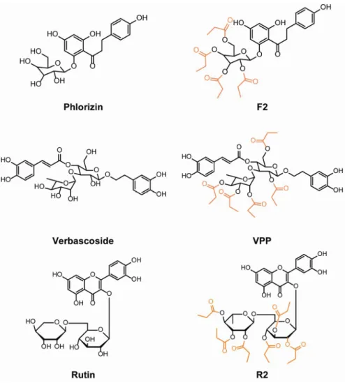
- Design Consideration of Glycosylated Polyphenols and Their Esterified
- Metal Binding Properties of Polyphenols
- Antioxidant Properties
- Regulating Toxicity Induced by Metal-Free and Metal-Associated Ab in Living
In order to understand the interaction of the compounds with metal ions, especially Cu(II) and Zn(II), their metal binding properties are compounds with metal ions, especially Cu(II) and Zn(II), their metal binding properties are studied by visible spectroscopy (UV-vis) or 1D 1H NMR. In the buffered aqueous solution (20 mM HEPES, pH 7.4), it was observed that the absorption spectra of the six polyphenols changed to different extents upon the titration of CuCl2. Addition of CuCl2 to solutions of either Phlorizin or F2 produced no significant change in spectral features, possibly due to the weak affinity of the phenol moiety in both compounds for Cu(II) in solution (Figure 3.2).
F2 showed a decrease in overall spectral intensity with CuCl2 titration; this may be the result of decomposition or oxidation catalyzed by the presence of Cu(II) in the solution.28 This tendency of Phlorizin and F2 can be understood as recognition of the lack of catechol function in their structure. This suggests that esterification of the Rutin framework may alter the interaction between the ligand and Cu(II). Both VPP and R2 were esterified from their parent compounds, but the degree of change in Cu-induced spectral variation was different, which may indicate the importance of the overall structure in the metal binding property of the compounds.
Overall, the results suggest that Verbascoside, VPP, Rutin and R2 may interact with Cu(II) and/or Zn(II), while Phlorizin and F2 did not, as evidenced by the metal-induced UV vis spectral shift of the ligands. indicating the necessity of the catechol group to interact with metal ions. This again indicates that esterification of the sugar moiety changes the function of the Verbascoside framework. This reduction in antioxidant capacity is surprising given the conservation of the catechol structures between Verbascoside and VPP, which are thought to be potentially responsible for the quenching of ABTS++ through the formation of semi-quinone and quinone26,27.
Conclusion
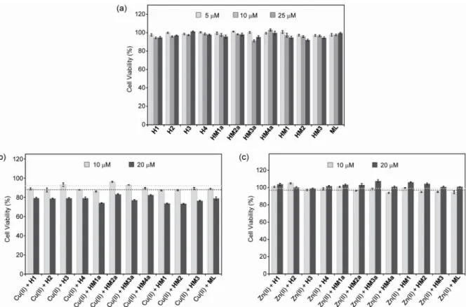
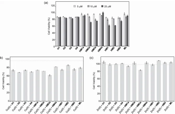
Effect of verbascoside, VPP, and rutin on Ab- or metalAb-induced toxicity in mouse neuroblastoma (N2a) neuro-2a cells. a) Cytotoxicity of compounds with or without metal ions. Cells treated with compounds [verbascoside, VPP and rutin (5-25 mM)] with or without different concentrations of metal chloride salts (CuCl2 or ZnCl2; 5-25 mM) were incubated for 24 hours at 37 °C. b) Effect of verbascoside, VPP and rutin on cytotoxicity induced by metal-free and metal Ab species in N2a cells.
Experimental Sections
- Metal Binding Studies
- Trolox Equivalent Antioxidant Capacity (TEAC) Assay
- Cell Viability Studies
Cells were treated with or without Ab and CuCl2 or ZnCl2, followed by addition of compounds (1% v/v final concentration of DMSO for Verbascoside, VPP and Rutin) and incubated for 24 h on cells. Cell-produced formazan was digested using an acidic solution of N,N-dimethylformamide (DMF, 50%, v/v aq) and sodium dodecyl sulfate (SDS, 20%, w/v) overnight at room temperature. the room in the dark.
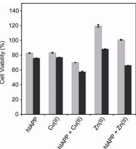
Investigations of human islet amyloid polypeptide-induced cytotoxicity in the absence and presence of metal ions. Furthermore, which hIAPP species (i.e., oligomers, fibrils) cause cytotoxicity is still unclear.8 Here, to better understand hIAPP-induced toxicity, results from cytotoxicity studies are described: investigations of (i) the impact of metals on hIAPP toxicity; (ii) toxicity of hIAPP mediators; (iii) hIAPP-induced apoptosis in the absence and presence of metal ions. Overall, hIAPP was shown to have different toxicities in the absence and presence of metal ions, particularly showing altered toxicity of Cu(II)-hIAPP depending on the peptide to metal ratio.
The lowest cell viability was observed in hIAPP-treated cells pre-incubated with Cu(II) for 3 hours (approximately 60%) (Figure 4.3b). In the case of Cu(II)-hIAPP, cell viability was proportionally reduced as a function of CuCl2 concentration (Figure 4.4a). After treatment with the inhibitor, calpeptin or trehalose, hIAPP-associated cells showed an increase in survival of about 10-20% in the absence and presence of Cu(II), consistent with their inhibitory property to different extents depending on their concentrations.
Furthermore, hIAPP/metal-hIAPP-mediated cytotoxicity was affected in the presence of an apoptosis inhibitor (calpeptin or trehalose) or enhancer (bafilomycin). The concentration of hIAPP peptides in the solution was determined by measuring the absorbance of the solution at 280 nm (e = 1740 M-1cm-1) and then diluted to the desired concentration in 20 mM HEPES, pH 7.5.15.16. peptide samples, hIAPP (200 mM) was incubated in the presence and absence of metal ions (CuCl2 and ZnCl2, 1.4 mM) at 37 °C with stirring at 300 rpm. The cells were treated with or without hIAPP and CuCl2 or ZnCl2 in the absence and presence of compounds (calpeptin, trehalose, and bafilomycin; 1% v/v final concentration of DMSO) and incubated in the cells for 24 h.
Investigations of Cytotoxicity Induced by the Human Islet Amyloid Polypeptide in
Results and Discussion
- Cytotoxicity of hIAPP in the Absence and Presence of Metal Ions
- Peptide Conformation-Dependent Cytotoxicity
- Investigation of Apoptosis Induced by Metal-Free hIAPP or Metal-hIAPP….…32
After addition of CuCl2 (70 or 140 mM) to hIAPP-treated cells, cell viability was reduced by approximately 15% compared to hIAPP alone (Figure 4.1). Treatment of cells with hIAPP (20 mM) and ZnCl2 (140 mM) showed a 10% reduced viability compared to hIAPP alone (Figure 4.1). Cell viability values (%) measured by the MTT assay were calculated relative to cells treated with H2O alone.
On the other hand, cells with hIAPP pre-incubated with Cu(II) exhibited changes in their viability after 24 hours of incubation, depending on the pre-incubation time points (Figure 4.3b). The values of cell viability (%), measured by the MTT assay after 24 h incubation, were calculated in comparison with cells treated only with HEPES. In order to further investigate toxicity induced by metal-hIAPP, cytotoxicity studies were performed with metal-hIAPP aggregates prepared after 3 hours of incubation [which indicated the lowest cell viability (Figure 4.3b)], at varying metal ion concentrations.
Cell viability values (%) obtained by the MTT assay were calculated relative to cells treated with HEPES alone. Bafilomycin reduced the viability of cells with hIAPP and Cu(II)-hIAPP regardless of its concentrations (about 40–50%) (Figures 4.5 and 4.6). Cell viability of hIAPP-treated cells after treatment with apoptosis inhibitors (calpeptin or trehalose) or enhancer (bafilomycin) with different compound concentrations.
Experimental Sections
- Sample Preparation
- Cell Viability Studies
Cell viability was calculated in percentage value compared to that of the untreated cells.