저작자표시-비영리-변경금지 2.0 대한민국 이용자는 아래의 조건을 따르는 경우에 한하여 자유롭게
l 이 저작물을 복제, 배포, 전송, 전시, 공연 및 방송할 수 있습니다. 다음과 같은 조건을 따라야 합니다:
l 귀하는, 이 저작물의 재이용이나 배포의 경우, 이 저작물에 적용된 이용허락조건 을 명확하게 나타내어야 합니다.
l 저작권자로부터 별도의 허가를 받으면 이러한 조건들은 적용되지 않습니다.
저작권법에 따른 이용자의 권리는 위의 내용에 의하여 영향을 받지 않습니다. 이것은 이용허락규약(Legal Code)을 이해하기 쉽게 요약한 것입니다.
Disclaimer
저작자표시. 귀하는 원저작자를 표시하여야 합니다.
비영리. 귀하는 이 저작물을 영리 목적으로 이용할 수 없습니다.
변경금지. 귀하는 이 저작물을 개작, 변형 또는 가공할 수 없습니다.
2 0 17 년 2 월
박 사 학 위 논 문 Ac
curacy and reproducibility of implant stability quotient accordingto various factors: in vitrostudy
김
년 월 2017 2 박사학위논문
Accuracy and reproducibility of implant stability quotient according to various factors:
in vitro study
조선대학교 대학원
치 의 학 과
김 홍 석
[UCI]I804:24011-200000265974
Accuracy and reproducibility of implant stability quotient according to various factors:
in vitro study
다양한 요인에 따른 임플란트 안정성 지수의 정확성과 재현성에 관한 in vitro 연구
년 월 일
2017 2 24
조선대학교 대학원
치 의 학 과
김 홍 석
Accuracy and reproducibility of implant stability quotient according to various factors:
in vitro study
지도교수 오 지 수
이 논문을 치의학 박사학위신청 논문으로 제출함
년 월
2016 10
조선대학교 대학원
치 의 학 과
김 홍 석
김홍석의 박사학위 논문을 인준함
위원장 원광대학교 교 수 권 경 환 ( 인 ) 위 원 조선대학교 교 수 김 수 관 ( 인 ) 위 원 조선대학교 교 수 임 성 철 ( 인 ) 위 원 조선대학교 교 수 방 일 수 ( 인 ) 위 원 조선대학교 교 수 오 지 수 ( 인 )
년 월
2016 12
조선대학교 대학원
™
™ ···
- ii -
™
- iv -
™
™
™
- vi -
Dental implant surgery has been acknowledged as a successful treatment method for edentulous or partially edentulous patients.[1] Osseointegration between an implant and surrounding bone is necessary for long-term success of implant, and securing primary stability is important to ensure osseointegration.[2] Primary stability is secured by the mechanical engagement between an implant fixture and the surrounding bone after implant placement. Therefore, it is of great clinical importance to measure implant stability, which can be measured via the percussion test, radiographic interpretation, insertion/removal torque measurement, Periotest® (Medizintechnik Gulden, Bensheim, Germany), and resonance frequency analysis (RFA). Among these, radiographic interpretation, Periotest®, and RFA are the most commonly used in clinic.
While radiography is non-invasive and can be performed at any time, it has an unsatisfactory resolution, poses standardization difficulties, and is insensitive with regard to detection.[3] Moreover, it has been reported that radiographic interpretation and bone-implant contact have low correlation.[4]
Although Periotest® was originally developed to measure the mobility of natural teeth,[5] it is now more commonly used for measuring implant stability. A tapping rod attached to the handle of a Periotest® is used to hit the implant, and the contact time between the rod and implant is converted and expressed as a Periotest® value. However, this method lacks reproducibility and sensitivity.[6]
- 2 -
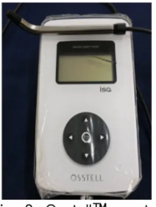
RFA is another method of measuring implant stability. A piezoceramic transducer is attached to an implant fixture, and a beam connected to another piezoceramic element generates vibration. The resonance frequency is affected by the contact stiffness between the implant and bone, and implant stability can be quantified. RFA has been acknowledged as a stable method of measurement with less than 1% error that can be used to evaluate osseointegration.[6] It has been commercialized as a product named Osstell™ (Osstell AB, Gothenburg, Sweden) that converts measurements to implant stability quotients (ISQ) reflecting implant stability.
The Osstell™ mentor is a recently developed device that wirelessly measures the ISQ of an implant attached to a Smartpeg™ (Osstell AB, Gothenburg, Sweden).
This study aimed to evaluate the accuracy of ISQ measurements using the Osstell™ mentor and Smartpeg™ in an in-vitro study, and identify factors that may affect measurements.
1. Material
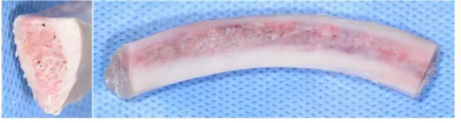
A pig rib bone that had been stored in a freezer was cut into segments 7 cm in length. A total of 20 bone specimens were prepared. For five of these specimens, cortical bone was eliminated to expose cancellous bone (Fig. 1). Each specimen was stored in a freezer. The specimens were thawed in a water bath before the experiment.
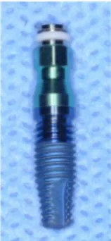
The implants used in this study were the staright type fixtures of USII system (Osstem Implant Co., Seoul, Korea) with 11.5 mm in length, 4 mm in diameter and surface treated with resorbable blast media (Fig. 2). The Osstell™ mentor was used as ISQ measurement device (Fig. 3). For the transducer attached to the top of an fixture, a type 1 (article No. 100353) Smartpeg™ was used (Fig. 4).
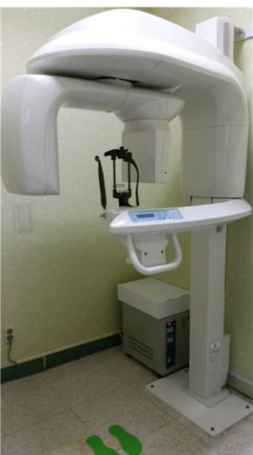
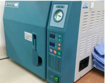
After drilling the bone specimen, a cone beam computerized tomography (CT) scanner (CS9300, Carestream Health, Inc., NY, USA) (Fig. 5) scanned the specimens at 90 kV and 4 mA using a 100 × 50 field of view. A high-pressure autoclaving sterilizer (3041VD, Hanshin Medical Co., Incheon, Korea) (Fig. 6) was used for Smartpeg™ sterilization.
2. Methods
(1) Correlation between bone density and ISQ
After drilling 10 holes into bone specimens with the cortical bone intact and exposing the cancellous bone respectively, a CT scan was performed
- 4 -
on the specimens for quantification of bone density. Hounsfield units (HU) of bone surrounding the drilled holes were then measured using software (OnDemand3D , Cybermed Inc., Tustin, USA) (Fig. 7). Following this, 20™ implants were placed to the bone specimens. And then, 20 Smartpegs™
were attached to the implants to measure the ISQ.
(2) Reliability of ISQ measurements according to Smartpeg™
attachment by different clinicians
Ten clinicians manually attached a Smartpeg™ to each of the 10 implants using a mount, and measured their ISQs. The Osstell™ mentor was placed at an identical location.
(3) Effect of repeated high-pressure autoclaving sterilization on Smartpeg™
Five Smartpegs™ were sterilized for 15 minutes at 132°C and 2 kg f/cm2 using a high-pressure autoclaving sterilizer. They were then attached to five implants to measure their ISQs. Sterilization was repeated 10 times, after which the Smartpegs™ were reattached to the implants, and their ISQs were remeasured.
(4) Changes in ISQ according to bone defect measurement direction around the implant
Bone surrounding the implant was divided into the front, back, mesial side, and distal side (Fig. 8). Bone in the front was then eliminated to
reproduce a peri-implant bone defect (Fig. 9). The thread of the implant fixture was exposed by 2 mm. A Smartpeg™ was then attached to the implant to measure ISQs in the four directions.
3. Statistical analysis
SPSS for Windows (v. 20.0, SPPS Inc., IBM) was used for statistical analysis. The correlation coefficient was determined using the Spearman rank test to assess the association between bone density and ISQ. Internal consistency reliability analysis using Cronbach's alpha ( ) coefficient wasα used to evaluate operator reliability. One-way analysis of variance was used to test the effect of the repeated sterilization on the Smartpeg , i.e.,™ to check if the ISQ values significantly changed after high-pressure autoclaving sterilization. Statistical significancy was accepted when P <
0.05.
- 6 -
1. Correlation between bone density and ISQ
HU and ISQ values were statistically significantly positively correlated (r=0.74, P<0.05) (Fig. 10).(Table 1)
HU 532 545 645 698 739 760 775 789 808 838 869 916 919 943 956 965 994 1000 1080 1132 ISQ 58 73 77 43 64 70 84 61 78 67 71 85 84 80 78 83 81 81 90 87
Table 1. Relationship between HU and ISQ
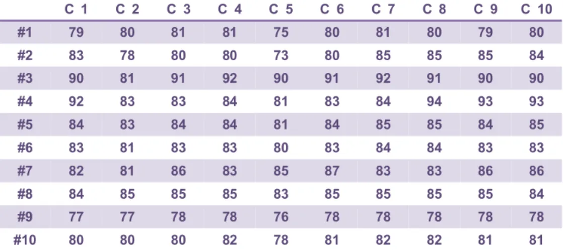
2. Reliability of ISQ measurements according to Smartpeg™
attachment by different clinicians
The ISQ measurements of the 10 implants after 10 different clinicians attached a Smartpeg™ to each were statistically consistent (Cronbach’s α
= 0.962).(Table 2)
C 1 C 2 C 3 C 4 C 5 C 6 C 7 C 8 C 9 C 10
#1 79 80 81 81 75 80 81 80 79 80
#2 83 78 80 80 73 80 85 85 85 84
#3 90 81 91 92 90 91 92 91 90 90
#4 92 83 83 84 81 83 84 94 93 93
#5 84 83 84 84 81 84 85 85 84 85
#6 83 81 83 83 80 83 84 84 83 83
#7 82 81 86 83 85 87 83 83 86 86
#8 84 85 85 85 83 85 85 85 85 84
#9 77 77 78 78 76 78 78 78 78 78
#10 80 80 80 82 78 81 82 82 81 81
Table 2. ISQ values of 10 implants after attaching Smartpeg™ by 10 clinicians
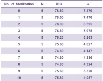
3. Effect of repeated high-pressure autoclaving sterilization on Smartpeg™
Although ISQ was slightly decreased as increase of the number of sterilization, there were no statistically significant changes recorded in ISQ values according to the number of high-pressure autoclaving sterilization procedures performed (P=0.957) (Fig.11).(Table 3)
- 8 -
No. of Sterilization N ISQ σ
0 5 78.60 7.470
1 5 78.60 7.470
2 5 78.00 6.595
3 5 76.80 5.975
4 5 76.20 5.263
5 5 75.60 4.827
6 5 74.80 4.147
7 5 74.60 4.336
8 5 74.80 4.324
9 5 75.60 5.320
10 5 75.80 4.087
Table 3. ISQ values after repeated sterilization
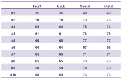
4. Changes in ISQ according to bone defect measurement direction around the implant
Overall, the ISQ measurements obtained from the front side and back side in the presence of the defect were lower than the measurements taken from the mesial and distal sides which were perpendicular to the defect.(Table 4)
Front Back Mesial Distal
#1 35 35 49 49
#2 70 70 73 73
#3 64 64 75 74
#4 61 61 78 78
#5 65 65 77 77
#6 64 64 67 68
#7 65 65 71 71
#8 65 65 73 73
#9 58 58 70 70
#10 58 59 73 73
Tabel 4. ISQ values of implant with bone defect in 4 directions
- 10 -
Adequate primary stability of bone implants plays an important role in the healing of bone surrounding implants by minimizing implant mobility and preventing invasion of soft tissues.[7] It is also important to accurately assess the primary stability of implants for prognosis and establishing a loading protocol.
Since the development of implant stability measuring devices that use resonant frequency in 1994, Meredith et al. validated the usefulness of RFA and also reported that devices using resonance frequency can detect small changes in implant stability.[8,9] Furthermore, bone quality, rather than bone quantity, has been shown to be greater importance for resonance frequencies,[10] and bone-implant contact is correlated with resonance frequency.[11] In addition, resonance frequencies increase as the surrounding bone of implant heals.[12,13] Based on these findings, an RFA can be considered an important objective method of implant stability measurements. However, when measuring implant stability using an RFA, a transducer has to be attached to the implant through a complex process, during which the measurement accuracy may be compromised.
Furthermore, different transducers have to be purchased for different implant systems, and each transducer is highly expensive. The Osstell™
mentor and Smartpeg™ are RFA devices that have recently been developed to overcome these shortcomings, and are currently used in clinics.
Lakholm et al. classified bone quality into cortical and cancellous types based on the morphological macrostructure.[14] Misch assessed bone
quality based on subjective tactile sense after drilling holes into different materials.[15] However, these methods of evaluation are limited in that they are neither objective nor quantitative. Recent research on bone quality classification using CT has enabled a more objective and quantitative evaluation of bone density around implants.[16] Manzano et al. reported a highly significant correlation between HUs measured by CT scans and ISQs measured using an RFA device.[17] In the present study, the HUs of the bone surrounding each hole were measured after holes were drilled into 20 specimens. ISQs were then measured after placing 20 implants into the holes. A statistically significant positive correlation was found between HU and ISQ (r=0.74, P<0.05), consistent with the results of previous studies.[17]
The manufacturer of Smartpeg™ recommends a single use of the product, and explains that ISQ values change as the measurement frequency increases. Aritza et al. reported that measuring ISQ with the Osstell™ device has high reproducibility and repeatability.[18] Won et al.
found that ISQ measurements do not change as long as screw pitches are not damaged and have not lost magnetism even after repeated use of Smartpeg .[19] Pattjin et al. reported that ISQ measurements made when™ a transducer was not completely fixed to an implant may be inaccurate.[20] In the present study, an experiment was conducted to evaluate the reliability of ISQ measurements according to attachment of a Smartpeg™ by different clinicians. We investigated the influence of the connection between an implant and a Smartpeg , which is usually made™
- 12 -
manually, based on ISQ measurements. Ten different clinicians with different clinical habits attached 10 Smartpegs™ to 10 implants respectively and measured their ISQs. Statistically significant measurements were found regardless of the clinician (Cronbach's α = 0.962). Therefore, ISQ measurements can be considered reliable even when a Smartpeg™ is attached to the implant using various degrees of manual torques.
The manufacturer of Smartpeg™ also noted that high-pressure autoclaving sterilization can reduce the stability of ISQ measurements by weakening the magnetic properties of the Smartpeg . However, Won et al.™ reported that sterilization performed at 138°C for 10 minutes did not affect ISQ measurements, while loss of magnetism was observed after sterilization at 150° for 5 minutes.[19] In the present study, sterilization was performed 10 times at 132°C and 2 kg f/cm2 for 15 minutes, and changes in ISQ measurements were analyzed. There was no statistically significant changes in ISQ measurements despite repeated high-pressure autoclaving sterilization (P=0.957). This was consistent with the results of previous studies,[19] confirming that sterilization conditions commonly used in clinics do not reduce the stability of ISQ measurements. However, since sterilization frequency was limited to 10 in the present study, additional research must be performed to determine the number of times sterilization can be repeated without affecting ISQ measurements.
Although numerous studies have been conducted on the primary stability of implants,[2,3,6,7] relatively little research has been performed on implant stability in cases of peri-implant bone defects. Stefan et al. described the
Osstell™ mentor device as apparatus that can reliably detect changes in implant stability according to bone defects.[21,22] In addition, Choi et al.
reproduced various bone defects on acrylic resin blocks, and analyzed changes in ISQ measurements using the Osstell™ mentor. The result was that, while prediction of circular bone loss is possible, it is difficult to obtain accurate information on partial bone loss using the Osstell mentor™
alone.[23] In this study, semi-circular bone defects were reproduced around the implants, and changes in ISQ measurements according to the direction of measurement by the Osstell mentor™ were analyzed. The results showed that ISQ measurements made in the direction perpendicular to the bone defect were smaller than those made parallel. This suggests that bone defects in the buccolingual direction, which are hidden by implants and are hard to observe on plain radiographs, may be detected using the Osstell™ mentor device. Further systematic research must be performed in this regard.
- 14 -
In this study, a RFA was performed after placing implants into a pig rib bone in which the bone quality had been controlled. For an RFA device, the Osstell™ mentor and Smartpeg™ were used. The accuracy of these devices, and factors affecting implant stability measurements were evaluated. The results were as follows:
1. A statistically significant correlation existed between bone density and ISQ.
2. ISQ measurements were consistent despite the attachment of Smartpegs by different clinicians.
™
3. No statistically significant differences were found in ISQ measurements following repeated high-pressure autoclaving sterilization of the Smartpegs
.
™
4. There were differences in ISQ measurements according to the measurement direction in the presence of peri-implant bone defects.
Based on these findings, RFA devices, the Osstell™ mentor, and Smartpegs , can measure implant stability with satisfactory accuracy and™ reliability. Furthermore, these devices may detect bone defects that are difficult to detect on plain radiographs.
1. Hussaini S, Weiner S, Ahmad M. Implant survival rates in a condensed surgical and prosthetic training program for general practitioners in dental implants. Implant Dent 2010;19:73-80.
2. Lioubavina Hack N, Lang NP, Karring T. Significance of primary stability‐ for osseointegration of dental implants. Clin Oral Implants Res 2006;17:244-250.
3. Oh J-h, Chang M. Comparison of initial implant stability measured by Resonance Frequency Analysis between different implant systems. J Kor Acad Periodontol 2008;38:529-534.
4. Sykaras N, Triplett RG, Nunn ME, Iacopino AM, Opperman LA. Effect of recombinant human bone morphogenetic protein 2 on bone regeneration‐ and osseointegration of dental implants. Clin Oral Implants Res 2001;12:339-349.
5. Meredith N, Friberg B, Sennerby L, Aparicio C. Relationship between contact time measurements and PTV values when using the Periotest to measure implant stability. Int J Prosthodont 1998;11:269-275.
6. Meredith N. On the clinical measurement of implant stability and osseointegration. PhD thesis, Göteborg University, Sweden, 1997.
7. Turkyilmaz I, Sennerby L, McGlumphy EA, Tözüm TF. Biomechanical aspects of primary implant stability: a human cadaver study. Clin Implant Dent Relat Res 2009;11:113-119.
8. Meredith N, Books K, Fribergs B, Jemt T, Sennerby L. Resonance frequency measurements of implant stability in vivo. A cross sectional‐
- 16 -
and longitudinal study of resonance frequency measurements on implants in the edentulous and partially dentate maxilla. Clin Oral Implants Res 1997;8:226-233.
9. Meredith N, Rasmussen L, Sennerby L, Alleyne D. Mapping implant stability by resonance frequency analysis. Med Sci Res 1996;24:191-193.
10. Bischof M, Nedir R, Szmukler Moncler S, Bernard JP, Samson J. Implant‐ stability measurement of delayed and immediately loaded implants during healing. Clin Oral Implants Res 2004;15:529-539.
11. Nkenke E, Hahn M, Weinzierl K, Radespiel Tröger M, Neukam FW,‐ Engelke K. Implant stability and histomorphometry: a correlation study in human cadavers using stepped cylinder implants. Clin Oral Implants Res 2003;14:601-609.
12. Park C, Lim JH, Cho IH, Lim HS. A study on the measurement of the implant stability using resonance frequency analysis. J Korean Acad Prosthodont 2003;41:182-206.
13. Huang HM, Chiu CL, Yeh CY, Lin CT, Lin LH, Lee SY. Early detection of implant healing process using resonance frequency analysis. Oral Implants Res 2003;14:437-443.
14. Lekholm U, Zarb GA. Patient selection and preparation. In: Branemark PI, Zarb GA, Albrektsson T, eds. Tissue-integrated prostheses:
osseointegration in clinical dentistry. Chicago, IL: Quintessence, 1985:199-209.
15. Misch CE. Density of bone: effect on treatment plans, surgical
approach, healing, and progressive boen loading. Int J Oral Implantol 1989;6:23-31.
16. Norton MR, Gamble C. Bone classification: an objective scale of bone density using the computerized tomography scan. Clin Oral Implants Res 2001;12:79-84.
17. Manzano-Moreno FJ, Herrera-Briones FJ, Bassam T, Vallecillo-Capilla MF, Reyes-Botella C. Factors affecting dental implant stability measured using the Ostell mentor device: a systematic review. Implant Dent 2015;24:565-577.
18. Brizuela-Velasco A, Fernández-González FJ, Martín-Blanco N, Chávarri-Prado D, Chento-Valiente Y, Dehesa-Ibarra B, et al.
Accuracy of resonance frequency analysis by third generation Osstell®.IntJOdontostomatol2015;9:489-492.
19. Won HY, Cho IH, Lee JS. A study of Smartpeg(TM)'s lifetime according to sterilization for implant stability. J Korean Acad Prosthodont 2008;46:42-52.
20. Pattijn V, Jaecques S, De Smet E, Muraru L, Van Lierde C, Van der Perre G, et al. Resonance frequency analysis of implants in the guinea pig model: influence of boundary conditions and orientation of the transducer. Med Eng Phys 2007;29:182-190.
21. Lachmann S, Jäger B, Axmann D, Gomez Roman G, Groten M, Weber‐ H. Resonance frequency analysis and damping capacity assessment.
Clin Oral Implants Res 2006;17:75-79.
22. Lachmann S, Laval J, Jäger B, Axmann D, Gomez-Roman G, Groten
- 18 -
M, et al. Resonance frequency analysis and damping capacity assessment. Part 2: peri-implant bone loss follow-up. An in vitro study with the Periotest and Osstell instruments. Clin Oral Implants Res 2006;17:80-84.
23. Choi HH, Chung CH, Kim SG, Son MK. Reliability of 2 implant stability measuring methods in assessment of various periimplant bone loss: an in vitro study with the Periotest and Osstell Mentor. Implant Dent 2014;23:51-56.
Fig. 1. Bone specimen exposing the medullary part
Fig. 2. Implant fixture (USII system)
Fig. 3. Osstell™ mentor
- 20 -
Fig. 4. Smartpeg™ (type 1)
Fig. 5. Cone beam computerized tomography scanner
Fig. 6. High-pressure autoclaving sterilizer
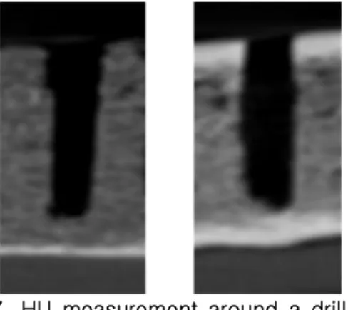
Fig. 7. HU measurement around a drill hole
Fig. 8. Four directions in measurement of ISQ
- 22 -
Fig. 9. Formation of peri-implant bone defect
Fig. 10. Correlation between HU and ISQ
Fig. 11. ISQ after repeated high-pressure autoclaving sterilization on Smartpeg™