68
Open Access
Periprocedural Hemoglobin Drop and Contrast-Induced
Nephropathy in Percutaneous Coronary Intervention Patients
Kang Hyu Lee, MD
1, Sang Rok Lee, MD
1, Kyung Pyo Kang, MD
2, Huy Jung Kim, MD
1, Sun Hwa Lee, MD
1, Kyoung-Suk Rhee, MD
1, Jei Keon Chae, MD
1, Won Ho Kim, MD
1and Jae Ki Ko, MD
11
Divisions of Cardiology and
2Nephrology, Department of Internal Medicine, Research Institute of Clinical Medicine, Chonbuk National University Hospital, Chonbuk National University Medical School, Jeonju, Korea
ABSTRACT
Background and Objectives : The development of contrast-induced nephropathy (CIN) is associated with an in- creased risk of death and late cardiovascular events after percutaneous coronary intervention (PCI). The relation- ship between CIN and hemoglobin drop has been controversial. The aim of this study was to evaluate the clinical usefulness of periprocedural hemoglobin drop as a nontraditional risk factor for CIN. Subjects and Methods : Five-hundred thirty-seven patients who underwent PCI were divided into 2 groups: Group I (486 patients: patients who did not develop CIN) and Group II (51 patients: patients who developed CIN). All patients were administered iodixanol as contrast media during coronary angiography. CIN is defined as a rise in serum creatinine of ≥25%
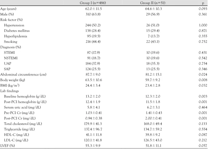
or ≥0.5 mg/dL above the baseline value within 48 hours after contrast administration. Results : Baseline clinical and cardiovascular risk factors were not significantly different between the two groups, except for low abdominal circumference (Group I : Group II=87.9±9.0 cm : 81.2±15.1 cm, p=0.024), body weight (Group I : Group II=
63.5±10.6 kg : 59.7±9.2 kg, p=0.008), body mass index (BMI) (Group I : Group II=24.4±3.4 kg/m
2: 23.4±
2.8 kg/m
2, p=0.032), pre-PCI hemoglobin (Group I : Group II=13.2±2.0 g/dL : 12.3±2.0 g/dL, p=0.003), and post-PCI hemoglobin (Group I : Group II=12.4±1.9 g/dL : 11.5±1.8 g/dL, p=0.001). Multiple logistic re- gression analysis showed that a periprocedural drop in hemoglobin (>1 g/dL) was an independent predictor of CIN, like other known risk factors. Conclusion : A periprocedural drop in hemoglobin of more than 1 g/dL is an- other important independent predictor for CIN, even in patients administered the lowest nephrotoxic contrast agent, iodixanol, during PCI. (Korean Circ J 2010;40:68-73)
KEY WORDS: Hemoglobin; Anemia; Contrast media; Renal insufficiency.
Introduction
Though percutaneous coronary intervention (PCI) is an effective treatment in the setting of coronary artery disease, contrast-induced nephropathy (CIN) is an im- portant complication of iodinated contrast media in-
fused during diagnostic or interventional procedures.
1)2)CIN is the third leading cause of acute renal failure in admitted patients, accounting for 10% of all cases.
3)This iatrogenic complication has been a concern to the interventional cardiologists in recent years because of its adverse effect on prognosis. The development of CIN is associated with an increased risk of death and late cardiovascular events after PCI such as myocardial in- farction and target vessel revascularization.
4-
8)CIN is commonly defined as a rise in serum creati- nine of ≥25% or ≥0.5 mg/dL above the baseline value within 48 hours after contrast administration.
On the basis of this definition, the overall incidence of CIN in the general population is reported to be 1.2 to 1.6%.
9-
11)A key step in minimizing CIN is to identify patients at high risk. Known risk factors for CIN include chro- nic kidney disease (CKD), diabetes mellitus (DM), vol-
Received: April 3, 2009 Revision Received: June 22, 2009 Accepted: July 1, 2009
Correspondence: Sang Rok Lee, MD, Division of Cardiology, Department of Internal Medicine, Research Institute of Clinical Medicine, Chonbuk National University Hospital, Chonbuk National University Medical School, 634-18 Geumam-dong, Deokjin-gu, Jeonju 561-712, Korea Tel: 82-63-250-2418, Fax: 82-63-250-1680
E-mail: [email protected]
○
ccThis is an Open Access article distributed under the terms of the Creative Commons Attribution Non-Commercial License (http://creativecommons.
org/licenses/by-nc/3.0) which permits unrestricted non-commercial use,
distribution, and reproduction in any medium, provided the original work is
properly cited.
ume depletion, nephrotoxic drug therapies, hemody- namic instability, and intra-aortic balloon pump treat- ment.
12-
14)The additive nature of risk has allowed for the development of prognostic scoring systems, but the type and dose of contrast administration have not been fully analyzed in these scoring systems.
15)16)Moreover, the American College of Cardiology/American Heart Association (ACC/AHA) guidelines for acute coronary syndromes in the setting of CKD list the use of isosmo- lal contrast media as a class I (Level A) recommenda- tion.
17)However, isosmolal contrast media is not admi- nistered for all patients in routine clinical practice, which creates discrepancies between the scoring systems and daily practice. To avoid such discrepancies, we investi- gated other possible risk factors for CIN in PCI patients who were administered an isosmolal contrast agent, io- dixanol, as the only contrast media.
Subjects and Methods
In order to minimize the potential role of hemody- namic instability as a cause of postprocedural renal fail- ure, patients were excluded who had ST-segment ele- vation myocardial infarction requiring primary PCI, or cardiogenic shock requiring treatment with an intra- aortic balloon pump (IABP) or inotropic agents. Patients with serum creatinine >4.0 mg/dL, dependence on dia- lysis, neoplasms, vascular malformations, thrombocy- topenia, or major bleeding (intracranial, intraocular, or retroperitoneal hemorrhage, clinical bleeding with he- moglobin drop >3.0 g/dL, or red cell transfusion ≥2 units) were also excluded. For patients who underwent more than one angiographic procedure during the st- udy period, only the first PCI was included.
There were 956 patients who underwent PCI at Ch- onbuk National University Hospital between January 2005 and December 2006. Of these, we excluded 23 pa- tients who had definite cause for renal dysfunction after PCI such as infection, renal artery thrombosis, hemoly- sis, rhabdomyolysis, renal stone, or prostate disease. We retrospectively analyzed the medical records of 537 pa- tients who underwent PCI and were administered io- dixanol (Visipaque
®, GE Health Care, Cork, Ireland) as contrast media. The patients were divided into two groups: Group I (486 patients who did not develop CIN) and Group II (51 patients who developed CIN).
Elective PCI patients received routine hydration with 0.9% normal saline >1 mL/kg/hour from 4 to 12 hours before PCI and continuing after PCI. However, some patients requiring urgent PCI could not receive suffi- cient hydration {79 (16.3%) in Group I vs. 10 (19.6%) in Group II}. N-acetylcysteine 600 mg was admini- stered twice on the day before the procedure and twice on the day after the procedure.
CIN was defined as a postprocedural increase in se-
rum creatinine of ≥0.5 mg/dL or an increase of 25%
from baseline. Absolute or relative increase in serum creatinine at 24 hours or 48 hours were compared to baseline serum creatinine, and CIN was diagnosed when alternative explanations for aggravation of renal dys- function were ruled out. Per study design, blood sam- ples were drawn before PCI and 24 hours after PCI to measure serum creatinine; further determinations of cre- atinine beyond 24 hours were done if clinically indicated.
Diagnostic angiography and PCI were performed after premedication with aspirin (at least 100 mg) and unfractionated or low molecular weight heparin. A load- ing dose of clopidogrel (between 300 mg and 600 mg) was administered before PCI. Coronary angiography was performed through the femoral or radial artery. Hepa- rin was infused throughout the procedure to maintain an activated clotting time of 250 seconds or longer. St- ents were deployed after prior balloon angioplasty, and administration of a platelet glycoprotein IIb/IIIa-recep- tor blocker was left to the decision of the surgeon. The appropriate length and diameter coronary stent was care- fully selected to properly cover the target lesion. Each individual physician decided on selection of the trans- radial or transfemoral approach, contrast dose, inter- ventional technique, supportive pharmacologic thera- pies, and post-dilatation with a non-compliant high- pressure balloon. Therapeutic decision-making and the need for interventional therapy in patients with unstable angina/non-ST-elevation myocardial infarction (NSTE- MI) were left to the physician’s choice according to the guidelines of the ACC/AHA/Society for Cardiovascu- lar Angiography and Interventions (SCAI).
18)19)The baseline clinical characteristics, laboratory char- acteristics, and coronary angiography findings {includ- ing ACC/AHA classification, and Thrombolysis In Myo- cardial Infarction (TIMI) flow grade before and after PCI} were analyzed for groups I and II. To examine a report that each 3% periprocedural drop in hematocrit (known to correspond approximately to 1 g/dL of he- moglobin) resulted in development of CIN, we also analyzed the effect of a decrease in hemoglobin by ≤ 1 g/dL, 1-2 g/dL, ≥2 g/dL on the development of CIN in our study population.
20)For continuous variables, comparisons between the
groups were done using Student’s t-test. Fischer’s exact
test was used to evaluate the categorical variables. To
test whether initial differences between the two groups
influenced the differing results, multiple logistic regres-
sion analysis was performed after controlling the vari-
ables that were significantly different at baseline. All
continuous variables are described as mean±standard
deviation. All analyses were 2-tailed, with clinical sig-
nificance defined as p<0.05. All statistical processing
was done using SPSS-PC 15.0 (Statistical Package for the
Social Sciences, SPSS-PC. Inc., Chicago, IL, USA).
Results
Baseline clinical and laboratory characteristics (Table 1) were not significantly different between the two gr- oups. However, patients who developed CIN (Group II) had lower abdominal circumference, body weight, body mass index (BMI), baseline hemoglobin, and post-PCI hemoglobin levels.
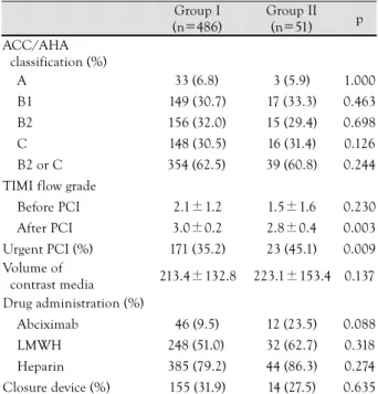
There was no significant difference between the two groups with regard to lesion characteristics by coronary angiography according to ACC/AHA classification or TIMI flow grade before PCI. Moreover, the drugs used for anticoagulation and the closure device were not dif- ferent between the two groups. However, the TIMI flow grade after PCI was higher in patients who did not de- velop CIN (Group I) compared to the patients who developed CIN (Group II) (3.0±0.2 vs. 2.8±0.4, p=
0.003) (Table 2). Indications for PCI were different bet- ween the two groups. The proportion of urgent PCIs (early invasive PCI rather than elective PCI) was higher in Group II (45.1%) than in Group I (35.2%) (p=0.009).
The proportion of patients who experienced a drop
in hemoglobin of more than 1 g/dL was higher among patients who developed CIN (Group II, 54.9%) com- pared with patients who did not develop CIN (Group I, 39.3%) (p=0.036) (Fig. 1).
Multivariate logistic regression analysis was done both by statistically significant variables (body weight, base- line hemoglobin, post-PCI hemoglobin, pre-PCI Cr, post-PCI Cr, and TIMI flow grade after PCI) and con- ventional risk factors for CIN (age, anemia, and urgent PCI). Multivariate logistic regression analysis showed that the strong predictors for the development of CIN (Table 3) were consistent with previous studies; age, body weight, anemia, pre-PCI creatinine, and urgent PCI. Like other known risk factors for CIN, a decrease in hemoglobin by >1 g/dL was a strong predictor for the development of CIN {adjusted odds ratio (aOR)=
2.238, p=0.012}.
Discussion
CIN is one of the most common causes of acute renal failure in hospitalized patients. The frequency of
Table 1. Baseline clinical characteristics
Group I (n=486) Group II (n=51) p
Age (years) 62.0±11.5 64.6±10.3 0.093
Male (%) 310 (63.8) 29 (56.9) 0.361
Risk factor (%)
Hypertension 244 (50.2) 26 (51.0) 1.000
Diabetes mellitus 138 (28.4) 15 (29.4) 0.871
Hyperlipidemia 95 (19.5) 7 (13.7) 0.355
Smoking 216 (44.4) 22 (43.1) 0.752
Diagnosis (%)
STEMI 87 (17.9) 10 (19.6) 0.431
NSTEMI 91 (18.7) 10 (19.6) 0.542
UAP 184 (37.9) 18 (35.3) 0.754
SAP 124 (25.5) 13 (25.5) 0.346
Abdominal circumference (cm) 87.7±9.0 81.2±15.1 0.024
Body weight (kg) 63.5±10.6 59.7±9.2 0.008
BMI (kg/m
2) 24.4±3.4 23.4±2.8 0.032
Lab findings
Baseline hemoglobin (g/dL) 13.2±2.0 12.3±2.0 0.003
Post-PCI hemoglobin (g/dL) 12.4±1.9 11.5±1.8 0.001
Serum uric acid (mg/dL) 5.8±4.1 6.2±3.1 0.464
Pre-PCI Cr (mg/dL) 1.03±0.41 1.41±0.43 0.001
Post-PCI Cr (mg/dL) 0.94±0.38 2.00±0.41 0.001
Total cholesterol (mg/dL) 179.9±41.3 168.0±49.4 0.133
Triglyceride (mg/dL) 137.4±96.7 134.7±59.2 0.554
HDL-C (mg/dL) 41.1±11.8 39.8±9.2 0.087
LDL-C (mg/dL) 120.1±41.8 126.5±43.0 0.212
LVEF (%) 55.3±9.9 51.8±11.1 0.057
STEMI: ST-elevation-myocardial infarction, NSTEMI: non-ST elevation myocardial infarction, UAP: unstable angina pectoris, SAP: stable angina pectoris, BMI: body mass index, PCI: percutaneous coronary intervention, HDL-C: high-density lipoprotein-cholesterol, LDL-C:
low-density lipoprotein-cholesterol, LVEF: left ventricular ejection fraction
CIN has decreased over the past decade from a general incidence of -15% to -7%.
15)CIN is associated with prolonged in-hospital stay and increased morbidity, mortality, and cost. In the Mayo Clinic registry, the in- hospital death rate was 22% among the patients who developed CIN compared to only 1.4% in patients who did not develop CIN. Among hospital survivors with CIN, 1- and 5-year estimated mortality rates were 12.1%
and 44.6%, respectively, much greater than the 3.7%
and 14.5% mortality rates noted in patients without CIN (p<0.0001).
1)Although the pathogenesis of CIN is not completely known, multiple mechanisms may be involved. Gener- ally, the pathophysiology of CIN assumes baseline re- duced nephron number, with superimposed acute va-
soconstriction caused by the release of adenosine, en- dothelin, and other renal vasoconstrictors triggered by iodinated contrast. After a very brief increase in renal blood flow, there is an overall -50% sustained reduc- tion in renal blood flow lasting for several hours. Pro- longed contrast transit time in the kidney increases the exposure of renal tubular cells to contrast. This stasis of contrast in the kidney allows for direct cellular in- jury and death in renal tubular cells. Furthermore, the sustained reduction in renal blood flow to the outer medulla leads to medullary hypoxia, ischemic injury, and death of renal tubular cells.
21)Many individual risk factors have been reported in relation to the development of CIN. The best recogniz- ed non-modifiable risk factors are older age, DM, pre- existent renal insufficiency, congestive heart failure, he- modynamic instability, and nephritic syndrome. Modi- fiable risk factors include volume depletion, volume of contrast media, nephrotoxic drug use, low serum albu- min level (<35 g/L), and anemia.
22)23)Dangas et al.
24)showed that the baseline hematocrit level is an independent predictor of CIN in patients with CKD (OR=0.95, p<0.00001). Nikolsky et al.
20)re- ported that lower baseline hematocrit was an indepen- dent predictor for CIN in 6,773 consecutive patients tr- eated with PCI. Moreover, the CIN rate steadily increas- ed with baseline hematocrit quintile decrements (from 10.3% in the highest quintile to 23.3% in the lowest qu- intile) (p<0.0001). Each 3% drop in hematocrit (known to correspond to approximately 1 g/dL of hemoglobin) resulted in a 30% increase in the risk of CIN (p<0.0001) in patients with baseline CKD and a 26% increase in the risk of CIN in patients without CKD (p<0.0001).
On multiple logistic regression analysis in the present study, traditional risk factors for CIN such as age, body weight, anemia, pre-PCI creatinine, and urgent PCI showed a significant association with CIN, consistent with previous studies. Interestingly, the present study indicates that a hemoglobin drop (>1 g/dL) during PCI is an important predicting factor for CIN, just like other known risk factors. Even though the association between hemoglobin drop and higher rates of CIN is not clearly understood, patients with a greater decrease in hemoglobin were older, had lower baseline hemo-
Table 3. Multiple logistic regression analysis
aOR 95% CI p
Age 1.560 1.151-1.980 0.007
Body weight (kg) 0.919 0.880-0.958 0.001
Anemia 1.554 1.145-1.972 0.042
Hemoglobin drop >1 g/dL 2.238 1.190-4.209 0.012 Pre-PCI Cr 4.672 2.393-9.121 0.001 Urgent PCI 2.688 1.425-5.072 0.002 PCI: percutaneous coronary intervention, aOR: adjusted odds ratio, CI: confidence interval
Table 2. Angiographic and anticoagulation characteristics Group I
(n=486)
Group II (n=51) p ACC/AHA
classification (%)
A 33 (6.8) 3 (5.9) 1.000
B1 149 (30.7) 17 (33.3) 0.463 B2 156 (32.0) 15 (29.4) 0.698 C 148 (30.5) 16 (31.4) 0.126 B2 or C 354 (62.5) 39 (60.8) 0.244 TIMI flow grade
Before PCI 2.1±1.2 1.5±1.6 0.230 After PCI 3.0±0.2 2.8±0.4 0.003 Urgent PCI (%) 171 (35.2) 23 (45.1) 0.009 Volume of
contrast media 213.4±132.8 223.1±153.4 0.137 Drug administration (%)
Abciximab 46 (9.5) 12 (23.5) 0.088 LMWH 248 (51.0) 32 (62.7) 0.318 Heparin 385 (79.2) 44 (86.3) 0.274 Closure device (%) 155 (31.9) 14 (27.5) 0.635 ACC/AHA: American College of Cardiology/American Heart As- sociation, TIMI: Thrombolysis In Myocardial Infarction, PCI: per- cutaneous coronary intervention, LMWH: low molecular weight heparin
70 60 50 40 30 20
191/486 (39.3%)
28/51 (54.9%) p=0.036
No CIN CIN
Fig. 1. There was a significant difference in the proportion of pa- tients with a drop in hemoglobin (>1 g/dL). There was higher pro- portion of patients with an drop in hemoglobin among patients who developed CIN. CIN: contrast-induced nephropathy.
Drop in hemoglobin (%)