염증성 가성종양은 일반적으로 비종양성 종괴로서, 다양한 염증세포와 다른 간엽 세포(mesenchymal cell)로 구성된다.
폐의 염증성 가성종양은 소아에 있어서 가장 흔한 흉부 종괴 이다(1). 그러나, 상대적으로 폐 실질 외에서 발생하는 경우는 드물다. 저자들은 성인에서 종격동과 폐동맥내 종괴로 발현한 염증성 가성종양의 증례를 보고하고자 한다.
증례 보고
48세 남자 환자가 흉부 단순촬영에서 발견된 이상 소견으로 인해 흉부외과로 전과되었다. 환자는 직장의 카르시노이드 종 양(carcinoid tumor)으로 외과에서 수술 예정이었다. 과거력 상 환자는 약 1년 동안의 노작성 호흡곤란이 있었다. 수술 전 촬영한 흉부 단순 촬영상 우측 폐문이 커져 있는 소견이 관찰 되었다. 또한 우측의 폐혈관 음영이 감소한 소견이 관찰되었다 (Fig. 1A).
흉부 전산화 단층 촬영(computed tomography, 이하 CT) 상 우폐동맥을 채우고 있는 혈전 혹은 종괴를 관찰할 수 있었 다. 조영 전 CT에서 혈전 혹은 종괴 내부에 작은 석회화가 보 였다(Fig. 1B). 또한 조영 후 CT에서 혈전 혹은 종괴의 부분 적인 조영증강이 관찰되었다(Fig. 1C, D). 주폐동맥은 확장되 어 있었으며 폐실질에 종괴나 다른 이상 소견은 보이지 않았 다. 종격동에는 단경 1 cm이하의 기관주위 림프절들이 관찰되 었다. 폐색전증 의심 하에 시행한 하지의 도플러(doppler) 초 음파 검사와 정맥조영술에서 심부정맥 혈전증의 소견은 보이
지 않았다.
환자는 종격동절개술(mediastinotomy)을 시행 받았다. 우측 폐문부의 종괴에서 생검을 시행하였고 조직 검사상 염증성 가 성종양(inflammatory pseudotumor)로 진단되었다(Fig. 1E).
환자는 우선 일반외과로 전과되어 직장의 카르시노이드 종양 과 대장의 용종에 대하여 대장절제술을 시행 받았다. 흉부외과 로 다시 입원하여 시행한 폐동맥 혈관조영술에서 우측 폐동맥 을 채우고 있는 종괴를 관찰할 수 있었으며, 도관(catheter)을 이용한 겸자 생검(forcep biopsy)에서도 염증성 가성종양으로 진단되었다(Fig. 1F). 환자에게 수술을 권유하였으나 환자는 수술을 거부하고 퇴원하였다.
고 찰
염증성 가성종양은 일반적으로 비종양성 종괴로서, 다양한 염증세포와 다른 간엽 세포(mesenchymal cell)로 구성된다 (1). Plasma cell granuloma, inflammatory myofibro- blastic tumor, fibroxanthoma, histriocystoma, xantho-granuloma, pseudosaromatous myofibroblastic prolifer-ation, inflammatory myofibroblastic proliferation 등의 다양한 명칭 으로 불린다(1-3).
염증성 가성종양은 흔하게는 폐와 안와를 침범하지만, 중추 신경계부터 위장관계까지 신체 어느 부위에서나 발생할 수 있 다(4).
폐의 염증성 가성 종양은 소아에 있어서 가장 흔한 폐 종괴 이다(1). 흉부 단순 촬영에서는 보통 경계가 좋은 단일폐결절 로 나타나며, 주로는 폐하엽에 호발한다. CT에서는 다양하고 비특이적인 소견을 보이는데 불균질한 음영과 조영을 보인다 (5).
─ 525 ─ 대한영상의학회지 2004;51:525-527
폐동맥을 침범한 염증성 가성종양: 증례 보고1
나형일・김양수・유승민・손동섭2・이화연・송인섭・이종범・김건상・유 현3
폐의 염증성 가성종양은 소아에 있어서는 가장 흔한 흉부 종괴이나, 상대적으로 폐 실질 외 에서 발생하는 경우는 드물다. 저자들은 폐동맥을 침범한 염증성 가성종양의 증례를 보고하고 자 한다. 48세 남자가 흉부 단순촬영 상 우측 폐문 비대의 소견을 보여 내원하였다. 과거력 상 환자는 1년간의 노작성 호흡곤란이 있었다. 전산화 단층 촬영에서 부분적인 조영 증강을 보이 는 종괴가 우측 폐동맥에서 관찰되었다. 종격동절개술과 폐동맥 혈관조영술을 통해 시행한 겸 자 생검에서 염증성 가성종양으로 진단되었다.
1중앙대학교 의과대학 방사선과학교실
2중앙대학교 의과대학 흉부외과학교실
3서울보훈병원 방사선과
이 논문은 2004년 7월 30일 접수하여 2004년 10월 11일에 채택되었음.
─ 526 ─
나형일 외: 폐동맥을 침범한 염증성 가성종양
A B
C D
E F
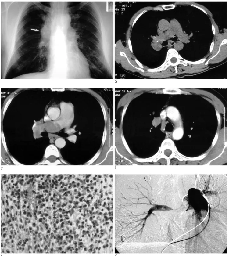
Fig. 1. A 48-year-old man with exertional dyspnea for 1 year.
A. Chest PA radiograph shows enlargement of the right hilum (white arrow). Pulmonary conus shows also bulging contour.
Pulmonary vascularity of the right lung is decreased.
B. Preenhanced CT scan shows an inhomogeneous hypoattenuating mass in the right pulmonary artery. Punctuate calcification is noted within the mass.
C. Contrast enhanced CT scan shows a hypoattenuating mass with mild heterogeneous enhancement in the right pulmonary artery.
D. At the more cranial level, right truncus anterior is also filled with mass.
E. Photomicrography of biopsy specimen shows dense infiltration of lymphocytes, plasma cells and histiocytes admixed with fi- broblasts.
F. Pulmonary artery angiography shows a well-defined filling defect in the origin of the right pulmonary artery. Main pulmonary trunk is also dilated.
흉부에서는 폐 실질 외에서 발생하거나 침범하는 경우는 상 대적으로 드물다. 폐문, 종격동, 기도 등을 침범할 수 있으며, 대부분 실질의 종괴가 이차적으로 침범한 경우가 대부분이다.
드물게 심장 혹은 혈관에서 발생하는 경우가 있으며 본 증례 와 같이 폐동맥에서 발생한 경우는 매우 드물다(3, 4).
본 증례와 같이 폐동맥 내 종괴가 있을 경우에는 우선 폐색 전증과 혈관육종의 감별이 필요하다. 두 질환을 임상 양상이나 방사선학적 소견으로 감별하는 것이 용이하지는 않으나, Tschirch 등(6)은 적절한 항응고제 치료에 반응이 없거나, 본 증례와 같이 폐색전증의 원인이 없는 경우, 추적 관찰 중 여러 개의 폐 실질 내 결절이 나타난 경우, 혈관 밖을 침범한 경우, 종격동 혹은 폐문부 림프절 비대의 소견을 보이는 경우, 자기 공명영상에서 조영 증강을 보이는 경우 등이 혈관 육종을 시 사하는 소견이라고 하였다.
본 증례는 CT상 부분적인 조영 증강을 보였다는 점과 서서 히 진행된 임상 양상이 폐색전증이나 혈관육종과의 감별점이 될 수 있겠으나, 수술 전 진단이 어려웠던 경우였다. 본 증례에 있 어서는 혈관 조영술을 통한 겸자 생검으로도 진단이 가능하였 다. 본 증례와 같은 혈관내 종괴에 있어서는 수술 전 혈관 조영 술과 도관을 통한 겸자 생검이 진단에 유용하리라고 생각한다.
참 고 문 헌
1. Pettinato G, Manivel JC, De Rosa N, Dehner LP. Inflammatory myofibroblastic tumor (plasma cell granuloma). Clinicopathologic study of 20 cases with immunohistochemical and ultrastructural observations. Am J Clin Pathol 1990;94:538-546
2. Coffin CM, Watterson J, Priest JR, Dehner LP. Extrapulmonary in- flammatory myofibroblastic tumor (inflammatory pseudotumor).
A clinicopathologic and immunohistochemical study of 84 cases.
Am J Surg Pathol 1995;19:859-872
3. Agrons GA, Rosado-de-Christenson ML, Kirejczyk WM, Conran RM, Stocker JT. Pulmonary inflammatory pseudotumor: radiolog- ic features. Radiology 1998;206:511-518
4. Narla LD, Newman B, Spottswood SS, Narla S, Kolli R. Inflamma- tory pseudotumor. Radiographics 2003;23:719-729
5. Copin MC, Gosselin BH, Ribet ME. Plasma cell granuloma of the lung: difficulties in diagnosis and prognosis. Ann Thorac Surg 1996;
61:1477-1482
6. Tschirch FT, Del Grande F, Marincek B, Huisman TA.
Angiosarcoma of the pulmonary trunk mimicking pulmonary thromboembolic disease. A case report. Acta Radiol 2003; 44:504- 507
─ 527 ─ 대한영상의학회지 2004;51:525-527
J Korean Radiol Soc 2004;51:525-527
Address reprint requests to : Hyoung Il Na, M.D., Department of Radiology, Chung-Ang University Yongsan Hospital 65-207, Hangangro-3-ga, Yongsan-gu, Seoul 140-757, Korea.
Tel. 82-2-748-9682 Fax. 82-2-798-4745 E-mail: [email protected]
Inflammatory Pseudotumor Involving the Pulmonary Artery: Case Report1
Hyoung Il Na, M.D., Yang Soo Kim, M.D., Seung Min Yoo, M.D., Dong Suep Sohn, M.D.2, Hwa Yeon Lee, M.D., In Sup Song, M.D., Jong Beum Lee, M.D.,
Kun Sang Kim, M.D., Hyoen Yu, M.D.3
1Department of Radiology, Chung-Ang University College of Medicine
2Department of Thoracic & Cardiovascular Surgery, Chung-Ang University College of Medicine
3Department of Radiology, Seoul Veterans Hospital
Pulmonary inflammatory pseudotumor is the most common primary lung mass seen in children, but extra- parenchymal involvement is relatively rare. We report here on a case of inflammatory pseudotumor involving the mediastinum and the pulmonary artery. A 48-year-old man presented with enlargement of the right hilum on a simple chest radiograph. He had a history of exertional dyspnea for 1 year. A non-homogeneous enhanc- ing mass was noted in the right pulmonary artery on computed tomography. Mediastinotomy and pulmonary artery angiography with a forcep biopsy revealed inflammatory pseudotumor of the mediastinum and pul- monary artery.
Index words :Pseudotumor, hepatic inflammatory Pulmonary arteries
Computed tomography (CT)