Aspiration Thrombectomy Using a Guiding Catheter in Acute Lower Extremity Deep Vein Thrombosis:
Usefulness of the Calf-Squeeze Technique
1Jae-A Lee, M.D., Hyo-Sung Kwak, M.D., Young-Min Han, M.D., Hee Chul Yu, M.D.
2Departments of 1Radiology and 2Surgery, Chonbuk National University Medical School, Chonbuk 561-712, Korea Received September 21, 2009 ; Accepted November 24, 2010
Address reprint requests to : Hyo-Sung Kwak, M.D., Department of Radiology, Chonbuk National University Medical School, 634-18 Keumam-dong, Deokjin-gu, Jeonju-shi, Chonbuk 561-712, Korea.
Tel. 82-63-250-2582 Fax. 82-63-272-0481 E-mail: [email protected]
Purpose: The effectiveness of the calf-squeeze technique during aspiration thrombec- tomy using guiding catheter in the treatment of an acute lower extremity deep vein thrombosis (DVT) was evaluated by the use of imaging and the clinical follow-up of pa- tients.
Materials and Methods: A prospective analysis of ten patients (seven women, three men; median age, 56.9 years) with common iliac vein (CIV) obstruction and ipsilateral DVT was performed for this study. All patients presented with leg edema or pain and were treated with catheter-directed thrombolysis via an ipsilateral popliteal vein ap- proach after insertion of a temporary inferior vena cava (IVC) filter. Subsequently, the patients were treated with by aspiration thrombectomy using a guiding catheter to re- move the residual thrombus. The calf-squeeze technique during aspiration thrombec- tomy can be used to induce the proximal migration of thrombi in the popliteal, tibial, and muscular veins were used to increase venous flow.
Results: The calf-squeeze technique was employed at mean of 1.3 times (range, 1-3 times). All patients showed proximal migration of a popliteal and muscular vein thrombus during the execution of the calf-squeeze technique. Successful recanaliza- tion was achieved in all patients (100%) without any complications. On duplex ultra- sonography, which was performed immediately after the aspiration thrombectomy, four patients had a residual thrombus in the soleal muscular veins. However, none of the patients had a thrombus in the popliteal and tibial veins; and, during follow-up, no DVT recurred in any patient.
Conclusion: The use of the calf-squeeze technique during aspiration thrombectomy af- ter catheter-directed thrombolysis can induce the proximal migration of thrombi in the popliotibial and muscular veins and is an effective method that can remove a throm- bus in calf veins.
Index words : Vein
Venous Thrombosis
Thrombolytic Therapy
Acute deep vein thrombosis (DVT) can result from common iliac vein (CIV) stenosis or occlusion.
Aggressive treatments have been proposed to alleviate pain, as well as avoid residual swelling and venous ulcer- ation (1-4). Most patients with acute DVT have extensive thrombosis in the popliteal vein and calf muscular veins, as depicted on contrast-enhanced computed tomography (CT). Treatment strategies for DVT include systemic thrombolytic therapy, surgical thrombectomy, catheter- directed thrombolysis (CDT) and percutaneous mechani- cal thrombectomy (PMT) (4-11). Recently, the use of thrombolytic therapy and PMT can result in the rapid re- duction of the thrombus burden and potentially preserve venous valvular function as well as prevent postthrom- botic syndrome (8-11). Percutaneous access to the thrombosed vessel for CDT and PMT can be achieved through popliteal vein access (4-11). Therefore, a venous thrombus below the popliteal vein access can remain af- ter treatment. Some studies have attempted treatments such as retrograde CDT, contralateral CDT, and CDT through posterior tibial vein access for a thrombus locat- ed below the popliteal vein (6, 12, 13).
The purpose of the present study was to use imaging and clinical follow-up of patients to evaluate the effec- tiveness of the use of the calf-squeeze technique during PMT in the treatment of an acute DVT.
Materials and Methods
Patients
Ten consecutive patients (7 male, 3 female; mean age 56.9 yrs; age range 35-72 yrs) with acute ipsilateral DVT were included in this study that took place between January 2007 and January 2008. The mean duration of DVT symptoms was 3.8 days (range, 2-6 days). The in- stitutional review board of our university and the ethics committee of the Institute of Medical Science approved this study. The procedure was explained in detail to all patients, and informed consent was obtained prior to performing the procedure.
Patients were included in the prospective study if they had documented CT confirmation of an acute DVT af- fecting the iliofemoral, popliteal, and calf muscular veins and common iliac vein obstruction. All patients presented with leg edema and pain.
Exclusion criteria for thrombolysis included the pres- ence of an isolated infrapopliteal thrombus, DVT with a duration of more than seven days (beyond this duration threshold, the condition is considered to be subacute),
and any contraindications for the use of anticoagulation therapy, contrast media, or thrombolytic agents.
Contraindications for thrombolytic agents include active internal bleeding, a recent cerebrovascular accident, al- lergy to thrombolytic agents and coagulopathy.
Endovascular Procedure: IVC filter, Thrombolysis, and Thrombectomy
To prevent a pulmonary thromboembolism (PTE) dur- ing or after thrombolysis and thrombectomy, an inferior vena cava (IVC) filter (OptEase; Cordis, Roden, The Netherlands) was placed in the IVC in all patients via a contralateral femoral approach. The attending physician made the retrieval decision in each individual case.
Percutaneous access to the thrombosed vessel was achieved via popliteal vein access. A 7-F vascular sheath (Cook, Bloomington, IN, USA) was inserted, through which all subsequent catheter and wire exchanges were performed. An ascending venography was performed using nonionic contrast material (320 mg/mL).
The infusion was performed using a 5-F multiple-side hole catheter system (65-100 cm whole catheter system, 10-15 cm infusion length; Boston Scientific, Watertown, MA, USA). The catheter was placed two-thirds of the distance into the thrombosed venous segment, and urokinase (Green Cross, Yongin, Korea) was adminis- trated continuously overnight at 30,000-60,000 U/h (1000-2000 U/kg/h) in split doses. One-third of the drug was administered through the 7-F vascular sheath and two-thirds of the drug was administered through the in- fusion catheter.
The thrombin and partial thromboplastin times were obtained every 4 hours after the initiation of throm- bolytic therapy. Clauss fibrinogen level, hemoglobin level, hematocrit, and platelet counts were measured every 6 hours. If the Clauss fibrinogen level decreased to less than 100 mg/dL, fresh frozen plasma or cryopre- cipitate was administered until the level was brought up to 150 mg/dL. If a patient had gum bleeding, throm- bolytic therapy was temporally stopped and restarted when the patient required additional thrombolysis.
After securing the catheters in place, patients were transferred to a standard inpatient hospital ward.
Repeated venography was performed at 6-15 hours in- tervals. Aspiration thrombectomy using guiding catheter in patients with residual thrombi after follow- up venography was performed with the use of an 8-F guiding catheter (Cordis) via a 10-F introducer sheath.
Fresh and resolving thrombi after thrombolysis were
easily removed with the use of a aspiration thrombecto- my catheter. The aspiration catheter was usually insert- ed through a guide wire to prevent venous rupture.
Aspirated thrombi contained fresh thrombi and white thrombi.
The Calf-Squeeze Technique
A venogram after aspiration thrombectomy was per- formed. If residual thrombi in the iliofemoral and popliteal segment were not present after aspiration thrombectomy, the vascular sheath after insertion of the guide wire was removed. A manual squeeze of the calf muscle was performed for migration of residual thrombi in the popliotibial and muscular veins. If a venogram af- ter calf-squeeze showed migration of residual thrombi, a repeated aspiration thrombectomy was performed. On ultrasonography of calf muscular veins or tibial veins, if there was remaining residual thrombus, the calf- squeeze repeated. If there is no apparent residual throm- bus in the calf veins on ultrasonography, the procedure was stopped.
Endovascular Procedure: Stent Deployment
Subsequent to the follow-up venography after throm- bolysis and aspiration thrombectomy, an angioplasty was performed whereby stents were inserted via the same route, assuming no evidence of fresh thrombi in the deep vein. After angioplasty, when the venography showed persistent stenosis in the common iliac vein, the lesion was treated by deployment of a self-expandable stent (SMART control; Cordis, Miami Lakes, FL, USA) with a diameter of 12-14 mm and length of 60-80 mm.
Deployed stents were fully expanded with the use of an angioplasty balloon of an appropriate diameter. After
endovascular therapy, anticoagulation therapy with sodium warfarin was initiated and continued for six months. Therapy was adjusted to attain an International National Ratio (INR) in the range of 2.0-3.0.
Follow- up and Complications
All patients underwent a color Doppler ultrasonogra- phy (US) examination to detect the presence of thrombi in the popliotibial and muscular veins before and after the use of the calf-squeeze technique. In addition, all pa- tients underwent a color Doppler US examination of the areas of the original DVT and contrast-enhanced chest CT for evaluation for a PTE and the IVC filter after the procedure one or two days later. Patients were followed up by clinic visits, and stent patency and recurrence of DVT were assessed by color Doppler US performed at 1, 3, 6, and 12 months after the procedure.
Minor complications were defined as those with no significant clinical sequelae and requiring minimal ther- apy, such as a local puncture site hematoma that did not require transfusion or other therapy. Major complica- tions were defined as those that necessitated a specific therapy and increased level of care, or resulted in a per- manent adverse event such as a symptomatic PTE or systemic bleeding requiring specific therapy, including a transfusion.
Results
Results are shown in Table 1. Causes of DVT and common iliac vein obstruction were May-Thurner syn- drome in all patients and the lesion was left-sided in all patients. In addition, all patients had a thrombus run- ning from the popliotibial and calf muscular vein to the
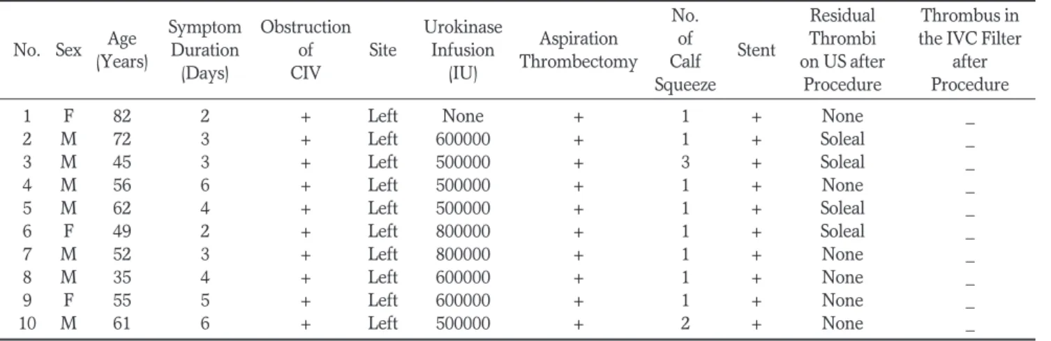
Table 1. Clinical Data and Results of 10 Patients
Symptom Obstruction Urokinase No. Residual Thrombus in No. Sex Age
Duration of Site Infusion Aspiration of
Stent Thrombi the IVC Filter (Years)
(Days) CIV (IU) Thrombectomy Calf on US after after Squeeze Procedure Procedure
1 F 82 2 + Left None + 1 + None _
2 M 72 3 + Left 600000 + 1 + Soleal _
3 M 45 3 + Left 500000 + 3 + Soleal _
4 M 56 6 + Left 500000 + 1 + None _
5 M 62 4 + Left 500000 + 1 + Soleal _
6 F 49 2 + Left 800000 + 1 + Soleal _
7 M 52 3 + Left 800000 + 1 + None _
8 M 35 4 + Left 600000 + 1 + None _
9 F 55 5 + Left 600000 + 1 + None _
10 M 61 6 + Left 500000 + 2 + None _
Note.
─CIV = common iliac vein
A B
C D
E F
Fig. 1. Images from an 82-year-old woman with acute left leg swelling.
A, B. A contrast-enhanced CT scan shows the acute thrombus (arrow) from the common iliac veins (A) to the popliteal vein (B) and the tibial veins.
C. Ascending venography in the prone position extensively shows an acute thrombus in the left leg veins.
D. After deployment of an IVC filter, aspiration thrombectomy using an 8-F guiding catheter shows good patency of the left leg veins with no evidence of thrombi in the femoral vein and ili- ac veins.
E. After removing the sheath with the inserted guide wire, a manual squeeze of the calf muscle is performed.
Venography shows the migrated thrombus in the superficial femoral vein (arrow). A repeated aspiration thrombectomy is performed.
F. A final direct venography shows
good patency of the left common iliac
vein after insertion of a 14-mm by 80-
mm self-expandable stent and no evi-
dence of thrombi in the entire vein.
common iliac vein.
Urokinase infusion was performed in nine patients with a dose of 500,000-800,000 IU (mean, 600,000 IU).
The duration of urokinase infusions was 5-14 hours (mean, 11 hours) in eight patients with overnight infu- sion and in one patient without overnight infusion. One patient did undergo direct aspiration thrombectomy us- ing a guiding catheter without UK infusions due to old- age and poor general condition.
Aspiration thrombectomy using a guiding catheter, was performed in all patients, including one patient where the procedure was performed without thrombol- ysis. Thrombus located above the access of the popliteal vein was successfully removed using a catheter in all pa- tients during aspiration thrombectomy (Fig. 1).
The mean frequency of the calf-squeeze technique was 1.3 times (range, 1-3 times). All patients showed proximal migration of the popliteal and muscular vein
A B C
D E
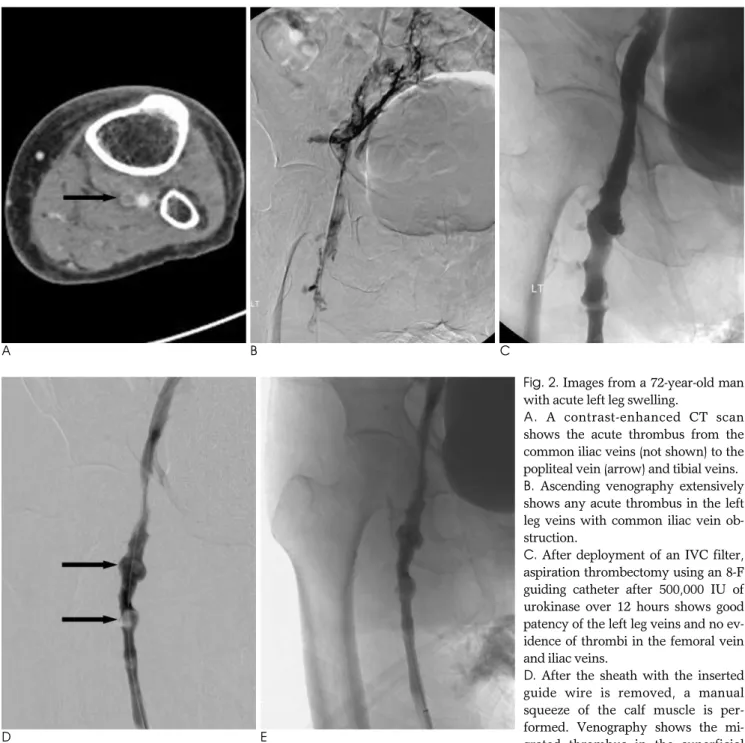
Fig. 2. Images from a 72-year-old man with acute left leg swelling.
A. A contrast-enhanced CT scan shows the acute thrombus from the common iliac veins (not shown) to the popliteal vein (arrow) and tibial veins.
B. Ascending venography extensively shows any acute thrombus in the left leg veins with common iliac vein ob- struction.
C. After deployment of an IVC filter, aspiration thrombectomy using an 8-F guiding catheter after 500,000 IU of urokinase over 12 hours shows good patency of the left leg veins and no ev- idence of thrombi in the femoral vein and iliac veins.
D. After the sheath with the inserted guide wire is removed, a manual squeeze of the calf muscle is per- formed. Venography shows the mi- grated thrombus in the superficial femoral vein (arrows). A repeated aspiration thrombectomy is performed.
E. Final direct venography shows good patency of the left common iliac vein after insertion of a 14-mm by 80-mm self-expandable
stent (not shown) and no evidence of thrombi in the entire vein.
thrombi during use of the calf-squeeze technique. On US that were immediately performed after the calf- squeeze, four patients had a residual thrombus in the soleal muscular veins (Fig. 2) and in six patients, a thrombus was not seen in the popliotibial and muscular veins.
Successful recanalization and clinical success was achieved in all patients (100%) without complications. A self-expandable stent in all patients were deployed into the stenosis of the common iliac vein after thrombolysis and aspiration thrombectomy. On contrast-enhanced chest CT that was performed after the procedure one or two days later, no thrombus was seen in the IVC filter and pulmonary artery (Fig. 2). Also, all patients did not show into the lower extremity portion of the femoral vein, and four patients had a residual thrombus in the soleal muscular veins on ultrasonography, one or two days later.
The clinical follow-up period was 1-12 months (mean, 6.5 months). For four patients with a thrombus in the soleal muscular veins depicted on US performed imme- diately after the calf-squeeze technique, three patients did not show the presence of any thrombus in the mus- cular veins. The fourth patient had chronic thrombi and collapse of the soleal vein. A follow-up Doppler US ex- amination showed intimal thickening without flow limi- tation in one patient. All patients had no recurrent DVT and valvular insufficiency for the valsalva maneuver.
Discussion
Nonsurgical treatment of an extensive iliofemoral DVT caused by May-Thurner syndrome is performed by CDT, mechanical thrombectomy with the use of a catheter and device, balloon dilatation, and stent de- ployment (4-11, 14-17).
CDT has been effective in the treatment of an il- iofemoral DVT (3-5, 18), but several disadvantages in- herent with this treatment have limited its widespread acceptance (6, 19, 20). First, complications have been recorded in as many as 25% of patients in a single-center series and in 11% of patients in a multicenter venous registry study (6, 19-21). Second, most patients require admission to a monitoring bed and have indwelling ve- nous sheaths for 1-3 days. Finally, the cost of providing a monitoring bed, thrombolytic infusions, multiple venogram procedures, and laboratory studies is consid- erable.
A mechanical thrombectomy not only physically mac-
erates the thrombus; it also exposes greater portions of the thrombus to the thromblytic agent (22). Several stud- ies have reported that CDT with PMT was associated with a reduction in treatment duration and total lytic agent dose when compared with the use of CDT alone, and does so with equivalent safety and efficacy (8-11).
Vedantham et al. (11) demonstrated a procedural suc- cess rate of 82% as well as a reduced thrombolytic agent dose requirement and infusion times compared with previously published series of patients treated with CDT alone. Kim et al. (9) reported that CDT with rheolytic PMT was associated with a 46% reduction in treatment duration and a 56% reduction in the total lytic agent dose compared to the use of urokinase CDT alone.
A thrombus can be cleared during mechanical thrombectomy with a catheter by using thrombolysis with transcatheter aspiration (8, 23). The potential disad- vantages of mechanical thrombectomy are theoretically increased risks of pulmonary embolism and venous valvular damage. In an animal study, the Amplatz me- chanical thrombectomy device did not cause physiologi- cally significant damage to valves 7 mm or larger in di- ameter (24). Also, an 8-F sheath dilator did not injure ve- nous structures with a mean diameter of 5.5 0.6 mm (24). In our study, we performed aspiration thrombecto- my with 8.0-F guiding catheters after thrombolysis in- stead of a mechanical thrombectomy with a device. All patients can remove the thrombus in veins of the lower extremity by aspiration thrombectomy using a guiding catheter.
Thrombi within in the femoropopliteal veins (above
knee), and less often those limited to the calf veins (be-
low knee), are more likely to cause pulmonary emboli
(25). Proximal spread from thrombi residing in the calf
veins is thought to occur in 20% of cases. In our study,
we performed the calf-squeeze technique for migration
of residual thrombi in the popliotibial and muscular
veins. If the venogram after the calf-squeeze showed mi-
gration of residual thrombi, a repeated aspiration
thrombectomy was performed. Migration of residual
thrombi occurred in all patients after employment of the
calf-squeeze technique. For US that was performed im-
mediately after calf-squeeze, four patients had a residual
thrombus in the soleal muscular veins and six patients
did not show a thrombus in the popliotibial and muscu-
lar veins. Patients with migrated thrombi underwent a
repeated aspiration thrombectomy. Some studies have
used treatments such as retrograde CDT, contralateral
CDT, and CDT via the posterior tibial vein access for a
thrombus below the popliteal vein (6, 12, 13).
Retrograde passage of a catheter-guide wire infusion system caused minimal to no damage to the venous valves in experimental models (12). However, repeated mechanical thrombectomy can cause injury to the ve- nous valves due to the use of a large device system.
Contralateral CDT and PMT for the treatment of a be- low the knee DVT cannot be applied, as our case, in pa- tients with CIV obstruction. In addition, an approach via the posterior tibial vein cannot be performed with a mechanical thrombectomy using a device or an 8-F guiding catheter due to a small vessel size. Calf-squeeze for the treatment of a below the knee DVT is a simple technique and can remove residual thrombi below the knee after CDT and PMT.
Treatment of common iliac vein obstruction caused by May-Thurner syndrome involves stent deployment after CDT or PMT (5-8, 17). The one-year patency rate with acute symptoms for patients who have received stents is above 90% (5-8). In our study, patients had good stent patency in the common iliac vein and no evi- dence of thrombi in the pulmonary artery, based on an immediately performed follow-up chest CT. The pa- tients were in good health and showed no evidence of DVT recurrence after clinical follow-up. In our study, a self-expandable stent in all patients were deployed into the stenosis of the common iliac vein after thrombolysis and aspiration thrombectomy.
This study has some limitations. First, we had a rela- tively small study population. Studies with a larger group of patients are needed to firmly establish the pro- tocol. Second, we did not report the risks of a pul- monary embolism occurring for a DVT in the popliotib- ial veins and muscular veins below the access of the popliteal vein after CDT and PMT. However, remaining thrombi in the popliotibial veins and muscular veins af- ter CDT and PMT can occur in a pulmonary embolism (25). Therefore, the simple calf squeeze technique can remove residual thrombi and decrease the risk of anoth- er pulmonary embolism.
In conclusion, endovascular management included stent deployment after CDT and PMT, with the use of a guiding catheter in patients with May-Thurner syn- drome. With this in mind, extensive iliofemoral DVT is an effective method for the restoration of venous paten- cy and relief of acute symptoms. Furthermore, the use of the calf-squeeze technique during aspiration thrombectomy after catheter-directed thrombolysis can induce proximal migration of a popliotibial and muscu-
lar vein thrombus and is an effective method that can remove a thrombus in calf veins. Further randomized studies are needed to confirm these promising results.
Acknowledgements
The authors thank Kevin Condren of the Harrisco Language Research Institute for his editorial assistance in the preparation and revision of the manuscript.
References
1. Markel A, Manzo RA, Bergelin RO, Strandness DE Jr. Valvular re- flux after deep vein thrombosis: incidence and time occurrence. J
Vasc Surg 1992;15:377-3842. Akessoon H, Brudin L, Dahlstom JA, Eklof B, Ohlin P, Plate G.
Venous function assessed during a 5 year period after cute il- iofemoral venous thrombosis treated with anticoagulation. Eur J
Vasc Surg 1990;4:43-483. Comerota AJ, Aldridge SC, Cohen G, Ball DS, Pliskin M, White JV.
A strategy of aggressive regional therapy for acute iliofemoral ve- nous thrombosis with contemporary venous thrombectomy or cateter-directed thrombolysis. J Vasc Surg 1994;20:244-254 4. Semb CP, Dake MD. Iliofemoral deep venous thrombosis: aggres-
sive therapy with catheter-directed thrombolysis. Radiology 1994;
191:487-494
5. O’Sullivan GJ, Semba CP, Bittner CA, Kee ST, Razavi MK, Sze DY, et al. Endovascular management of iliac vein compression (May- Thurner) syndrome. J Vasc Interv Radiol 2000;11:823-826
6. Kwak HS, Han YM, Lee YS, Jin GY, Chung GH. Stents in com- mon iliac vein obstruction with acute ipsilateral deep vein throm- bosis: early and late results. J Vasc Interv Radiol 2005;16:815-822 7. Patel NH, Stookey KR, Ketcham DB, Cragg AH. Endovascular
management of acute extensive iliofemoral deep venous thrombo- sis caused by May-Thurner syndrome. J Vasc Interv Radiol 2000;
11:1297-1302
8. Vedantham S, Vesely TM, Sicard GA, Brown D, Rubin B, Sanchez LA, et al. Pharmacomechanical thrombolysis and early stent place- ment for iliofemoral deep vein thrombosis. J Vasc Interv Radiol 2004;15:565-574
9. Kim HS, Patra A, Paxton BE, Khan J, Streiff M. Adjunctive percu- taneous mechanical thrombectomy for lower-extremity deep vein thrombosis: clinical and economic outcomes. J Vasc Interv Radiol 2006;17:1099-1104
10. Kasirajan K, Gray B, Ouriel K. Percutaneous angioJet thrombecto- my in the management of extensive deep venous thrombosis. J
Vasc Interv Radiol 2001;12:179-18511. Vedantham S, Vesely TM, Parti N, Darcy M, Hovseian DM, Picus D. Lower extremity venous thrombolysis with adjunctive mechan- ical thrombectomy. J Vasc Interv Radiol 2002;13:1001-1008 12. Jaffe JW, Newcomb JA, York T, Matulewicz TJ. Venous valvular
assessment after retrograde catheterization. J Vasc Intev Radiol 1996;7:595-597
13. Armon MP, Whitaker SC, Tennant WG. Catheter-directed throm- bolysis of iliofemoral deep vein thrombosis. A new approach via the posterior tibial vein. Eur J Endovasc Surg 1997;13:413-416 14. Enden T, Sandvik L, Klow N, Hafsahl G, Holme PA, Holmen LO,
et al. Catheter-directed venous thrombolysis in acute iliofemoral
vein thrombosis-the CaVenT study: rationale and design of a mul-
ticenter, randomized, controlled, clinical trial. Am Heart J 2007;
154:808-814
15. Lin PH, Zhou W, Dardik A, Mussa F, Kougias P, Hedayati N, et al.
Catheter-directed thrombolysis versus pharmacomechanical thrombectomy for treatment of symptomatic lower extremity deep venous thrombosis. Am J Surg 2006;19:782-788
16. O’Sullivan G, Lohan DG, Gough N, Cronin CG, Kee ST.
Pharmacomechanical thrombectomy of acute deep vein thrombo- sis with the Trellis-8 isolated thrombolysis catheter. J Vasc Interv
Radiol 2007;18:715-72417. Ko ¨lbel T, Lindh M, Holst J, Uher P, Eriksson KF, Sonesson B, et al.
Extensive acute deep vein thrombosis of the iliocaval segment:
midterm results of thrombolysis and stent placement. J Vasc Interv
Radiol 2007;18:243-5018. Elsharawy M, Elzayat E. Early results of thrombolysis vs anticoag- ulation in ilofemoral venous thrombosis: a randomized clinical tri- al. Eur J Vasc Endovasc Surg 2002;24:209-214
19. Watson LI, Armon MP. Thrombolysis for acute deep vein throm- bosis. Cochrane Library 2005;1:1-66
20. Wells PS, Forster AJ. Thrombolysis in deep vein thrombosis: is there still an indication. Thromb Haemost 2001;86:499-508 21. Mewissen WM, Seabrook GR, Meissner MH, Cynamn J,
Labroonlos N, Haughton SH, et al. Catheter-directed thrombolysis for lower extremity deep venous thrombosis: report of national multicenter registry. Radiology 1999;211:39-49
22. McLennan G, Trerotola SO, Davidson E, Rhodes CA, Lazzaro C, Dreesen J, et al. The effects of a mechanical thrombolytic device on normal canine vein valves. J Vasc Interv Radiol 2001;12:89-94 23. Roy S, Laerum F. Transcatheter aspiration: the key to successful
percutaneous treatment of deep venous thrombosis? Acad Radiol 1999;6:730-735
24. Sharafuddin MJ, Gu X, Han YM, Urness M, Gunther R, Amplatz K. Injury potential to venous valve from the Amplatz thrombecto- my device. J Vasc Interv Radiol 1999;10:64-69
25. Polak JF. Peripheral vascular sonography: a practical guide, 2nd ed.
Philadelphia: Lippincott Williams and Wilkins, 2004:168-220
대한영상의학회지 2011;64:231-238
급성하지정맥혈전증에서 유도카테터를 이용한 흡입제거술:
종아리 압박술의 유용성1