Veterinary Science
3
0
0
전체 글
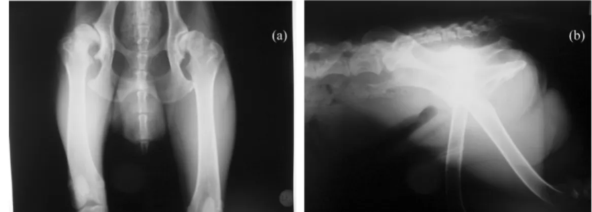
(2) 170. Nam-Soo Kim et al.. Fig. 1. Ventrodorsal (a) and lateral (b) radiographs of the pelvis revealed dysplasia and subluxation of the right coxofemoral joint.. Fig. 2. A test implantation of the acetabular cup.. Fig. 3. Reaming of the femoral canal.. prepared and when it reached a doughy state, it was thumb packed into the acetabular bed filling the anchor holes and lining the surface. The acetabular cup was then seated and held in place with an acetabular positioner until the cement became polymerized and hardened. The trial endoprosthesis was inserted into the femoral shaft, and a test reduction was performed and it was seemed to be proper fit. The trial endoprosthesis was removed. The femoral canal was then flushed with saline solution and a suction tube was placed. The polymethyl methacrylate cement mixture was injected. Fig. 4. Photograph of the coxofemoral joint after implantation of the artificial prosthesis.. into the medullary canal. When the canal became filled the tube was removed, the femoral stem was inserted until the collar rested on the bone, and held in position until the cement became hardened. Then the head of the endoprosthesis was reduced into the acetabular component (Fig. 4). The joint capsule was securely closed in an overlapping pattern. The muscles, subcutaneous tissues and skin were closed in a routine manner. Postoperative care consisted of limiting the degree of exercise by compartmental confinement for the first week followed by 3 weeks of very limited activity on a leash and continual restriction at cage. After the first operative month exercise was gradually increased. The out side activity was allowed only on a leash with no running, jumping or playing and minimal use of restrain. Postoperative oral cephalexin therapy (Youngil Pharmaceutical, Korea, 25 mg/kg, qid) was continued for 1 week. Physical, radiographic examinations and medical history were used in pre- and post-operative patient evaluation. Immediately after surgery, radiographs of the hip (Figs. 5a and b) were taken to evaluate the position of the prosthesis and how completely the cement filled the area. The radiographs revealed a well polymerization and appropriate placement of the prosthesis. The optimal angle of the hip.
(3) Total hip replacement in a dog. 171. Fig. 5. Ventrodorsal (a) and lateral (b) radiographs of the pelvis taken immediately after THR.. joint following THR appeared to be 135°. For evaluating the function of THR, orthopedic and radiographic examinations of the hip were performed at every 2 weeks for 3 months. The dog began to bear weight with a normal range of motion in the hip and slowly returned to a normal exercise pattern 2 months after the implantation of the prosthesis. THR becomes one of the most successful procedures performed today, with predictably excellent and reproducible results [3]. The minimum age 10 months and body weight 35 pounds reported to be suitable for THR in the dog [10,13]. There is no upper age or size limit for THR. Dogs as old as 14 years have had successful THR [9]. In our patient, the age, size, signalment and outcomes were in consistent with the previous reports [9-11]. In this patient, we used modular prosthesis which is better than the fixedhead prosthesis [11]. A minimum period of 4 weeks after the surgical correction is reported to be adequate for evaluating the function of THR [8]. In our case, however, we followed up for 12 weeks for the evaluation of the functional outcome of the THR. Complications associated with THR include luxation, aseptic loosening, sciatic neurapraxia, infection, femur fracture, patellar luxation, pulmonary embolism etc [4,5,6]. The reported complication rate for THR was 8.7% for dislocation, 7.7% for infection of all origin, 3.2% for fracture and noninfected loose acetabular cups, and 2.2% for sciatic neuroapraxia [8]. However, in our patient THR resulted in satisfactory clinical functions and no complications were observed during 6-month follow up period. The dog began to bear weight with a normal range of motion in the hip and slowly returned to a normal exercise pattern. The findings of this study suggest that canine modular THR could be a successful treatment modality for the management of disabling conditions of the coxofemoral joint and could provide a pain-free normal function for the joint.. Acknowlegments This work was supported by the Korea Science and. Engineering Foundation Grant (No. P01-2004-000-10459-0).. References 1. Bergh MS, Muir P, Markel MD, Manley PA. Femoral bone adaptation to stable long-term cemented total hip arthroplasty in dogs. Vet Surg 2004, 33, 214-220. 2. Bergh MS, Muir P, Markel MD, Manley PA. Femoral bone adaptation to unstable long-term cemented total hip arthroplasty in dogs. Vet Surg 2004, 33, 238-245. 3. Chung WK, Liu D, Foo LSS. Mini-incision total hip replacement-surgical technique and early results. J Orthop Surg 2004, 12, 19-24. 4. Konde LJ, Olmstead ML, Hohn RB. Radiographic evaluation of total hip replacement in the dog. Vet Radiol 1982, 20, 98-106. 5. Liska WD. Femur fractures associated with canine total hip replacement. Vet Surg 2004, 33, 164-172. 6. Montgomery RD, Milton JL, Pernell R, Aberman HM. Total hip arthroplasty for treatment of canine hip dysplasia. Vet Clin North Am Small Anim Pract 1992, 22, 703-719. 7. Olmstead ML, Hohn BH, Turner TM. Technique for canine total hip replacement. Vet Surg 1981, 10, 44-50. 8. Olmstead ML, Hohn BH, Turner TM. A five-year study of 221 total hip replacement in the dog. J Am Vet Med Assoc 1983, 183, 1991-1994. 9. Olmstead ML. Total hip replacement in the dog. Semin Vet Med Surg (Small Anim) 1987, 2, 131-140. 10. Olmstead ML. Total hip replacement. Vet Clin North Am Small Anim Pract 1987, 17, 943-955. 11. Olmstead ML. The canine cemented modular total hip prosthesis. J Am Anim Hosp Assoc 1995, 31, 109-124. 12. Olmstead ML. Canine cemented total hip replacement: State of the art. J Small Anim Pract 1995, 36, 395-399. 13. Tomlinson J, McLaughlin RJr. Total hip replacement: The best treatment for dysplastic dogs with osteoarthrosis. Symposium on CHD: Surgical Management. Vet Med 1996, 91, 118-124..
(4)
수치

관련 문서
