ABSTRACT
Previous pathologic, intravascular imaging, and clinical studies have investigated the association between adverse cardiac events and stent malapposition, including acute stent malapposition (ASM, that is detected at index procedure) and late stent malapposition (LSM, that is detected during follow-up) that can be further classified into late-persistent stent malapposition (LPSM, ASM that remains at follow-up) or late-acquired stent malapposition (LASM, newly developed stent malapposition at follow-up that was not present immediately after index stent implantation). ASM has not been associated with adverse cardiac events compared with non-ASM, even in lesions with large-sized malapposition. The clinical outcomes of LSM may depend on its subtype. The recent intravascular ultrasound studies with long-term follow-up have consistently demonstrated that LASM steadily increased the risk of thrombotic events in patients with first-generation drug-eluting stents (DESs). This association has not yet been identified in LPSM. Accordingly, it is reasonable that approaches to stent malapposition should be based on its relationship with clinical outcomes. ASM may be tolerable after successful stent implantation, whereas prolonged anti-thrombotic medications and/or percutaneous interventions to modify LASM may be considered in selected patients with first-generation DESs. However, these treatments are still questionable due to lack of firm evidences.
Keywords: Coronary artery disease; Percutaneous coronary intervention; Stents
INTRODUCTION
Stent apposition refers to the proximity of struts to the vascular wall.
1-3)Good stent apposition is sufficiently close contact to preclude blood flow between any strut and the underlying artery
2)while stent malapposition is separation of any strut from the intimal surface of the arterial wall that is not overlapping a side branch.
2)The frequency of stent- vessel wall malapposition after percutaneous coronary intervention varies with the clinical scenario, lesion morphology, and the type of stent implanted. Unfortunately, there is a lack of agreement as to its importance and clinical impact.
1)Early short-term intravascular ultrasound (IVUS) studies in small numbers of patients have given way to both long-term IVUS studies in larger numbers of patients as well as to detailed, higher resolution optical
Review Article
Received: May 7, 2020 Revised: Jun 12, 2020 Accepted: Jul 14, 2020 Correspondence to
Myeong-Ki Hong, MD, PhD Division of Cardiology, Severance Cardiovascular Hospital, Yonsei University Health System, 50-1, Yonsei-ro, Seodaemun-gu, Seoul 03722, Korea.
E-mail: [email protected]
Copyright © 2020. The Korean Society of Cardiology
This is an Open Access article distributed under the terms of the Creative Commons Attribution Non-Commercial License (https://
creativecommons.org/licenses/by-nc/4.0) which permits unrestricted noncommercial use, distribution, and reproduction in any medium, provided the original work is properly cited.
ORCID iDs
Seung-Yul Lee
https://orcid.org/0000-0002-9039-9806 Gary S. Mintz
https://orcid.org/0000-0003-3296-8705 Jung-Sun Kim
https://orcid.org/0000-0003-2263-3274 Byeong-Keuk Kim
https://orcid.org/0000-0003-2493-066X Yangsoo Jang
https://orcid.org/0000-0002-2169-3112 Myeong-Ki Hong
https://orcid.org/0000-0002-2090-2031
FundingThis work was supported by grants from the Korea Health Technology Research &
Development Project through the Korea Health
Seung-Yul Lee , MD
1, Gary S. Mintz , MD
2, Jung-Sun Kim , MD
3,
Byeong-Keuk Kim , MD
3, Yangsoo Jang , MD
3, and Myeong-Ki Hong , MD, PhD
31
Regional Cardiocerebrovascular Center, Wonkwang University Hospital, Iksan, Korea
2
Cardiovascular Research Foundation, New York, NY, USA
3
Division of Cardiology, Severance Cardiovascular Hospital, Yonsei University Health System, Seoul, Korea
Long-term Clinical Outcomes of Drug-
Eluting Stent Malapposition
Industry Development Institute, funded by the Ministry of Health & Welfare, Republic of Korea (No. HI17C0882, HI16C2211, and HI15C2782);
the Bio & Medical Technology Development Program of the National Research Foundation funded by the Korean government (No.
2015M3A9C6031514); the Cardiovascular Research Center, Seoul, Korea; and the Wonkwang University in 2020.
Conflict of Interest
Mintz has received honoraria from Boston Scientific, Philips/Volcano, Medtronic, and Terumo. The remaining authors have no disclosures to report.
Author Contributions
Conceptualization: Lee SY, Hong MK;
Supervision: Hong MK; Writing - original draft:
Lee SY, Mintz GS, Hong MK; Writing - review &
editing: Mintz GS, Kim JS, Kim BK, Jang Y.
coherence tomography (OCT) studies. This review provides a comprehensive summary of the relationship between coronary stent malapposition and its long-term clinical outcomes based on both IVUS and OCT studies.
ACUTE STENT MALAPPOSITION
Definition, prevalence, and mechanisms
The prevalence of acute stent malapposition (ASM), occurring immediately after
implantation, is higher when assessed using OCT than with IVUS. On IVUS, ASM has been reported from 7.3% to 38.5% (averaging approximately 13%); on OCT, the prevalence varies from 39.1% to 72.3% (averaging approximately 51%) (Table 1).
There are 2 main mechanisms of ASM.
1)4)5)The most common is a stent whose cross-sectional area is smaller than that of the surrounding lumen due to an undersized stent or an intra- stent aneurysmal/ectatic segment or at the proximal edge because of lumen tapering (for example, proximal to a side branch when treating a bifurcation lesion). The lesser cause of ASM is the transition from non-calcified to calcified plaque (especially a calcified nodule) because the expanded stent cannot conform to abrupt changes in lumen geometry.
Clinical outcomes
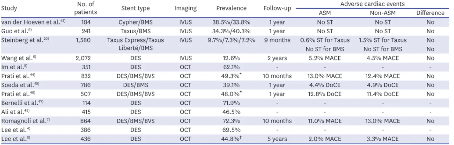
Table 1 summarizes the clinical outcomes of ASM. To date there has been no dedicated, prospective study; and most studies are based on retrospective or sub-group analyses.
Nevertheless, despite the heterogeneity of study design, patient characteristics, stent types, and imaging modalities, studies consistently show that adverse cardiac events do not differ between patients with vs without ASM.
The IVUS study by Wang et al.
6)included 2,072 patients with 2,446 lesions from ADAPT- DES (Assessment of Dual Antiplatelet Therapy With Drug-Eluting Stents) study which was a prospective, multicenter registry designed to assess the relationship between platelet reactivity and other clinical and procedural variables vs subsequent stent thrombosis (ST) and adverse clinical events in patients successfully treated with drug-eluting stent
Table 1. Studies investigating ASM
Study No. of
patients Stent type Imaging Prevalence Follow-up Adverse cardiac events
ASM Non-ASM Difference
van der Hoeven et al.
43)184 Cypher/BMS IVUS 38.5%/33.8% 1 year No ST No ST No
Guo et al.
9)241 Taxus/BMS IVUS 34.3%/40.3% 1 year No ST No ST No
Steinberg et al.
20)1,580 Taxus Express/Taxus
Liberté/BMS IVUS 9.7%/7.3%/7.2% 9 months 0.6% ST for Taxus 1.5% ST for Taxus No No ST for BMS No ST for BMS No
Wang et al.
6)2,072 DES IVUS 12.6% 2 years 5.2% MACE 4.5% MACE No
Im et al.
5)351 DES OCT 62.1% - - - -
Prati et al.
44)832 DES/BMS/BVS OCT 49.3%
*10 months 13.0% MACE 12.4% MACE No
Soeda et al.
45)786 DES/BMS OCT 39.1% 1 year 4.4% DoCE 4.9% DoCE No
Prati et al.
46)507 DES/BMS/BVS OCT 48.0%
*1 year 12.8% DoCE 11.4% DoCE No
Bernelli et al.
47)114 DES OCT 71.9% - - - -
Ali et al.
48)415 DES OCT 46.5% - - - -
Romagnoli et al.
7)864 DES/BMS/BVS OCT 72.3% 10 months 11.0% MACE 13.0% MACE No
Lee et al.
4)386 DES OCT 69.5% - - - -
Lee et al.
8)436 DES OCT 44.8%
†5 years 2.0% MACE 3.3% MACE No
ASM = acute stent malapposition; BMS = bare metal stent; BVS = bioresorbable vascular scaffold; DES = drug-eluting stent; DoCE = device-oriented clinical end point; IVUS = intravascular ultrasound; MACE = major adverse cardiac events; OCT = optical coherence tomography; ST = stent thrombosis.
*
Lesions with >200 μm of maximum malapposed distance;
†Lesions with ≥400 μm of maximum malapposed distance or ≥1 mm of maximum malapposed length.
(DES). At 2-year follow-up, there was no significant difference in the incidence of cardiac death; myocardial infarction; early, late, or very late ST; or clinically driven target lesion revascularization in patients with ASM vs those without ASM.
6)In fact, the largest areas of ASM were not associated with events. These results were consistent even when ASM was forced into the multivariable model.
6)The study by Romagnoli et al.
7)was the most detailed quantitative OCT analysis to date. It analyzed post-procedural OCT findings in 864 patients undergoing percutaneous coronary intervention, assessing prevalence and magnitude of ASM and exploring correlation with clinical outcomes.
7)At a median follow-up of 302 days, ASM did not affect risk of major adverse cardiac events (MACE) regardless of its size or length; residual ASM was comparable in terms of size (median 210 μm vs 200 μm distance) and length (2.0 mm vs 2.2 mm) in patients with vs without MACE.
7)The OCT study by Lee et al.
8)was a pooled analysis from 6 small randomized trials and included 436 patients with 444 non-complex lesions treated with DES. Adverse cardiac events at 5-year follow-up were compared according to the severity, not simply the presence of ASM.
The rate of MACE was 3.3% in patients with ASM ≥400 μm of maximum malapposed strut distance vs 3.1% in those with no ASM or ASM <400 μm (p=0.89) and 1.2% in patients with ASM ≥1mm of maximum malapposed strut length vs 4.6% in those with no ASM or ASM
<1mm of maximum malapposed strut length (p=0.06).
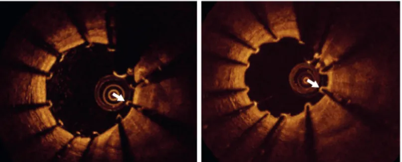
8)Approximately half of ASM resolve at follow-up (Figure 1). Guo et al.
9)reported that 40%
of ASM identified by IVUS resolved at follow-up. Im et al.
5)similarly reported that 69% of OCT-defined ASM resolved spontaneously. Resolution of ASM depends on its size, especially when the distance from the stent to the vessel wall is less than 350–400 μm.
5)10)11)Conversely, Kolandaivelu et al.
12)examined the thrombogenicity of malapposed struts via in vitro experiments. In single-strut 2-dimensional simulations with various detachment distances, stent-wall recirculation zones, highly thrombogenic areas around struts, first grew in size, shifted down-stream, and then lost stent communication.
12)However, when the distance between stent strut and wall was greater than 320 μm, the recirculation zones became smaller and eventually disappeared.
12)Thus, smaller ASM resolve while larger ASM may not affect the blood flow adjacent to arterial wall and, instead, just float in the lumen like struts across the ostium of a side branch.
13)Figure 1. Resolution of acute stent malapposition assessed by optical coherence tomography. The malapposed
strut (arrow) at index procedure (left) was resolved at 1 year of follow-up (right) through neointimal integration
between stent strut and vessel wall.
LATE STENT MALAPPOSITION
Definition, prevalence, and mechanisms
Late stent malapposition (LSM) refers to stent malapposition that is identified at follow-up using IVUS or OCT. LSM can be further classified into late-persistent stent malapposition (LPSM) or late-acquired stent malapposition (LASM). LPSM is ASM that remains visible at follow-up while LASM is newly developed stent malapposition at follow-up that was not present immediately after stent implantation.
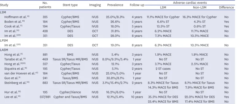
1)2)Table 2 summarizes the prevalence of LSM (combining LPSM and LASM) or just LASM or LPSM in IVUS or OCT studies: 8.3% to 31.8% for LSM and 2.7% to 30.8% for LASM.
Importantly, LSM or LASM was higher in first-generation DES compared with bare metal stents (BMSs). The differentiation between LASM and LPSM requires IVUS or OCT at baseline and at follow-up. However, coronary artery aneurysms can be observed at follow- up angiography.
14)Because late aneurysm formation represents a type of large-sized LASM, a comparison of angiography between post-intervention and follow-up may be sometimes useful to discriminate between LPSM and LASM in patients who did not have serial intravascular imaging.
Positive vessel remodeling is the leading mechanism for LASM. In the IVUS report by Mintz et al.,
15)there was an increase in external elastic membrane radius within the region of LASM, but no change in plaque mass. Besides regional positive vascular remodeling, abluminal thrombus dissolution can contribute to LASM, especially in acute coronary syndromes.
1)Post-intervention and follow-up IVUS or OCT is required to differentiate LASM caused by positive remodeling vs LASM caused by thrombus dissolution.
Table 2. Studies investigating clinical outcomes of LSM
Study No.
patients Stent type Imaging Prevalence Follow-up Adverse cardiac events
LSM Non-LSM Difference
LSM
Hoffmann et al.
16)325 Cypher/BMS IVUS 25.0%/8.3% 4 years 11.1% MACE for Cypher 16.3% MACE for Cypher No
Boden et al.
49)184 Cypher/BMS IVUS 26.6% 5 years 6.6% ST 4.3% ST Yes
Cook et al.
17)194 Cypher/Taxus IVUS 18.0% 5 years 13.5% ST 0.6% ST Yes
Im et al.
50)428 DES OCT 31.8% 6 years 6.2% MACE 11.7% MACE No
Im et al.
5)33)351 DES OCT 28.0% 8 years 7.3% MACE 10.5% MACE No
LPSM
Im et al.
5)33)351 DES OCT 19.0% 8 years 6.3% MACE 10.5% MACE No
LASM
Hong et al.
21)881 BMS IVUS 5.4% 3 years 1.9% MACE 1.8% MACE No
Tanabe et al.
19)469 Taxus SR/Taxus MR/BMS IVUS 8.0%/9.5%/5.4% 1 year No ST No ST No
Hong et al.
22)23)557 Cypher/Taxus IVUS 12.1% 3 years 3.7% MACE 2.5% MACE No
Siqueira et al.
18)195 Cypher/Taxus IVUS 5.1% 2 years 2 ST cases No ST No
van der Hoeven et al.
43)184 Cypher/BMS IVUS 25.0%/5.0% 1 year No ST No ST No
Guo et al.
9)241 Taxus/BMS IVUS 30.8%/8.1% 1 year No ST No ST No
Steinberg et al.
20)883 Taxus SR/Taxus MR/BMS IVUS 3.1%/15.4%/2.7% 2 years 8.3% MACE for Taxus 8.1% MACE for Taxus No 14.3% MACE for BMS 7.9% MACE for BMS No
Hur et al.
39)195 Cypher/Xience IVUS 16.2%/6.0% 1 year No ST No ST No
Lee et al.
24)557/881 Cypher and Taxus/BMS IVUS 12.1%/5.4% 10 years 35.5% MACE for DES 22.0% MACE for DES Yes
23.4% MACE for BMS 17.4% MACE for BMS No
BMS = bare metal stent; DES = drug-eluting stent; IVUS = intravascular ultrasound; LASM = late-acquired stent malapposition; LPSM = late-persistent stent
malapposition; LSM = late stent malapposition; MACE = major adverse cardiac events; MR = moderate release; OCT = optical coherence tomography; SR = slow
release; ST = stent thrombosis.
Clinical outcomes
Table 2 summarizes the clinical outcomes of LSM, LASM, or LPSM. Similar to ASM, a dedicated prospective study is absent; and most studies have been based on retrospective or sub-group analyses from randomized trials or registries. Unlike studies of ASM, results from studies of LSM are not consistent.
The study by Hoffmann et al.
16)was a pooled analysis from the RAVEL (RAndomised study with the sirolimus-eluting Bx VELocity-stent), E-SIRIUS (European, multicentre, randomised, double-blind trial of the SIRolImUS- coated Bx VELOCITY stent in the
treatment of patients with de novo coronary artery lesions), and SIRIUS (SIRolImUS-eluting stent in de novo coronary lesions) comparing sirolimus-eluting stents vs bare metal stents) studies. It included a total of 325 patients that had follow-up IVUS at 6 to 8 months after stent implantation after which 4-year clinical follow-up was available in all included patients.
16)The frequency of MACE was 11.1% for patients with LSM vs 16.3% for those without LSM (p=0.48).
16)In contrast to this study, Cook et al.
17)reported a sub-group analysis of SIRTAX (Sirolimus-Eluting Versus Paclitaxel-Eluting Stents for Coronary Revascularization) showing that very late ST out to 4 years occurred more frequently among patients with vs without incidentally-detected IVUS LSM at 8 months (13.5% vs 0.6%, respectively, p<0.001).
However, the type of LSM (LPSM vs LASM) could not be differentiated in the previous 2 studies because of the lack of post-intervention IVUS.
16)17)One registry with a small number of patients with LASM (n=10 patients) raised the possibility of a link between LASM and poor clinical outcomes during median 24.3 months follow- up.
18)Conversely, an IVUS study from TAXUS II showed that no ST occurred in patients with LASM over a period of 12 months.
19)Furthermore, an integrated IVUS analysis of the TAXUS IV, V, and VI and TAXUS ATLAS Workhorse, Long Lesion, and Direct Stent studies reported LASM in 7 BMS patients, 17 patients with slow-release TAXUS, and 12 patients with moderate-release TAXUS.
20)Over the 2 ensuing years, MACE rates were similar in patients with vs without LASM for each of these 3 stent types.
20)Two studies by Hong et al.
21-23)using a large retrospective IVUS registry initially reported that incidentally detected LASM after implantation of first-generation DES or BMSs was not associated with MACE during the first 3-year follow-up period. However, with extended follow-up out to 10 years, LASM was related to a greater risk of MACE (hazard ratio [HR], 1.67; 95% confidence interval [CI], 1.04–2.67;
p=0.03) and very late ST (HR, 3.53; 95% CI, 1.15–10.80; p=0.03) vs non-LASM in patients
treated with first-generation DESs, but not in patients treated with bare-metal stents.
24)Virmani et al.
25)reported the first case of fatal acute myocardial infarction and cardiac
rupture as a result of late thrombosis of a first-generation sirolimus-eluting stent deployed
18 months previously. This patient underwent IVUS immediately after stent implantation
and at 8-month follow-up when angiographic and IVUS demonstrated enlargement of the
stented arterial segments with LASM, indicating positive vessel remodeling.
25)Autopsy
showed aneurysmal dilatation of these segments with severe localized hypersensitivity
consisting predominantly of T-lymphocytes and eosinophils.
25)Cook et al.
26)analyzed
thrombus aspirates in patients presenting with very late DES thrombosis and demonstrated
that eosinophils were more common in thrombi harvested from very late DES thrombosis
vs aspirates from spontaneous acute myocardial infarction, early bare-metal ST, early DES
thrombosis, and late bare-metal ST. The eosinophil counts also correlated with extent of
LSM.
26)Thus, chronic inflammation and hypersensitivity can weaken the vascular wall, lead
to LASM, and induce local stasis of blood flow within the positively remodeled segments.
27)These inflammatory responses have been observed most frequently in the first-generation sirolimus-eluting stent among all coronary stents, including BMSs, other first-generation DES, new-generation DES, and polymer-free DESs, highlighting the clinical significance of polymers and drugs in DES system design.
28-32)There is little data regarding LPSM. One OCT study including 351 patients with 356 DES- treated lesions reported that 31% of ASM remained malapposed at 6-month follow-up, and LPSM was not associated with 8-year MACE.
5)33)CLINICAL APPROACHES TO DRUG-ELUTING STENTS MALAPPOSITION
Clinical approach to acute stent malapposition
The recent consensus paper from the European Association of Percutaneous Cardiovascular Interventions recommended treating ASM having an axial distance ≥400 μm or longitudinal length ≥1 mm.
34)This recommendation was based on OCT studies showing that ASM ≥400 μm usually persists and 3 OCT-based ST registry studies showing that stent malapposition was one of leading mechanisms of (very) late ST.
35-37)However, these registries did not have control groups, and OCT was performed only at the time of ST. ASM can usually be corrected using a balloon sized to the diameter of the arterial lumen at the site of malapposition inflated to nominal (not high) pressures. For percutaneous coronary intervention of bifurcation lesions, the recent consensus document from the European Bifurcation Club suggested that a stent optimization (the so-called POT technique) should be performed routinely during the bifurcation procedure before side branch rewiring as it facilitated access towards the side branch and reduced the possibility that the wire might cross into the side branch behind the main-branch stent along with main branch stent crush.
38)Clinical approach to late stent malapposition
There is no recommendation or consensus on proper managements of patients with incidentally detected LSM. However, fortunately, rapid advances of DES technology have made stent struts thinner and have applied biocompatible or biodegradable polymers to metallic struts. This progress has reduced the thrombogenicity of new-generation DES.
From the autopsy study by Otsuka et al.,
31)second-generation cobalt-chromium everolimus- eluting stents (Xience
®; Abbott Vascular, Santa Clara, CA, USA or Promus
®; Boston Scientific, Marlborough, MA, USA) demonstrated greater strut coverage with less inflammation, less fibrin deposition, and less late and very late ST compared with first-generation DES.
Clinically, these everolimus-eluting stents showed a lower frequency of LASM than first- generation sirolimus-eluting stents at 8-month follow-up IVUS
39)and were associated with a significant reduction of ST compared with first-generation DES.
40)When the type of LSM is discriminated as LASM rather than LPSM, the long-term clinical outcomes appear to be worse. Thus, prolonged dual antiplatelet or anticoagulation therapy may be considered in patients having LASM. The study from Doi et al.
41)showed that patients with coronary artery ectasia receiving optimal anticoagulation therapy did not experience the occurrence of MACE. Surgical resection or percutaneous angioplasty with covered stents may be considered for multiple or giant coronary artery aneurysms.
42)In contrast to LASM, the studies by Im et
al.
5)33)suggest that treatments may not be necessary for patients with LPSM.
CONCLUSION
The effects of DES malapposition on long-term clinical outcomes have been controversial.
Regardless of its severity, ASM has not been associated with adverse cardiac events across all IVUS and OCT studies. In contrast to ASM, cumulative evidences have demonstrated that LASM of first-generation DES is likely to cause (very) late ST through chronic inflammatory reactions.
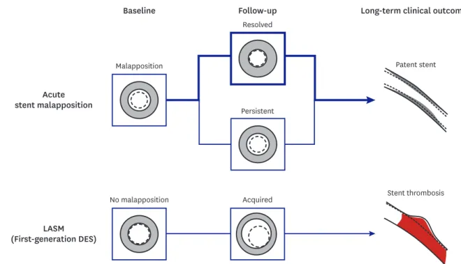
This association is not clear in LPSM and may be attenuated in recent DESs which have thin struts and biocompatible/biodegradable polymers. Figure 2 provides the illustrative overview about the association between DES malapposition and long-term clinical outcomes. Although prolonged anti-thrombotic therapy and/or percutaneous intervention using large-sized balloon or covered stent may be considered in patients with LASM, the treatments should be made considering the clinical situations of individual patient due to lack of firm evidences.
REFERENCES
1. Attizzani GF, Capodanno D, Ohno Y, Tamburino C. Mechanisms, pathophysiology, and clinical aspects of incomplete stent apposition. J Am Coll Cardiol 2014;63:1355-67.
PUBMED | CROSSREF
2. Mintz GS, Nissen SE, Anderson WD, et al. American College of Cardiology clinical expert consensus document on standards for acquisition, measurement and reporting of intravascular ultrasound studies (IVUS). a report of the American College of Cardiology task force on clinical expert consensus documents. J Am Coll Cardiol 2001;37:1478-92.
PUBMED | CROSSREF
3. Tearney GJ, Regar E, Akasaka T, et al. Consensus standards for acquisition, measurement, and reporting of intravascular optical coherence tomography studies: a report from the International Working Group for Intravascular Optical Coherence Tomography Standardization and Validation. J Am Coll Cardiol 2012;59:1058-72.
PUBMED | CROSSREF
Acute stent malapposition
(First-generation DES) LASM
Baseline Follow-up Long-term clinical outcomes
Resolved
Patent stent
Stent thrombosis Persistent
Malapposition
Acquired No malapposition
Figure 2. Overview of stent malapposition assessed by intravascular imaging modality. Even though malapposed struts immediately after stent implantation
remain occasionally during follow-up, they usually do not link to ST. However, LASM increases the risk of ST through chronic inflammatory reactions especially in first-generation DESs.
DES = drug-eluting stent; LASM = late-acquired stent malapposition; ST = stent thrombosis.
4. Lee SY, Ahn CM, Yoon HJ, et al. Early follow-up optical coherence tomographic findings of significant drug-eluting stent malapposition. Circ Cardiovasc Interv 2018;11:e007192.
PUBMED | CROSSREF
5. Im E, Kim BK, Ko YG, et al. Incidences, predictors, and clinical outcomes of acute and late stent malapposition detected by optical coherence tomography after drug-eluting stent implantation. Circ Cardiovasc Interv 2014;7:88-96.
PUBMED | CROSSREF
6. Wang B, Mintz GS, Witzenbichler B, et al. Predictors and long-term clinical impact of acute stent malapposition: an assessment of dual antiplatelet therapy with drug-eluting stents (ADAPT-DES) intravascular ultrasound substudy. J Am Heart Assoc 2016;5:e004438.
PUBMED | CROSSREF
7. Romagnoli E, Gatto L, La Manna A, et al. Role of residual acute stent malapposition in percutaneous coronary interventions. Catheter Cardiovasc Interv 2017;90:566-75.
PUBMED | CROSSREF
8. Lee SY, Im E, Hong SJ, et al. Severe acute stent malapposition after drug-eluting stent implantation:
effects on long-term clinical outcomes. J Am Heart Assoc 2019;8:e012800.
PUBMED | CROSSREF
9. Guo N, Maehara A, Mintz GS, et al. Incidence, mechanisms, predictors, and clinical impact of acute and late stent malapposition after primary intervention in patients with acute myocardial infarction:
an intravascular ultrasound substudy of the harmonizing outcomes with revascularization and stents in acute myocardial infarction (HORIZONS-AMI) trial. Circulation 2010;122:1077-84.
PUBMED | CROSSREF
10. Kawamori H, Shite J, Shinke T, et al. Natural consequence of post-intervention stent malapposition, thrombus, tissue prolapse, and dissection assessed by optical coherence tomography at mid-term follow- up. Eur Heart J Cardiovasc Imaging 2013;14:865-75.
PUBMED | CROSSREF
11. Shimamura K, Kubo T, Akasaka T, et al. Outcomes of everolimus-eluting stent incomplete stent apposition: a serial optical coherence tomography analysis. Eur Heart J Cardiovasc Imaging 2015;16:23-8.
PUBMED | CROSSREF
12. Kolandaivelu K, Swaminathan R, Gibson WJ, et al. Stent thrombogenicity early in high-risk interventional settings is driven by stent design and deployment and protected by polymer-drug coatings. Circulation 2011;123:1400-9.
PUBMED | CROSSREF
13. Sawaya FJ, Lefèvre T, Chevalier B, et al. Contemporary approach to coronary bifurcation lesion treatment.
JACC Cardiovasc Interv 2016;9:1861-78.
PUBMED | CROSSREF
14. Hong SJ, Kim H, Ahn CM, et al. Coronary artery aneurysm after second-generation drug-eluting stent implantation. Yonsei Med J 2019;60:824-31.
PUBMED | CROSSREF
15. Mintz GS, Shah VM, Weissman NJ. Regional remodeling as the cause of late stent malapposition.
Circulation 2003;107:2660-3.
PUBMED | CROSSREF
16. Hoffmann R, Morice MC, Moses JW, et al. Impact of late incomplete stent apposition after sirolimus- eluting stent implantation on 4-year clinical events: intravascular ultrasound analysis from the multicentre, randomised, RAVEL, E-SIRIUS and SIRIUS trials. Heart 2008;94:322-8.
PUBMED | CROSSREF
17. Cook S, Eshtehardi P, Kalesan B, et al. Impact of incomplete stent apposition on long-term clinical outcome after drug-eluting stent implantation. Eur Heart J 2012;33:1334-43.
PUBMED | CROSSREF
18. Siqueira DA, Abizaid AA, Costa JR, et al. Late incomplete apposition after drug-eluting stent implantation: incidence and potential for adverse clinical outcomes. Eur Heart J 2007;28:1304-9.
PUBMED | CROSSREF
19. Tanabe K, Serruys PW, Degertekin M, et al. Incomplete stent apposition after implantation of paclitaxel- eluting stents or bare metal stents: insights from the randomized TAXUS II trial. Circulation 2005;111:900-5.
PUBMED | CROSSREF
20. Steinberg DH, Mintz GS, Mandinov L, et al. Long-term impact of routinely detected early and late incomplete stent apposition: an integrated intravascular ultrasound analysis of the TAXUS IV, V, and VI and TAXUS ATLAS workhorse, long lesion, and direct stent studies. JACC Cardiovasc Interv 2010;3:486-94.
PUBMED | CROSSREF
21. Hong MK, Mintz GS, Lee CW, et al. Incidence, mechanism, predictors, and long-term prognosis of late stent malapposition after bare-metal stent implantation. Circulation 2004;109:881-6.
PUBMED | CROSSREF
22. Hong MK, Mintz GS, Lee CW, et al. Late stent malapposition after drug-eluting stent implantation: an intravascular ultrasound analysis with long-term follow-up. Circulation 2006;113:414-9.
PUBMED | CROSSREF
23. Hong MK, Mintz GS, Lee CW, et al. Impact of late drug-eluting stent malapposition on 3-year clinical events. J Am Coll Cardiol 2007;50:1515-6.
PUBMED | CROSSREF
24. Lee SY, Ahn JM, Mintz GS, et al. Ten-year clinical outcomes of late-acquired stent malapposition after coronary stent implantation. Arterioscler Thromb Vasc Biol 2020;40:288-95.
PUBMED | CROSSREF
25. Virmani R, Guagliumi G, Farb A, et al. Localized hypersensitivity and late coronary thrombosis secondary to a sirolimus-eluting stent: should we be cautious? Circulation 2004;109:701-5.
PUBMED | CROSSREF
26. Cook S, Ladich E, Nakazawa G, et al. Correlation of intravascular ultrasound findings with
histopathological analysis of thrombus aspirates in patients with very late drug-eluting stent thrombosis.
Circulation 2009;120:391-9.
PUBMED | CROSSREF
27. Karalis I, Ahmed TA, Jukema JW. Late acquired stent malapposition: why, when and how to handle? Heart 2012;98:1529-36.
PUBMED | CROSSREF
28. Lüscher TF, Steffel J, Eberli FR, et al. Drug-eluting stent and coronary thrombosis: biological mechanisms and clinical implications. Circulation 2007;115:1051-8.
PUBMED | CROSSREF
29. Wilson GJ, Nakazawa G, Schwartz RS, et al. Comparison of inflammatory response after implantation of sirolimus- and paclitaxel-eluting stents in porcine coronary arteries. Circulation 2009;120:141-9.
PUBMED | CROSSREF
30. Nakazawa G, Finn AV, Vorpahl M, Ladich ER, Kolodgie FD, Virmani R. Coronary responses and differential mechanisms of late stent thrombosis attributed to first-generation sirolimus- and paclitaxel- eluting stents. J Am Coll Cardiol 2011;57:390-8.
PUBMED | CROSSREF
31. Otsuka F, Vorpahl M, Nakano M, et al. Pathology of second-generation everolimus-eluting stents versus first-generation sirolimus- and paclitaxel-eluting stents in humans. Circulation 2014;129:211-23.
PUBMED | CROSSREF
32. Rizas KD, Mehilli J. Stent polymers: do they make a difference? Circ Cardiovasc Interv 2016;9:e002943.
PUBMED | CROSSREF
33. Im E, Hong SJ, Ahn CM, et al. Long-term clinical outcomes of late stent malapposition detected by optical coherence tomography after drug-eluting stent implantation. J Am Heart Assoc 2019;8:e011817.
PUBMED | CROSSREF
34. Räber L, Mintz GS, Koskinas KC, et al. Clinical use of intracoronary imaging. Part 1: guidance and optimization of coronary interventions. An expert consensus document of the European Association of Percutaneous Cardiovascular Interventions. Eur Heart J 2018;39:3281-300.
PUBMED | CROSSREF
35. Souteyrand G, Amabile N, Mangin L, et al. Mechanisms of stent thrombosis analysed by optical coherence tomography: insights from the national PESTO French registry. Eur Heart J 2016;37:1208-16.
PUBMED | CROSSREF
36. Taniwaki M, Radu MD, Zaugg S, et al. Mechanisms of very late drug-eluting stent thrombosis assessed by optical coherence tomography. Circulation 2016;133:650-60.
PUBMED | CROSSREF
37. Adriaenssens T, Joner M, Godschalk TC, et al. Optical coherence tomography findings in patients with coronary stent thrombosis: a report of the PRESTIGE consortium (prevention of late stent thrombosis by an interdisciplinary global European effort). Circulation 2017;136:1007-21.
PUBMED | CROSSREF
38. Banning AP, Lassen JF, Burzotta F, et al. Percutaneous coronary intervention for obstructive bifurcation lesions: the 14th consensus document from the European Bifurcation Club. EuroIntervention 2019;15:90-8.
PUBMED | CROSSREF
39. Hur SH, Lee BR, Kim SW, et al. Late-acquired incomplete stent apposition after everolimus-eluting stent versus sirolimus-eluting stent implantation in patients with non-ST-segment elevation myocardial infarction and ST-segment elevation myocardial infarction. EuroIntervention 2016;12:e979-86.
PUBMED | CROSSREF
40. Palmerini T, Kirtane AJ, Serruys PW, et al. Stent thrombosis with everolimus-eluting stents: meta-analysis of comparative randomized controlled trials. Circ Cardiovasc Interv 2012;5:357-64.
PUBMED | CROSSREF
41. Doi T, Kataoka Y, Noguchi T, et al. Coronary artery ectasia predicts future cardiac events in patients with acute myocardial infarction. Arterioscler Thromb Vasc Biol 2017;37:2350-5.
PUBMED | CROSSREF
42. Kawsara A, Núñez Gil IJ, Alqahtani F, Moreland J, Rihal CS, Alkhouli M. Management of coronary artery aneurysms. JACC Cardiovasc Interv 2018;11:1211-23.
PUBMED | CROSSREF
43. van der Hoeven BL, Liem SS, Dijkstra J, et al. Stent malapposition after sirolimus-eluting and bare-metal stent implantation in patients with ST-segment elevation myocardial infarction: acute and 9-month intravascular ultrasound results of the MISSION! intervention study. JACC Cardiovasc Interv 2008;1:192-201.
PUBMED | CROSSREF
44. Prati F, Romagnoli E, Burzotta F, et al. Clinical impact of OCT findings during PCI: the CLI-OPCI II study.
JACC Cardiovasc Imaging 2015;8:1297-305.
PUBMED | CROSSREF
45. Soeda T, Uemura S, Park SJ, et al. Incidence and clinical significance of poststent optical coherence tomography findings: one-year follow-up study from a multicenter registry. Circulation 2015;132:1020-9.
PUBMED | CROSSREF
46. Prati F, Romagnoli E, Gatto L, et al. Clinical impact of suboptimal stenting and residual intrastent plaque/
thrombus protrusion in patients with acute coronary syndrome: the CLI-OPCI ACS substudy (Centro per la Lotta Contro L'Infarto-Optimization of Percutaneous Coronary Intervention in Acute Coronary Syndrome). Circ Cardiovasc Interv 2016;9:e003726.
PUBMED | CROSSREF
47. Bernelli C, Shimamura K, Komukai K, et al. Impact of culprit plaque and atherothrombotic components on incomplete stent apposition in patients with ST-elevation myocardial infarction treated with everolimus-eluting stents - an OCTAVIA substudy. Circ J 2016;80:895-905.
PUBMED | CROSSREF
48. Ali ZA, Maehara A, Généreux P, et al. Optical coherence tomography compared with intravascular ultrasound and with angiography to guide coronary stent implantation (ILUMIEN III: OPTIMIZE PCI): a randomised controlled trial. Lancet 2016;388:2618-28.
PUBMED | CROSSREF
49. Boden H, van der Hoeven BL, Liem SS, et al. Five-year clinical follow-up from the MISSION! intervention study: sirolimus-eluting stent versus bare metal stent implantation in patients with ST-segment elevation myocardial infarction, a randomised controlled trial. EuroIntervention 2012;7:1021-9.
PUBMED | CROSSREF
50. Im E, Lee SY, Hong SJ, et al. Impact of late stent malapposition after drug-eluting stent implantation on long-term clinical outcomes. Atherosclerosis 2019;288:118-23.
PUBMED | CROSSREF