549 Original Article
Korean Circulation J 2006;36:549-552
ISSN 1738-5520
ⓒ 2006, The Korean Society of Circulation CASE REPORT
Unruptured Aneurysm of the Left Sinus of Valsalva Presenting as Acute Coronary Syndrome: A Case Report
So-Ra Park, MD, Jin-Yong Hwang, MD, Yong-Ran Kang, MD, Min-Gyeong Kang, MD, Mung-Gi Seo, MD, Sung-Ji Park, MD, Bong-Ryong Choi, MD and Choong-Hwan Kwak, MD Department of Internal Medicine, Gyeongsang National University College of Medicine, Jinju, Korea ABSTRACT
We report here on a case of a 34-year-old man with unruptured aneurysm of the left sinus of Valsalva, and he presented with acute coronary syndrome due to the putative dynamic compression of both the left main co- ronary artery and the left circumflex coronary artery. The cardiac multislice computed tomography scanning and coronary angiogram revealed the compression of the two coronary arteries by the aneurysm of the left sinus of Valsalva. Aneurysmectomy was performed for surgical repair. After the surgery, the patient stayed asymptomatic during the 6-months of follow-up. (Korean Circulation J 2006;36:549-552)
KEY WORDS:Aneurysm;Sinus of Valsalva;Acute coronary syndrome.
Introduction
Although acute coronary syndrome(ACS) is gene- rally caused by coronary arterial diseases, ACS in a few patients results from compression of the coronary art- ery by adjacent extra-coronary arterial structures such as aneurysm and tumor in the aorta or the heart. An aneurysm of the sinus of Valsalva is a rare congenital malformation.1) This unruptured aneurysm is usually asymptomatic;1) however, the aneurysm of the left sinus of Valsalva(ALSV) can compress the left coronary art- ery because of its adjacent location, which can result in myocardial ischemia.5-10) Unlike the previously reported cases that presented with effort angina and myocardial infarction, we report here on a 34-year-old man with unruptured ALSV, and he presented with resting chest pain and syncope that were probably due to the dyna- mic compression of the left main coronary artery and the left circumflex artery by the aneurysm.
Case
A 34-year-old man was admitted to the cardiology
department due to recurrent resting chest pain and syncope that occurred in the middle of night. Three weeks previously, he developed substernal chest pain at midnight; the pain’s nature was squeezing and its duration was several minutes. One week ago, the chest pain and syncope recurred at midnight. He was a non- smoker and had no history of hypertension or diabetes mellitus. On admission, the physical examination and laboratory data, including the cardiac enzyme levels, did not showed any abnormalities. The chest radiograph delineated a normal cardiothoracic ratio and there was no focal lung lesion. The electrocardiogram showed a rS pattern of the QRS, T-wave inversion in the inferior leads and superior axis deviation of the QRS, which all suggested left anterior fascicular block. The transtho- racic and transesophageal echocardiograms(Fig. 1) de- monstrated a large unruptured ALSV and normal left ventricular systolic function in the absence of any re- gional wall motion abnormality. The exercise treadmill test didn’t give rise to angina or ischemic ST change during maximal exercise. The coronary angiography proved that the left main coronary artery(LMCA) and the proximal left circumflex artery(LCX) were stret- ched and displaced upward(Fig. 2). Ergonovine ch- allenge testing in the right coronary artery was negative.
The multislice spiral computed tomography(CT) and aortography revealed that an isolated aneurysm of the left sinus of Valsalva was compressing the LMCA and proximal LCX(Fig. 2, 3). An operation was performed using standard cardiopulmonary bypass. The aortic
Received:June 9, 2006 Accepted:July 19, 2006
Correspondence:Jin-Yong Hwang, MD, Department of Internal Medicine, Gyeongsang National University College of Medicine, Chiram-dong 90, Jinju 660-702, Korea
Tel: 82-55-750-8064, Fax: 82-55-758-9122 E-mail: [email protected]
550·Korean Circulation J 2006;36:549-542
valve was normal. The unruptured saccular aneurysm
(2.5×2.4 cm in diameter) arose from the central part of the left coronary sinus and the size of the opening of the aneurysm was 4 mm(Fig. 4). The LMCA and proximal LCX were compressed and superiorly dis- placed by the aneurysm(Fig. 4A). The opening of an- eurysm was closed by continuous suture with using double velour Dacron patch, and the aneurysm’s sac was removed. Postoperative multislice spiral CT showed good coronary flow without compression(Fig. 2B). The patient was discharged without complications and he was asymptomatic during 6-months of follow-up.
Discussion
Aneurysms of the right and non-coronary sinuses occur more frequently than those of the left sinus, and
Fig. 2. The coronary angiography showed that the left main coronary artery and proximal left circumflex artery were stretched and displaced upward. The aortography revealed that an isolated aneurysm of the left sinus of Valsalva compressed the left main coronary artery and the proximal left circumflex artery. The black arrows indicate the border of the unruptured aneurysm of the left sinus of Valsalva, and the white arrow indicates the orifice of aneurysm of the left sinus of Valsalva. LMCA: left main coronary artery, LCX: left circumflex artery.
Fig. 3. Preoperative multislice CT scanning revealed that an isolated aneurysm of the left sinus of Valsalva compressed the left main coronary artery and the proximal left circumflex artery (Fig. 2A). Postoperative multislice CT scanning showed good coronary flow without compression (Fig. 2B).
The black arrows indicate that the left main coronary artery and proximal left circumflex artery were compressed by the aneurysm. Ao: aorta, An:
aneurysm, LMCA: left main coronary artery, LAD: left descending artery, LCX: left circumflex artery.
B A
Fig. 1. The transesophageal echocardiography showed an approxi- mately 2 cm-sized echo-lucent sac adjacent to the left coronary cusp of the aortic valve. AV: aortic valve, ALSV: aneurysm of the left sinus of Valsalva.
So-Ra Park, et al:Unruptured Aneurysm of the Left Sinus of Valsalva Presenting as Acute Coronary Syndrome·551
these aneurysms usually protrude and rupture into the right ventricle and right atrium.1) The ALSV is an extremely rare anomaly.2) The unruptured aneurysm of the sinus of Valsalva does not cause any symptoms, but it can be potentially fatal if it ruptures into the cardiac chambers, the pulmonary artery or outside of the heart.3)4) It may be detected in the form of myocardial infarction or ischemia owing to partial or complete compression of the left main coronary artery.5-10)
Not only noninvasive diagnostic tools such as echo- cardiography, multislice CT and magnetic resonance imaging, but also invasive diagnostic tools such as car- diac catheterization aortography and selective coronary arteriography are helpful to demonstrate coronary artery compression.5) A clear and satisfactory image that re- vealed the dynamic obstruction of the coronary artery in this patient was obtained via multislice CT scann- ing and aortography.
An aggressive surgical approach is recommended in order to prevent coronary artery obstruction that can lead to myocardial infarction or severe left ventricular ischemia.8) The surgical methods to treat an unrup- tured aneurysm of the left sinus of Valsalva consist of closure of the opening of the aneurysm, replacement of the aortic valve, coronary artery bypass grafting or a combination of these.8-10) Because this patient had a normal aortic valve and no visible atherosclerosis in his coronary artery, simple closure of the opening of the aneurysm and aneurysmectomy were performed.
After the operation, the patient’s chest pain and syn- cope disappeared.
Although we could not document the solid evidence of myocardial ischemia that was caused by dynamic compression of the left coronary artery, there were several possibilities that his ALSV might have caused the syncope and ischemic chest pain. First, vasospastic angina with and/or without the influence of an ALSV might cause a similar clinical situation. However, he
had no risk factors for variant angina. Particularly, ergonovine testing in the right coronary artery was negative. Although we did not exclude spasm of the left coronary artery, variant angina was unlikely to be a diagnosis in our opinion. Second, we hypothesized that transient compression of the left coronary artery, including the left main coronary artery, might have caused myocardial ischemia and/or an undetected serious ventricular arrhythmia because the ALSV was a very compliant, thin membrane-walled sac whose vo- lume was dependent on the intravascular volume via the opening of ALSV. It was reported that ALSV also might expand very rapidly and influence the coronary circulation and consequently lead to death.11) ALSV has more risks than does an aneurysm of the right sinus of Valsalva.6)8) Therefore, we should consider making emergency surgical repair, especially in the situation where the ALSV may disturb the coronary circulation.
Disappearance of the patient’s chest pain and syncope for six months after removal of ALSV supported this hypothesis
Acute coronary syndrome is mostly caused by ath- erosclerotic coronary artery disease. However, rare congenital anomalies like ALSV are able to cause life- threatening myocardial ischemia. Clinical cardiologist should keep in mind congenital abnormalities as a cause of myocardial ischemia, and especially for the younger patients who are without coronary risk factors.
REFERENCES
1) Guo DW, Cheng TO, Lin ML, Gu ZQ. Aneurysm of the sinus of valsalva: a roentgenologic study of 105 Chinese patients. Am Heart J 1987;114:1169-77.
2) Tahir MZ, Rustom M, al Ebrahim K. Left coronary sinus of valsalva aneurysm: an extremely rare malformation: a case re- port. Angiology 1995;46:753-8.
3) Chakfe N, Kretz JG, Nicolini P, et al. Triple aneurysm of the valsalva sinus complicated by right coronary occlusion: apropos of a case and review of the literature. Ann Chir 1994;48:825-31.
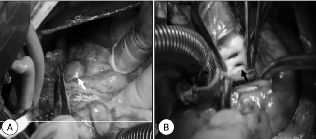
Fig. 4. The unruptured saccular aneurysm (2.5×2.4 cm in diameter) arose from the central part of the left coronary sinus. The left main coronary artery and the proximal left circumflex artery were compressed by the aneurysm(Fig. 4A). The opening of the aneurysm was located in the inferior coronary ostium and the size was 4 mm(Fig. 4B). The white arrow indicates the unruptured aneurysm of the left sinus of Valsalva and the black arrow indicates the orifice of the aneurysm of the left sinus of Valsalva.
B A
552·Korean Circulation J 2006;36:549-542
4) Moon KS, Choi RK, Lim DS, et al. Clinical characteristics in patients with ruptured aneurysm of sinus of valsalva. Korean Circ J 2000;30:183-90.
5) Shin JK, Jung JP, Park CR, et al. Acute myocardial infarction due to unruptured aneurysm of left sinus of valsalva with aortic valve regurgitation. J Card Surg 2005;20:545-8.
6) Bashour TT, Chen F, Yap A, Mason DT, Baladi N. Fatal myo- cardial ischemia caused by compression of the left coronary system by a large left sinus of valsalva aneurysm. Am Heart J 1996;132:1050-2.
7) Ferreira AC, de Marchena E, Mayor M, Bolooki H. Sinus of valsalva aneurysm presenting as myocardial infaction during Dobutamine stress test. Cathet Cardiovasc Diagn 1996;39:400-2.
8) Lijol A, Parodi E, Passerone GC, Scarano F, Caruso D, Iannetti MV. Unruptured of the left sinus of valsalva causing coronary insufficiency. Tex Heart Inst J 2002;29:40-4.
9) Koike S, Takayama S, Furihata A, et al. Infective endocarditis causing acute myocardial infaction by compression of the proxi- mal left coronary artery due to a mycotic aneurysm of the sinus of valsalva. Jpn Cir J 1991;55:1228-32.
10) Takahara Y, Sudo Y, Sunazawa T, Nakajima N. Aneurysm of the left sinus of valsalva producing aortic valve regurgitation and myocardial ischemia. Ann Thorac Surg 1998;65:535-7.
11) Faillace RT, Greenland P, Nanda NC. Rapid expansion of a saccular aneurysm on the left coronary sinus of valsalva: a role for early surgical repair? Br Heart J 1985;54:442-4.