An orthodontic approach for Class III malocclusion in a pediatric cancer patient: A case report
7
0
0
전체 글
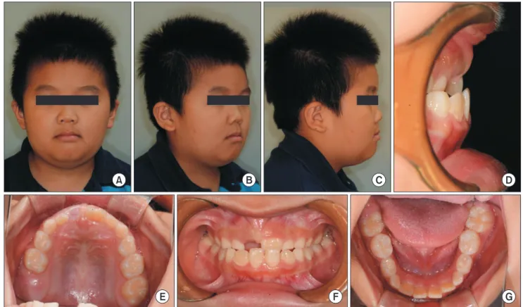
(2) Orthodontic approach in a pediatric cancer patient. training programs often do not emphasize information on. cal history included ALL. Treatment for leukemia was initi-. orthodontic treatment for patients with a childhood cancer. ated at the age of 2 years, and the duration of treatment was. history [5].. 1 year.. The American Academy of Pediatric Dentistry (AAPD) [6]. Clinical and radiographic examinations revealed a Class. recommends that orthodontic care be started or resumed after at least two disease-free years post-completion of cancer therapy, after which the risk of relapse is lower. However, detailed guidelines for orthodontic care in pediatric cancer patients, including the optimal force and the pace of orthodontic therapy, remain undefined [7]. The purpose of this case report was to delineate dental developmental complications of chemotherapy and document outcomes of first-phase orthodontic treatment in a pediatric patient with a history of ALL.. CASE A 7-year-old boy visited the Department of Pediatric Dentistry at Chonnam National University Dental Hospital with the chief complaint of anterior crossbite. The patient’s medi-. A. Fig. 2. Lateral cephalogram of the patient at first visit for the orthodontic consultation.. B. E. C. F. D. G. Fig. 1. Extra-oral (A-C) and intra-oral (D-G) photos of the patient at the first visit for the orthodontic consultation. Note concave profile and anterior crossbite.. 104. www.chosunobr.org.
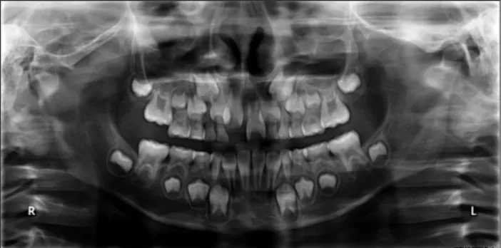
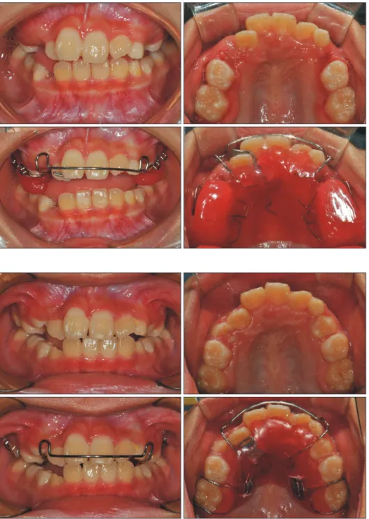
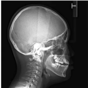
(3) Jae-Hwan Kim and Myeongkwan Jih. III molar relationship, an overbite of 3.5 mm, and an overjet. mask were delivered to the patient (Fig. 4). Conventional. of –2 mm. The lateral profile was concave with anterior. instructions regarding the use of the appliances were pro-. crossbite (Fig. 1, 2). The patient’s uncle on the mother’s side. vided following: turning the expansion screw once a day. had undergone a two-jaw surgery, and his mother’s facial pro-. for 2 weeks and wearing the face mask every day for a. file was also concave. At the age of 5 years, he had received. minimum of 10 hours.. orthodontic treatment using a face mask with a rapid pala-. Eight months later, the RPE was removed due to mobility. tal expander (RPE) for 9 months, and anterior crossbite had. in the maxillary primary first molars (Fig. 5). The anterior. been resolved. Nonetheless, anterior crossbite had reap-. crossbite had resolved except for the maxillary right lateral. peared during the period of a long-term broken appoint-. incisor. A removable appliance with a finger spring was. ments. No negative complication resulting from cancer. fabricated to correct the tooth position (Fig. 6).. therapy was seen in the oral cavity, but the lower second. A sagittal II appliance with a spring was fabricated and. premolars and the second molars were smaller in coronal. delivered to acquire space for the upper canines. Sagittal. size in comparison to the first molars and the adjacent pre-. II appliances were changed four times over a 16-month. molars (Fig. 3).. period in order to replace the screw, to replace a lost ap-. The treatment plan was to use the face mask with RPE,. pliance, and to provide a better fit of the appliance (Fig. 7).. and then to use a full-bonding bracket system. However,. At the end of the first-phase orthodontic treatment with. the patient was diagnosed with a relapse of leukemic ma-. removable appliances, the patient’s profile had improved,. lignancy, and orthodontic treatment was delayed until the. and anterior crossbite as well as crowding had resolved (Fig.. patient’s condition was stable.. 8, 9). Several cephalometric indicators were improved (Fig.. After 4 months of cancer therapy and a 5-month main-. 10, 11). The A point, nasion, B point angle (ANB) had in-. tenance period, laboratory results returned to normal and. , and Wits appraisal had changed creased from –0.5°to 1.5°. a good systemic condition was established. RPE and face. from –6.0 mm to –4.0 mm. Anterior dysplasia indicator. Fig. 3. Panorama image of the patient at first visit for the orthodontic consultation.. Fig. 5. Panorama image of the patient after rapid palatal expander removal. Space for the maxillary canines is insufficient.. Fig. 4. Intra-oral view of the rapid palatal expander appliance with hooks from face mask.. 105.
(4) Orthodontic approach in a pediatric cancer patient. Fig. 6. Delivery of removable spring appliance.. Fig. 7. Delivery of sagittal appliance with finger spring.. (ADPI) values had improved from 90.0°to 86.0°(Table 1). Oral complications from chemotherapy in this patient in-. patient did not have any discomfort wearing removable appliances and showed good compliance.. cluded microdontia and root thinning. The coronal widths of the maxillary second premolars and the mandibular second molars were far less than the average according to. DISCUSSION. Korean standards [8]. Fortunately, the patient’s systemic. Patients with a history of pediatric cancer require ex-. medical status was fine, and side effects on oral tissues. tensive consideration when dental treatment is planned.. were not seen during the orthodontic interventions. The. Complications found in pediatric cancer patients can result. 106. www.chosunobr.org.
(5) Jae-Hwan Kim and Myeongkwan Jih. A. B. E. C. F. D. G. Fig. 8. Extra-oral (A–C) and intra-oral (D–G) photos of the patient at the end of the first-phase orthodontic treatment. Anterior crossbite has been resolved, and a space required for the maxillary canines is sufficient.. Fig. 9. Panorama image of the patient at the end of the first-phase orthodontic treatment.. directly from the disease itself or from the effects of radiation and chemotherapy [9]. Complications of cancer therapy related to dental development include arrested root development, disturbances. Fig. 10. Lateral cephalogram of the patient at the end of the firstphase orthodontic treatment.. in enamel mineralization, microdontia, hypodontia, and premature apical closure [5]. The patient’s age at the time. [10]. Long-term survivors have a particular risk of compli-. of the initial treatment is an important factor in the devel-. cations during the course of orthodontic treatment due to. opment and the degree of adverse effects in the oral cavity. disturbances in dental development and craniofacial skel-. 107.
(6) Orthodontic approach in a pediatric cancer patient Table 1. Comparison of the cephalometric values before and after the treatment. Fig. 11. Superimposition of lateral cephalometric measurements. Anterior crossbite has been resolved with improved esthetic line (black: before, red: after).. eton growth. As knowledge of these risk factors is essential, a dental clinician must be aware of the previous treatment history and its effects on the orthodontic treatment plan [5].. Measurement. Norm. SNA (°) SNB (°) ANB (°) FMA (°) SN-MP (°) U1/SN (°) U1/NA (°) L1/NB (°) IMPA (°) Interincisal angle (°) UL to E-line (mm) LL to E-line (mm) L1 to Pog (mm) U1 to A-Pog (mm) L1 to A-Pog (mm) A-B plane angle (°) APDI (°) Wits (mm). 81.0 78.0 3.5 26.5 36.0 105.5 24.5 30.0 96.0 122.0 2.5 4.0 3.0 7.0 3.5 –6.0 81.5 –2.0. Treatment Before. After. 80.0 80.5 –0.5 26.0 29.5 106.0 22.0 22.5 90.0 134.5 0.0 2.0 7.5 2.0 5.0 –1.0 90.0 –6.0. 82.5 81.0 1.5 29.5 30.5 119.5 34.0 25.0 91.5 118.5 1.0 2.0 4.5 7.5 6.5 –4.0 86.0 –4.0. ANB, A point, nasion, B point angle; ADPI, anterior dysplasia indicator.. da Fonseca [7] has argued that fixed orthodontic appliances or space maintainers must be removed if the oral. a comparably short time. Since no major cancer treatment. hygiene in the patient is compromised and if the treatment. was administered and no negative hematologic signs were. has a high risk of development of mucositis. To provide. observed, planned orthodontic treatment began with mini-. further orthodontic care for these children and adolescents,. mal delay. A face mask with RPE was successful in improv-. the orthodontist must check for signs of gingival oozing,. ing maxillary retrusion. Sequentially removable appliances. hematomas, petechiae, ulcerations, gingival hypertrophy,. were used, primarily to resolve a lack of space and second-. pain, and inflammation in the pharynges and lymph nodes. arily to guide single teeth to normal positions. Adequate. [11]. Barbería et al. [12] have suggested that, in a patient. space for canines was acquired successfully. No compli-. preparing for an active cancer treatment, fixed appliances. cation or discomfort due to orthodontic devices or treat-. with brackets, bands, or lingual arches can be applied if the. ment processes were observed. The AAPD recommends. patient tolerates the equipment and if no sign of irritation. waiting for a period of two disease-free years before initial. of the mucous membrane is seen. Additional recommended. orthodontic treatment. Our patient received orthodontic. strategies in the orthodontic treatment of pediatric cancer. intervention after only 5 months of disease-free period but. patients are as follows: (1) use appliances that cause the. did not show any negative effects of the use of orthodontic. least risk of root resorption, (2) use light forces, (3) finish. appliances. Treatment with removable appliances showed. treatment earlier than usual, (4) adapt the simplest method,. reasonable treatment outcomes in this patient with no side. and (5) do not treat the mandible [13].. effects on the roots of teeth and soft tissue. This may be. The patient in the present case received additional che-. due to limited growth of cancer cells and to less extensive. motherapy after the orthodontic diagnosis was made due. cancer therapy. However, clinicians must be aware of the. to a relapse of leukemic malignancy. It was fortunate that. extent of cancer growth and the course and intensity of the. the extent of relapse was very limited, allowing him to re-. cancer treatment.. ceive limited chemotherapy only. The patient recovered in. 108. www.chosunobr.org. Before beginning any therapeutic intervention, pediatric.
(7) Jae-Hwan Kim and Myeongkwan Jih. dental specialists should gather information on the underlying disease, time of the diagnosis, treatment modality, and complications, including cancer relapse. Providing optimal care and proper adjustment during orthodontic management can lead to successful treatment and improve quality of life in cancer survivors. Pediatric cancer patients have a better chance of survival due to recent advances in cancer therapy. Survivors of antineoplastic treatment often suffer from side effects in the oral cavity. Despite possible complications, the firstphase treatment of a dental Class III patient with ALL was successful via the use of face mask with RPE, spring appliances, and sagittal appliances. Before beginning the actual intervention, clinicians must examine the medical records of the patient thoroughly and special care must be taken to prevent any further discomfort or negative consequences due to the side effects of cancer treatment.. CONFLICTS OF INTEREST The authors declare that they have no competing interests.. ORCID Jae-Hwan Kim https://orcid.org/0000-0001-8088-6216 Myeongkwan Jih https://orcid.org/0000-0001-9579-076X. REFERENCES 1. Amin MB, Edge S, Greene F, Byrd DR, Brookland RK, Washington MK, Gershenwald JE, Compton CC, Hess KR, Sullivan DC, Jessup JM, Brierley JD, Gaspar LE, Schilsky RL, Balch CM, Winchester DP, Asare EA, Madera M, Gress DM, Meyer LR. AJCC cancer staging manual. 8th ed. Chicago: Springer; 2017;979-997. 2. Xavier AM, Hegde AM. Preventive protocols and oral management in childhood leukemia--the pediatric specialist's role. Asian Pac J Cancer Prev 2010;11:39-43.. 3. Simioni C, Martelli AM, Zauli G, Vitale M, McCubrey JA, Capitani S, Neri LM. Targeting the phosphatidylinositol 3-kinase/Akt/mechanistic target of rapamycin signaling pathway in B-lineage acute lymphoblastic leukemia: an update. J Cell Physiol 2018;233:6440-6454. doi: 10.1002/ jcp.26539. 4. Kumar S, Valiathan A, Jayaswal P, Sivakumar A. Class II treatment of an adolescent patient with a history of acute lymphocytic leukemia. J Clin Orthod 2011;45:661-665; quiz 691-692. 5. Neill CC, Migliorati C, Trojan T, Kaste S, Karydis A, Rowland C, Parris W. Experience and expertise regarding orthodontic management of childhood and adolescent cancer survivors. Am J Orthod Dentofacial Orthop 2015;148:765770. doi: 10.1016/j.ajodo.2015.05.027. 6. American Academy of Pediatric Dentistry. Guideline on dental management of pediatric patients receiving chemotherapy, hematopoietic cell transplantation, and/or radiation. Pediatr Dent 2013;35:E185-E193. 7. da Fonseca MA. Dental care of the pediatric cancer patient. Pediatr Dent 2004;26:53-57. 8. Baik BJ, Park JY, Kim JG, Lee DC. A study on the size of the permanent teeth. J Korean Acad Pediatr Dent 2003;30:502509. 9. Valéra MC, Noirrit-Esclassan E, Pasquet M, Vaysse F. Oral complications and dental care in children with acute lymphoblastic leukaemia. J Oral Pathol Med 2015;44:483-489. doi: 10.1111/jop.12266. 10. Kaste SC, Goodman P, Leisenring W, Stovall M, Hayashi RJ, Yeazel M, Beiraghi S, Hudson MM, Sklar CA, Robison LL, Baker KS. Impact of radiation and chemotherapy on risk of dental abnormalities: a report from the Childhood Cancer Survivor Study. Cancer 2009;115:5817-5827. doi: 10.1002/ cncr.24670. 11. Sheller B, Williams B. Orthodontic management of patients with hematologic malignancies. Am J Orthod Dentofacial Orthop 1996;109:575-580. doi: 10.1016/S08895406(96)70068-9. 12. Barbería E, Hernandez C, Miralles V, Maroto M. Paediatric patients receiving oncology therapy: review of the literature and oral management guidelines. Eur J Paediatr Dent 2008;9:188-194. 13. Dahllöf G, Jönsson A, Ulmner M, Huggare J. Orthodontic treatment in long-term survivors after pediatric bone marrow transplantation. Am J Orthod Dentofacial Orthop 2001;120:459-465. doi: 10.1067/mod.2001.118102.. 109.
(8)
수치
+2
관련 문서
The following treatment objectives were planned: (1) the periodic follow-up of the hypertrophic alveolar bone area, (2) an improvement in the skeletal Class III anteroposterior
This report presents a rare case of a child with G6PD deficiency and discuss the main considerations and precautions to take into account during dental treatment
Class III 악간관계와 교합평면의 심한 부조화를 보이는 환자 에서 심미적, 해부학적 기준으로 교합평면을 재설정하여 상악 은 총의치로, 하악은 치아 및
Dental manage- ment of PWS patients consists of active preventive treatment, along with dietary consultation according to the patient’s nutritional phase.. Behavior management
Do-Sup Kim et al: Bisphosphonate-related osteone- crosis of the jaw in a patient with osteoporosis fol- lowing treatment of testicular cancer: a case report.. J Korean Assoc
Objective: The purpose of this study was to compare the longitudinal treatment effects of facemask with rapid maxillary expansion (FM/RME) and chincup (CC) therapy followed by
Objective: The purpose of this study was to report the clinical effectiveness of Korean medicine treatment on FOLFOX-induced symptoms such as nausea and dizziness in a
Objectives : To observe the mitigating effects of a Traditional Korean Medicine treatment program, called Wheel Balanced Cancer Therapy (WBCT), with Adriamycin and 5-FU
