Acute Appendicitis in Children with Acute Leukemia:
Experiences of a Single Institution in Korea
Eun Young Kim,
1Jae Wook Lee,
2Nak-Gyun Chung,
2Bin Cho,
2Hack-Ki Kim,
2and Jae Hee Chung
11Department of Surgery, Yeouido St. Mary’s Hospital, College of Medicine, The Catholic University of Korea, Seoul;
2Department of Pediatrics, Seoul St. Mary’s Hospital, College of Medicine, The Catholic University of Korea, Seoul, Korea.
Received: June 24, 2011 Revised: October 3, 2011 Accepted: October 4, 2011
Corresponding author: Dr. Jae Hee Chung, Department of Surgery, Yeouido St. Mary’s Hospital, College of Medicine,
The Catholic University of Korea, 63-ro 10, Yeongdeungpo-gu, Seoul 150-713, Korea.
Tel: 82-2-3779-1031, Fax: 82-2-786-0802 E-mail: [email protected]
∙ The authors have no financial conflicts of interest.
© Copyright:
Yonsei University College of Medicine 2012 This is an Open Access article distributed under the terms of the Creative Commons Attribution Non- Commercial License (http://creativecommons.org/
licenses/by-nc/3.0) which permits unrestricted non- commercial use, distribution, and reproduction in any medium, provided the original work is properly cited.
Purpose: Acute appendicitis has been reported to be relatively rare in pediatric leukemia patients but there is no official data for this in Korea. And there is no consensus for its treatment in this population. Materials and Methods: We con- ducted a retrospective study of 7 patients diagnosed with appendicitis among 1209 pediatric patients who were diagnosed with leukemia from 1996 to 2008 at a sin- gle institution in Korea. Results: The median age at the time of the diagnosis of appendicitis was 12 years (range: 3-15 years), and 3 of the patients were male. The median absolute neutrophil count (ANC) at the time of diagnosis was 0.99×10⁹/L (range: 0-3×10⁹/L). The mean time from the onset of symptoms to the diagnosis was 4 days. All 7 leukemia patients with appendicitis underwent surgery and they demonstrated a survival of 100% without significant complications. Conclusion:
The incidence of appendicitis in pediatric leukemia patients was 0.57% in our study. Early diagnosis with abdominal ultrasound or computed tomography and early surgical resection in leukemic patient with acute appendicitis may be a safer and more effective treatment option. Even when perforation has already occurred and when the patient has an ANC of 0×109/L, surgical treatment may improve overall survival without incurring significant complications.
Key Words: Acute appendicitis, leukemia, neutropenia, appendectomy
INTRODUCTION
Chemotherapy-related toxicity can lead to neutropenia, undermining a patient’s immune system, and, in turn, leaving the patient highly susceptible to a plethora of infections. In such patients, the most common causes of right lower quadrant ab- dominal pain include appendicitis and typhlitis, and the physician’s ability to dif- ferentiate between these etiologies is critical. Typhlitis, also known as neutropenic colitis, is an inflammation of the ileocecal region and ascending colon, and may be observed after chemotherapy. The primary therapy thereof is conservative and sur- gery may be considered when the condition is aggravated or when complications begin to occur.1,2 In contrast, appendicitis is a localized inflammation of the appen- dix, which is usually treated with surgery in immune competent patients. Such sur- gical treatment is used with caution in leukemia patients, who are at an increased
period of neutropenia that occurred after intensive chemo- therapy: 3 and 1 patients developed acute appendicitis dur- ing the period of neutropenia that occurred after consolida- tion chemotherapy and reinduction chemotherapy following post-transplantation relapse, respectively (Table 1). None of them presented an extramedullary lesion.
The median absolute neutrophil count (ANC) at the time of diagnosis and operation were 0.99×10⁹/L (range: 0-3×10⁹/ L) and 1.32×10⁹/L (range: 0-18.64×10⁹/L), respectively. The median platelet count at the time of diagnosis was 160×109/L (range: 17-225×109/L) (Table 1). C-reactive proteins were above normal levels in 4 of the 7 patients, but the others were not checked. Lactate dehydrogenase increased in 3 patients.
Among the leukemia patients, the median length of the duration of total neutropenia at the time of symptom onset was 20 days (range: 11-71 days). One patient was on oral maintenance chemotherapy and the rest were on either in- travenous chemotherapy or they had completed chemother- apy within the past 30 days. Four patients were in a neutro- penic state at the start of symptoms (range: 2-47 days). The remaining 3 patients also were in a neutropenic state at less than 1 week before the start of symptoms (1-6 days). For the leukemia patients, the mean time from the onset of symptoms to the diagnosis of acute appendicitis was 4 days (range: 1-9 days) (Table 1).
All the patients displayed abdominal pain except for one (86%), and other symptoms included fever in 5 patients (71%), vomiting in 3 patients (43%), diarrhea in 1 patients (14%) and abdominal distension in 1 patient (14%). On physical examination, 6 patients (86%) showed signs of peritoneal irritation such as direct tenderness or rebound tenderness.
Perforated appendicitis was diagnosed in 4 patients and appendicitis without perforation was diagnosed in 3 pa- tients. In 2 patients subphrenic free air was evident on plain film, which obviated the need for further diagnostic proce- dures before surgery, and of the remaining 5 patients, 1 un- derwent abdominal sonography, 2 underwent CT imaging and 2 underwent both procedures (Table 1). On the preop- erative radiologic study, 1 patient was tentatively diagnosed with acute appendicitis without appendicolith, 2 patients were tentatively diagnosed with acute appendicitis with ap- pendicolith and 2 patients were tentatively diagnosed with periappendiceal abscess due to appendicitis. All 5 patients were differentiated from having typhlitis.
All 7 leukemia patients received surgery, 2 with laparoto- my and 5 with laparoscopic appendectomy. Six patients re- risk of complications and death. As a result, there is cur-
rently little consensus as to the optimum treatment method for appendicitis in these immune compromised patients.2-6 This report is a single institution study that spanned 12 years of experience with leukemia patients who developed appendicitis; we report the disease incidence, clinical char- acteristics and treatment methods thereof.
MATERIALS AND METHODS
The study population consisted of 7 patients who were di- agnosed with appendicitis out of 1209 patients who were treated for leukemia between 1996 and June, 2008 at Saint Mary’s Hospital, The Catholic University of Korea. The pa- tients’ records were retrospectively reviewed for the patient’s age, gender, underlying disease, clinical hematologic status at the onset of symptoms, signs and symptoms, laboratory and radiologic data, treatment modalities and outcomes.
Neutropenia was defined as an absolute neutrophil count below 1×10⁹/L. The ultrasound (US) or computed tomogra- phy (CT) criteria for the diagnosis of appendicitis included a blind-ending incompressible tubular structure in the right lower quadrant of the abdomen, an overall diameter of the appendix >6 mm and increased echogenicity of the sur- rounding mesenteric tissues with or without free fluid or abscess in the right lower quadrant of the abdomen. The criterion for typhlitis was a certain circumferential segmen- tal cecal or terminal ileal wall thickness greater than 3 mm.3
RESULTS
In the 12 year period between 1996 and June, 2008 a total of 1209 pediatric patients were treated for acute leukemia, and seven of these patients developed appendicitis for an inci- dence of 0.57%. Of the 1209 patients, 845 had acute lym- phocytic leukemia (ALL) and 364 had acute myelocytic leu- kemia (AML) with appendicitis developing in 3 (0.35%) and 4 (1.09%) patients, respectively, in each disease group.
The median age at the time of the diagnosis of appendici- tis was 12 years old (range: 3-15 years); 3 patients were male and 4 were female. Of the 3 ALL patients, 1 patient had just completed remission induction chemotherapy, 1 pa- tient was receiving maintenance chemotherapy and the other patient had completed delayed intensification chemotherapy.
All 4 AML patients developed acute appendicitis during the
cal wound infection was observed in 1 patient, and this re- sponded well to supportive care (Table 2). All the cases were confirmed to have appendicitis with neutrophil infil- tration by surgical pathology. None of the appendices showed evidence of leukemic infiltration on H & E stain and immunohistochemical study (Table 2).
DISCUSSION
Appendicitis is the most common ailment requiring surgery in the pediatric population, with an incidence of 1% in chil- dren below the age of 15. However, the only studies of ap- pendicitis in patients with underlying hematologic illnesses such as leukemia are limited to case reports, and only a few single institution studies with a large study population have been conducted over a substantial period of time.2,3,6
The incidence of appendicitis among the patients with hematologic malignancy has been reported to be 0.5-4.4%.
In 1992, Angel, et al.6 reported on 6099 patients with leuke- mia and other malignancies. The patients were accrued over a span of 27 years with 16 patients experiencing appendici- tis (0.5%). In 2005, Hobson, et al.2 reported on 7 patients with appendicitis (1.5%) out of 464 patients with hemato- logic malignancies in a study that compared appendicitis and typhlitis. In 2007, Alioglu, et al.8 reported on 2 patients who were diagnosed with appendicitis out of 118 leukemia ceived surgery either on the day of diagnosis or one day after.
The ANCs assessed on the day of surgery in 6 patients were almost the same as the ANCs assessed at the time of symp- tom presentation. Only one patient among these 6 patients preoperatively received transfusions with 1 unit of packed red blood cells and 1 unit of single donor platelets. One pa- tient received laparoscopic interval appendectomy 6 weeks after the diagnosis. This patient presented pancytopenia that did not respond well to transfusions for preoperative man- agement, and the patient was started on conservative care that included broad spectrum antibiotics. We decided to per- form the interval appendectomy 6 weeks later. The symp- toms had subsided and the pancytopenia recovered 9 days after the diagnosis; however, despite the usage of antibiotics for 6 weeks, follow-up CT showed a remaining 3.5×2.4×4.2 cm sized pelvic abscess that necessitated laparoscopic ap- pendectomy and drainage. On the operation day, this pa- tient’s ANC was 2.78×10⁹/L. All the patients received pre and post-operative antibiotics as indicated under the Infec- tious Diseases Society of America guidelines.7 G-CSF (granu- locyte colony stimulating factor) was injected into 5 pa- tients, of whom 2 were further treated with antithrombin III and intravenous immune globulin because of sepsis with abnormal levels of antithrombin III, D-Dimer, fibrinogen degradation products and fibrinogen. All 7 patients recov- ered well after surgery. An oral diet was commenced at a mean of 5.1 days after surgery (range: 2-10 days). Umbili-
Table 1. Demographic Characteristics, Symptoms and Laboratory Findings in Appendicitis Patients with Acute Leukemia Patient
No. Age
(yrs) Sex Diagnosis & disease status Neutropenia and onset of symptoms
Duration from onset of symptoms
to diagnosis of appendicitis (days)
Duration from diagnosis to operation of appendicitis
(days)
ANC/platelet count at presentation of
appendicitis (×109/L)
ANC at operation
(×109/L)
1 3 F ALL, remission induction 3 days after recovery
from neuropenia 6 0 3/225 18.64
2 14 M ALL, 1st CR, maintenance
chemotherapy 2 days after
neutropenia 1 1 0.99/159 0.99
3 15 F ALL, 1st CR, intensification
chemotherapy 6 days after recovery
from neuropenia 2 0 1.32/160 1.32
4 6 M AML (M2), 1st CR,
consolidation chemotherapy 1 day after recovery
from neuropenia 2 1 1.54/255 1.71
5 15 F AML (M2), 1st CR,
consolidation chemotherapy 10 days after
neutropenia 5 42 (interval
appendectomy) 0/18 2.78
6 6 M AML (M2), 1st CR,
consolidation chemotherapy 26 days after
neutropenia 9 0 0/101 0
7 12 F AML (M6), post-BMT,
1st relapse, reinduction 47 days after
neutropenia 3 0 0/17 0
Median 12 4 (mean) 0.99/160 1.32
ALL, acute lymphocytic leukemia; AML, acute myelocytic leukemia; ANC, absolute neurophil count; CR, complete remission; BMT, bone marrow transplan- tation.
gins with periumbilical pain that migrates to the right lower quadrant (RLQ). Appendiceal distension leads to nausea and vomiting in 85% of these patients and continued in- flammation eventually leads to fever.12 These signs may not be so evident in neutropenic or immune compromised pa- tients and for whom fever may be the first sign13 and also for whom RLQ pain and the signs of peritoneal irritation may not be so obvious.6 Hobson, et al.2 indicated that diar- rhea and a high fever above 38.5°C are more characteristic of typhlitis than appendicitis. In our group, abdominal pain and peritoneal irritation of the RLQ were the most com- monly reported symptoms of the patients (86%), and fever was a presenting symptom in 2 patients. Symptoms of vomiting and diarrhea (43% and 14% respectively) were not as frequent in our patients and 1 patient complained only of diarrhea and abdominal distension without abdomi- nal pain. Free air in this patient was evident on simple X- ray leading up to the operation.
So often, the gastrointestinal complications seen in neu- tropenic patients are difficult to diagnose on the basis of clinical findings alone. The average time from the onset of symptoms to diagnosis was 4 days for acute leukemia pa- tients. Difficulty with differentiating abdominal pain due to patients (1.7%), and in 2008 Wiegering, et al.3 reported on
5 patients who were diagnosed with appendicitis out of a group of 113 leukemia patients (4.4%). In our study, 7 pa- tients were diagnosed with appendicitis out of a group of 1209 pediatric acute leukemia patients, representing an in- cidence of 0.57%.
The etiology of appendiceal disease is luminal obstruc- tion and the most common cause of obstruction is a feca- lith. Not all cases of appendicitis are associated with a feca- lith, but in most, some form of obstruction occurs. Lymphoid tissure, which is found in the wall of the appendix, may be- come hyperplastic in response to viral infections of the gut or respiratory tract, resulting in obstruction of the lumen of the appendix. Extramedullary involvement in leukemia is a rare complication, and only a few reports have demonstrat- ed leukemic cell infiltration of appendix. Acute appendicitis can be presented as the initial manifestation or the relapse of acute myelogenous leukemia.9-11 However, such cases have only been recorded in adults and there is no case report of leukemic cell infiltration of the appendix in pediatric he- matologic malignancies in the literature. In our study, none showed leukemic cell infiltration of the appendix.
In immune competent children, appendicitis usually be-
Table 2. Operative Findings, Pathologic Findings, Treatment and Outcome of the Acute Leukemia Patients with Appendicitis Patient
No. Radiologic
diagnosis Operative
findings Pathologic findings Operative treatment
Conservative treatment (except IV antibiotics)
Duration from treatment to soft
diet start (days) Outcome
1 Abdominal
X-ray (free air) Perforated
appendicitis Acute appendicitis
with perforation Open
appendectomy G-CSF, FOY,
transfusion 10 Uneventful
recovery
2 US, CT Non-perforated
appendicitis Acute appendicitis Lap-appendectomy - 2 Uneventful recovery
3 US, CT Perforated
appendicitis Acute gangrenous
appendicitis Lap-appendectomy G-CSF 3 Uneventful
recovery
4 Abdominal
X-ray (free air) Perforated appendicitis
Acute suppurative appendicitis with periappendiceal abscess formation
Open
appendectomy Transfusion 3 Uneventful
recovery
5 CT Perforated
appendicitis Acute appendicitis with perforation
Lap-interval appendectomy (6 wks later)
G-CSF, ATIII, IVGV, FOY, transfusion, (1st Tx)
15 (1st Tx)
2 (POD) Uneventful recovery
6 CT Non-perforated
appendicitis Acute gangrenous
appendicitis Lap-appendectomy G-CSF, ATIII, IVGV, transfusion 10 Umbilical wound infection, complete recovery
7 US Non-perforated
appendicitis
Chronic inflammation with granulation tissue formation, colonization of fungal organism
Lap-appendectomy G-CSF, transfusion 6 Uneventful recovery
G-CSF, granulocyte colony stimulating factor; FOY, gabexate mesilate; ATIII, antithrombin III; IVGV, human immunoglobulin; POD, postoperative day; US, ultrasound; CT, computed tomography.
cecal wall of 39 mm in length was present.
The optimum method of treatment remains a controver- sy. The reasons why some authors have a negative opinion about early appendectomy revolve around postoperative complications and mortality. Sbragia, et al.5 recommended an initial nonoperative approach in which surgery is only considered after a rise in neutrophil count or with an aggra- vation of clinical symptoms when these patients are man- aged by conservative care alone. Wiegering, et al.3 noted that even perforated appendicitis may improve with conser- vative care alone. They reported that 1 case was complicated by perforation during the conservative care and the median time to normalization of the US findings was 14 days. More- over, although imaging evidence for recurrence of appendi- citis was absent, 3 cases experienced recurrent right lower abdominal pain during a subsequent episode of febrile neu- tropenia after the following cycle of chemotherapy. Not- withstanding, conservative care alone requires a lengthy pe- riod for full recovery and incurs risks of progression, perforation and recurrence. In immune competent children, an inflamed appendiceal mass may be successfully treated with percutaneous drainage and antibiotics in 90-97% of the cases with a recurrence of symptoms only happening in 5-14%, so interval appendectomy may not be required in all patients.16-20 However, immune compromised children may tend towards a different clinical course. In our study, 1 AML patient who presented with a perforated appendicitis along with a 5 cm sized pelvic abscess with bilateral obstructive ureteral dilatations clinically improved after conservative management alone and was started on a diet on the 15th day after the appearance of the symptoms, even as the patient’s ANC rose to 1×10⁹/L on the 7th day after the start of symp- toms. Despite the usage of antibiotics for 6 weeks, follow- up CT showed a remaining right pelvic abscess 3.5×2.4×4.2 cm in size without ureteral dilatation, and this required lap- aroscopic appendectomy and drainage. Furthermore, delayed recovery from infection leads to delay in the continuation of chemotherapy, which could possibly lead to aggravation of the patient’s underlying illness and escalation of medical costs. Therefore, conservative care alone is not the most ef- fective method of treatment.
Since the initial reports on surgical treatment for pediatric leukemia patients who present with an acute abdomen were put forward by Björnsson, et al.21 and Exelby, et al.22 nu- merous studies have reported on improved outcomes result- ing from surgery (Table 3).2,3,6,13,23 Exelby, et al.22 reported that the conditions that led to a 100% mortality when rely- appendicitis from the pain resulting from chemotherapy
toxicity, and the use of corticosteroids, which mask the pre- cise presentation of symptoms, delayed the accurate diag- nosis of these patients.2,6,13 Also, in our study, over 50% of the cases were perforated upon diagnosis.
Imaging may aid in forming a diagnosis in these cases.
Although Hobson, et al.2 noted that the diagnostic accuracy of CT for appendicitis is only about 33%, Stroman, et al.14 reported that the sensitivity and overall accuracy of CT for appendicitis in immune competent patients was about 92%
and 90% respectively. Other authors have also noted that either CT or ultrasound is appropriate for diagnosing ap- pendicitis in neutropenic patients.2,3,6,13 However, it remains unclear what criteria were used to make the differentiation of acute appendicitis from typhlitis. Typhlitis is a poorly un- derstood gastrointestinal complication of neutropenia. Patho- logically, typhlitis is described as a compromisation of bow- el wall integrity with subsequent bacterial or fungal invasion.
Although pathologic abnormalities can involve any segment of the small or large bowel, the cecum is the most common location of abnormality. CT findings of typhlitis include right-sided colon wall thickening, pericolonic stranding, as- cites, and cecal pneumatosis. Kirkpatrick and Greenberg15 compared mean bowel wall thickening for the differentia- tion of specific gastrointestinal complications including C difficile colitis, neutropenic colitis and graft versus-host dis- ease in neutropenic patients, and found that bowel wall thickening was significantly more prominent in C difficile colitis (mean wall thickness, 12 mm; range, 8-20 mm) than in neutropenic enterocolitis (mean thickness, 7 mm; range, 4-15 mm; p<0.01) and graft-versus-host disease (mean thickness, 5 mm; range, 3-7 mm; p<0.01).
In our study, the criterion for typhlitis was a cecal or termi- nal ileal wall thickness greater than 3 mm.3 However, de- pending on the severity or portion of the inflammation of the appendix, the cecal wall around the base of the appendix can be thickened by inflammation. So, although wall thickness is important, the circumferential thickened length of the cecum and ascending colon wall could be a more important finding.
However, it remains unclear as to how long of a segment would have to be involved for the diagnosis of typhlitis be- cause large periappendiceal abscess formation can cause the circumferential thickening of certain segmental cecal walls, too. In 3 of 4 of our cases wall thickness greater than 3 mm was present only around the appendix base of the cecum. In one patient, who underwent the interval appendectomy with a large periappendiceal abscess, a circumferential thickened
when perforation has occurred and the patient has an ANC of 0×109/L, surgical treatment may improve overall surviv- al without incurring significant complications.
REFERENCES
1. Schlatter M, Snyder K, Freyer D. Successful nonoperative man- agement of typhlitis in pediatric oncology patients. J Pediatr Surg 2002;37:1151-5.
2. Hobson MJ, Carney DE, Molik KA, Vik T, Scherer LR 3rd, Rouse TM, et al. Appendicitis in childhood hematologic malignancies:
analysis and comparison with typhilitis. J Pediatr Surg 2005;40:
214-9.
3. Wiegering VA, Kellenberger CJ, Bodmer N, Bergstraesser E, Nig- gli F, Grotzer M, et al. Conservative management of acute appen- dicitis in children with hematologic malignancies during chemo- therapy-induced neutropenia. J Pediatr Hematol Oncol 2008;30:
464-7.
4. Schaller RT Jr, Schaller JF. The acute abdomen in the immunolog- ically compromised child. J Pediatr Surg 1983;18:937-44.
5. Sbragia Neto L, Oliveira-Filho AG, Epelman S, Koeller HF, Bus- torff-Silva JM, Brandalise SR. Selective surgical indication in the management of neutropenic children presenting with acute abdo- men. Pediatr Hematol Oncol 2000;17:483-7.
6. Angel CA, Rao BN, Wrenn E Jr, Lobe TE, Kumar AP. Acute ap- pendicitis in children with leukemia and other malignancies: still a diagnostic dilemma. J Pediatr Surg 1992;27:476-9.
7. Hughes WT, Armstrong D, Bodey GP, Bow EJ, Brown AE, Ca- landra T, et al. 2002 guidelines for the use of antimicrobial agents in neutropenic patients with cancer. Clin Infect Dis 2002;34:730-51.
ing on non-surgical methods improved to 50% overall sur- vival when these patients were given surgical treatment, while Angel, et al.6 reported on a series of 16 leukemia pa- tients with appendicitis, of whom 14 were treated with sur- gery with only 1 death (7.1%). Hobson, et al.2 reported 100% overall survival after appendectomy for leukemia pa- tients. In our study, the mortality was 0% and the only com- plication was a minor wound infection. All the patients who received surgery recovered rapidly, despite an ANC of 0×109/L and they were started on an oral diet at a mean of 5.1 days, and no later than 10 days. The relationship be- tween ANC on the operation day and postoperative recov- ery time was obscure, but a long duration from onset of symptoms to diagnosis of appendicitis tended to show lon- ger durations from treatment to start of a soft diet.
Laparoscopic appendectomy in children is a procedure that we have been performing in our institution since 2000.
This procedure has the advantages of a small wound, rapid recovery, decreased pain and fewer wound infections, and these qualities may gain importance in immune compro- mised pediatric leukemia.
In conclusion, when right lower quadrant abdominal pain occurred in pediatric leukemia patients, early diagnosis of acute appendicitis by US and/or CT and early surgical re- section before perforation may be the more effective treat- ment option for leukemia patients with appendicitis. Even
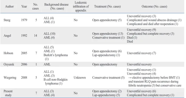
Table 3. Acute Appendicitis in Pediatric Hematologic Malignancies in the Literatures
Author Year No.
cases Background disease (No. cases)
Leukemic infiltration of
appendix Treatment (No. cases) Outcome (No. cases)
Steeg 1979 5 ALL (4)
AML (1) No Open appendectomy (5) Uneventful recovery (3)
Complicated and wound abscess drainage (1) Complicated and died after reoperation (1)
Angel 1992 14 ALL (10)
AML (4) No Open appendectomy (13)
Conservative treatment (1)
Uneventful recovery (9)
Complicated but complete recovery (3) Died (1)
Died
Hobson 2005 7
ALL (5) AML (1)
Burkitt’s lymphoma (1)
No Open appendectomy (6)
Lap-appendectomy (1) Uneventful recovery (7)
Ozyurek 2006 1 AML No Open appendectomy Uneventful recovery
Wiegering 2008 5
ALL (1) AML (3)
B cell non-Hodgkin lymphoma (1)
Unknown Conservative treatment (5)
Uneventful recovery (1) Uneventful recovery (4)
→ elective appendectomy before BMT (1) and transient RLQ pain recurrence during febrile neutropenia (3) but conservative care Present
study 7 ALL (3)
AML (4) No Open appendectomy (2)
Lap-appendectomy (5) Uneventful recovery (6)
Complicated but complete recovery (1) ALL, acute lymphocytic leukemia; AML, acute myelocytic leukemia; BMT, bone marrow transplantation; RLQ, right lower quadrant.
the neutropenic patient: characterization and differentiation with abdominal CT. Radiology 2003;226:668-74.
16. Deakin DE, Ahmed I. Interval appendicectomy after resolution of adult inflammatory appendix mass--is it necessary? Surgeon 2007;5:45-50.
17. Styrud J, Eriksson S, Nilsson I, Ahlberg G, Haapaniemi S, Neovi- us G, et al. Appendectomy versus antibiotic treatment in acute ap- pendicitis. A prospective multicenter randomized controlled trial.
World J Surg 2006;30:1033-7.
18. Jeffrey RB Jr, Tolentino CS, Federle MP, Laing FC. Percutaneous drainage of periappendiceal abscesses: review of 20 patients. AJR Am J Roentgenol 1987;149:59-62.
19. Yamini D, Vargas H, Bongard F, Klein S, Stamos MJ. Perforated appendicitis: is it truly a surgical urgency? Am Surg 1998;64:970-5.
20. Kaminski A, Liu IL, Applebaum H, Lee SL, Haigh PI. Routine in- terval appendectomy is not justified after initial nonoperative treat- ment of acute appendicitis. Arch Surg 2005;140:897-901.
21. Björnsson S, Yates JW, Mittelman A, Holland JF. Major surgery in acute leukemia. Cancer 1974;34:1272-5.
22. Exelby PR, Ghandchi A, Lansigan N, Schwartz I. Management of the acute abdomen in children with leukemia. Cancer 1975;35:
826-9.
23. Ver Steeg K, LaSalle A, Ratner I. Appendicitis in acute leukemia.
Arch Surg 1979;114:632-3.
8. Alioglu B, Avci Z, Ozcay F, Arda S, Ozbek N. Neutropenic en- terocolitis in children with acute leukemia or aplastic anemia. Int J Hematol 2007;86:364-8.
9. Papageorgiou SG, Foukas P, Economopoulou C, Tsirigotis P, Sa- korafas G, Georgakopoulos N, et al. Acute apenditis in patient with acute promyelocytic leukemia. Leuk Res 2011;35:e4-5.
10. Hsiao PJ, Kuo SM, Chen JH, Lin HF, Chu PL, Lin SH, et al.
Acute myelogenous leukemia and acute leukemic appendicitis: a case report. World J Gastroenterol 2009;15:5624-5.
11. Toubai T, Kondo Y, Ogawa T, Imai A, Kobayashi N, Ogasawara M, et al. A case of leukemia of the appendix presenting as acute appendicitis. Acta Haematol 2003;109:199-201.
12. Swain RS. Appendix and Meckel Diverticulum. In: Oldham KT, Colombani PM, editors. Surgery of infants and children: scientific principles and practice. Philadelphia: Lippincott-Raven; 1997.
p.1215-24.
13. Ozyurek E, Arda S, Ozkiraz S, Alioglu B, Arikan U, Ozbek N. Fe- brile neutropenia as the presenting sign of appendicitis in an ado- lescent with acute myelogenous leukemia. Pediatr Hematol Oncol 2006;23:269-73.
14. Stroman DL, Bayouth CV, Kuhn JA, Westmoreland M, Jones RC, Fisher TL, et al. The role of computed tomography in the diagno- sis of acute appendicitis. Am J Surg 1999;178:485-9.
15. Kirkpatrick ID, Greenberg HM. Gastrointestinal complications in