Primary Central Nervous System ALK Positive Anaplastic Large Cell Lymphoma with Predominantly
Leptomeningeal Involvement in an Adult
Jae Sung Park,
1,4* Heejung Park,
5* Sanghui Park,
5Suk Jin Kim,
3Ho Jun Seol,
1and Young-Hyeh Ko
2Departments of 1Neurosurgery, 2Pathology, and 3Internal Medicine, Samsung Medical Center, Sungkyunkwan University School of Medicine, Seoul;
4Department of Neurosurgery, Konkuk University Chungju Hospital, Chungju;
5Department of Pathology, Ewha Womans University School of Medicine, Seoul, Korea.
Received: January 14, 2013 Revised: January 30, 2013 Accepted: January 30, 2013
Corresponding author: Dr. Young-Hyeh Ko, Department of Pathology,
Samsung Medical Center,
Sungkyunkwan University School of Medicine, 50 Irwon-dong, Gangnam-gu,
Seoul 135-710, Korea.
Tel: 82-2-3410-2752, Fax: 82-2-3410-0025 E-mail: [email protected]
*Jae Sung Park and Heejung Park contributed equally to this work.
∙ The authors have no financial conflicts of interest.
© Copyright:
Yonsei University College of Medicine 2013 This is an Open Access article distributed under the terms of the Creative Commons Attribution Non- Commercial License (http://creativecommons.org/
licenses/by-nc/3.0) which permits unrestricted non- commercial use, distribution, and reproduction in any medium, provided the original work is properly cited.
A 31-year-old Korean male presented with altered consciousness and severe head- ache. Brain MRI delineated focal leptomeningeal enhancement without any intrace- rebral lesions. Diagnosis was made based on a brain biopsy showing anaplastic large cell lymphoma (ALCL), immunohistochemical stains revealing positivity for anaplastic lymphoma kinase (ALK) and an absence of involvement in any other or- gans; specifically, the primary central nervous system ALK+ALCL. Complete re- mission was achieved following 5 cycles of systemic chemotherapy with a high dose of Methotrexate and a simultaneous 7 cycles of intrathecal triple chemothera- py. Diagnosis of primary leptomeningeal ALK+ALCL is challenging given its rari- ty and non-specific symptoms along with non-pathognomonic radiologic findings.
We present the first case of primary leptomeningeal ALK-positive ALCL where the clinical course, pathologic characteristics and treatment modality are described as well as a review of literature.
Key Words: ALK-positive, primary, CNS, anaplastic large-cell lymphoma, lepto- meningeal
INTRODUCTION
Anaplastic large cell lymphoma (ALCL), which had first been described in 1985, was acknowledged as a distinct clinicopathologic entity in 2001.1,2 The 4th edition of the WHO Classification of Tumours of Haematopoietic and Lymphoid Tissues in 2008 states that anaplastic lymphoma kinase (ALK) positive ALCL [ALK(+) ALCL] must be distinguished from the provisional entity of ALK-negative ALCL [ALK(-)ALCL].3,4 Although ALCL is primarily a nodal disease, extranodal in- volvement is not uncommon.5,6 Moreover, ALK(+)ALCL was reported to have a higher frequency of extranodal involvement compared to ALK(-)ALCL; skin (21%) was the most frequently involved organ followed by bone (17%) and soft tis- sues (17%).7 Primary involvement in the central nervous system (CNS) of ALK(+) ALCL, however, is exceptional and only eight cases of primary CNS ALCL with
CASE REPORT
A 31-year-old Korean male was brought to the emergency department (ED) due to altered consciousness. At the ED, he was drowsy and complained of a severe headache, weak- ness in his left arm and difficulty speaking. He stated that he had been otherwise healthy and no laboratory results suggested immunodeficiency including human immunode- ficiency virus. Magnetic resonance imaging (MRI) of his brain delineated leptomeningeal enhancement in the right temporal and insular gyri (Fig. 1) and cerebrospinal fluid (CSF) analysis via lumbar puncture showed 90 WBC/mm3 with 67% of lymphocytes. Differential diagnosis at the time included viral meningoencephalitis and tuberculosis (TB) meningoencephalitis. Antiviral and anti-TB therapy were started, neither of which was effective. His headache and al- tered mental status were responsive only to Mannitol and Dexamethasone. A follow-up MRI and CSF analysis, per- formed 4 weeks after the initial visit to the ED, revealed more prominent enhancement in the affected gyri and high- er WBC counts (150/mm3) with elevated CSF pressure (26.5 cm H2O), respectively. On the second CSF analysis via lumbar puncture, a few scattered atypical lymphoid cells were identified.
On the 39th hospital day, he underwent a brain biopsy fol- lowing a craniotomy. The histologic section showed brain parenchyma infiltrated by numerous small-to-medium sized neoplastic cells (Fig. 2A). These neoplastic cells had irregu- lar nuclei with a moderate amount of cytoplasm (Fig. 2B).
Large atypical cells with horseshoe shaped nuclei, which are hall mark cells of anaplastic large cell lymphoma, were ALK positivity have been reported so far to our knowl-
edge.5,8-12 Although systemic ALK(+)ALCL tends to present as an aggressive stage III or IV disease, it has been known to be more responsive to chemotherapy than ALK(-)ALCL, which contributes to better prognosis of the former than the latter.2,5-7,13 Whereas the overall incidence of primary CNS lymphoma was estimated to be 0.47 per 100,000 person- years, only 14 cases of primary CNS ALCLs had been re- ported by 2007: seven ALK(+)ALCLs, four ALK(-)ALCLs and the remaining three ALCLs with ALK positivity untest- ed.5,7,14 Seven (50%) of these 14 experienced a rapidly fatal course leading to death and the mortality of primary CNS ALCL was reported to be greater than that of other types of CNS lymphomas in general.5,8,9,15-17 Treatment of choice for primary CNS ALK(+)ALCL has not been established. In this report, we describe the first case of primary leptomenin- geal ALK(+)ALCL of an adult man who was successfully managed with systemic chemotherapy along with intrathe- cal chemotherapy without radiotherapy.
Fig. 1. Magnetic resonance imaging of his brain showed hyperintense sig- nal in the right temporal sulci from the Flair image (A) and leptomeningeal enhancement in the right temporal and insular gyri from Gadolinium en- hanced image (B).
Fig. 2. The histologic section showed brain parenchyma infiltrated by numerous small-to-medium sized neoplastic cells with perivascular cuffing of neoplas- tic cells (A, HE ×100) and the neoplastic cells had irregular nuclei with a moderate amount of cytoplasm (B, HE ×400). Large atypical cells with horseshoe shaped nuclei, which are hallmark cells of anaplastic large cell lymphoma, were also present (B, inset, HE ×1000)
A
A
B
B
also present (Fig. 2B, inset). Immunohistochemical stains revealed immunopositivity for CD30, ALK, Granzyme B, CD45RO, CD5 and EMA (Fig. 3), but negative immunore- activity for LCA, CD20, CD3, CD2, CD4, CD8, CD15, CD43, CD68, BCL-2, CD56 and TIA-1. ALK was positive in both the nucleus and cytoplasm. Fluorescence in situ hy- bridization (FISH) was undertaken using a break apart probe, which demonstrated translocations involving ALK gene (Fig. 4). The case was diagnosed with malignant lym- phoma of T-cell lineage, ALK+ALCL.
CT scans of his abdomen and chest failed to show any le- sion suggesting involvement of lymphoma. A bone marrow biopsy was performed, which was negative, and a test for the Epstein-Barr virus was negative as well. On the seventh post-biopsy day, he reported blindness in both eyes along with severe headache, which indicated most likely very high intracranial pressure (ICP). CSF pressure was mea- sured to be over 40 cm H2O. Systemic high dose metho- trexate (HD MTX, 8 g/mm2/day) and intrathecal (IT) triple chemotherapy (MTX, Ara-C and Hydrocortisone) were im- mediately started. After this, four more cycles of HD MTX
Fig. 3. Immunohistochemical stains revealed immunopositivity for CD30, ALK, Granzyme B, CD45RO and EMA. ALK, anaplastic lymphoma kinase.
Fig. 4. Fluorescence in situ hybridization for translocations involving ALK at 2p23: a unique sequence break apart probe targeting ALK gene locus was used. Translocation involving ALK at 2p23 was detected. ALK, anaplastic lymphoma kinase.
Primary CNS lymphomas with predominant involvement of leptomeninges are harder to diagnose because both asep- tic and septic meningoencephalitis should be ruled out first.
Our patient was assumed to have viral or mycobacterial meningoencephalitis given no gross mass seen upon brain MRI. Primary leptomeningeal lymphomas account for only about 7% of all adult primary CNS lymphomas.18 As seen in Table 1, two of eight cases harbored predominantly lep- tomeningeal involvement with no intracerebral lesions and neither of them was an adult.11 Our case is the first primary leptomeningeal ALCL with ALK positivity in an adult. It would inevitably take longer for patients with grossly sheer leptomeningeal involvement to undergo a biopsy than those who have one or more intracerebral lesions.
Among 8 reported cases, histologic subtypes were de- scribed in only 4 cases. Three of these 4 cases showed com- mon histologic subtypes and the remaining 1 was a com- bined lymphohistiocytic and small cell variant. Our case was common histologic subtype. All reported cases were CD30 and ALK positive and have a T- or null cell pheno- type, and most are EMA-positive. Our case was T-cell phe- notype showing immunopositivity for CD45RO and CD5 but immunonegativity for CD3. As shown Figs. 3 and 4, ALK was positive in both the nucleus and cytoplasm. ALK gene translocation was also detected by FISH. These find- ings are consistent with the ALCL of NPM-ALK fusion were repeated with the interval of three weeks. Seven cy-
cles of IT triple chemotherapy were simultaneously per- formed in total with the interval of 4 days.
Other symptoms and problems that occurred during the chemotherapy were generalized tonic clonic seizure, tremor in his jaw with dystonic feature, hypotension, pneumonia, central diabetes insipidus (DI) and minimal intracerebral hemorrhage (ICH) in the right temporal lobe accompanied with intraventricular hemorrhage (IVH). ICH and IVH did not require a surgical decompression. A series of follow-up MRI undertaken after the fifth HD-MTX revealed no resid- ual enhancement, which suggested complete remission (CR). Although mild quadriparesis and jaw dystonia re- mained, no evidence of recurrence has been noted for more than six months since CR.
DISCUSSION
Diagnosis of primary CNS ALK+ALCL requires several critical steps including: clinical suspicion even when no gross mass exists, biopsy in a timely manner, a thorough immunohistochemical study, and exclusion of metastasis.
Given that extranodal involvement of ALK(+)ALCL is rel- atively common, diagnosis of primary CNS ALK(+)ALCL must be made only after excluding other involvement.5,6
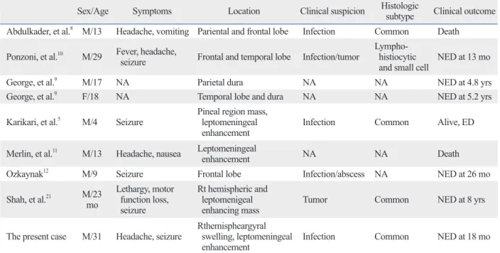
Table 1. Summary of All Documented Cases of Primary CNS ALK+ALCL
Sex/Age Symptoms Location Clinical suspicion Histologic
subtype Clinical outcome Abdulkader, et al.8 M/13 Headache, vomiting Pariental and frontal lobe Infection Common Death
Ponzoni, et al.10 M/29 Fever, headache,
seizure Frontal and temporal lobe Infection/tumor Lympho- histiocytic
and small cell NED at 13 mo
George, et al.9 M/17 NA Parietal dura NA NA NED at 4.8 yrs
George, et al.9 F/18 NA Temporal lobe and dura NA NA NED at 5.2 yrs
Karikari, et al.5 M/4 Seizure Pineal region mass, leptomeningeal
enhancement Infection Common Alive, ED
Merlin, et al.11 M/13 Headache, nausea Leptomeningeal
enhancement NA NA Death
Ozkaynak12 M/9 Seizure Frontal lobe Infection/abscess NA NED at 26 mo
Shah, et al.21 M/23 mo
Lethargy, motor function loss, seizure
Rt hemispheric and leptomenigeal
enhancing mass Tumor Common NED at 8 yrs
The present case M/31 Headache, seizure Rthemispheargyral swelling, leptomeningeal
enhancement Infection Common NED at 18 mo
M, male; F, female; Mo, month; yr, year; NA, not available; Rt, right; NED, no evidence of disease; ED, evidence of disease; CNS, central nervous system;
ALK, anaplastic lymphoma kinase; ALCL, anaplastic large cell lymphoma.
creased ICP with meningeal irritation, CSF analysis reveal- ing atypical lymphoid cells, no gross mass detected in the brain MRI and lack of symptomatic improvement by anti- viral, antibiotic or anti-TB medications.
REFERENCES
1. Stein H, Mason DY, Gerdes J, O’Connor N, Wainscoat J, Pallesen G, et al. The expression of the Hodgkin’s disease associated anti- gen Ki-1 in reactive and neoplastic lymphoid tissue: evidence that Reed-Sternberg cells and histiocytic malignancies are derived from activated lymphoid cells. Blood 1985;66:848-58.
2. Jaffe ES, Harris NL, Stein H, Wardiman JW. Tumors of haemato- poietic and lymphoid tissues. Lyon: IARC Press; 2001.
3. Campo E, Swerdlow SH, Harris NL, Pileri S, Stein H, Jaffe ES.
The 2008 WHO classification of lymphoid neoplasms and beyond:
evolving concepts and practical applications. Blood 2011;117:
5019-32.
4. Swerdlow SH, Campo E, Harris NL, Jaffe ES, Pileri SA, Stein H, et al. WHO Classification of Tumours of Haematopoietic and Lymphoid Tissues. 4th ed. Lyon, France: IARC Press; 2008.
5. Karikari IO, Thomas KK, Lagoo A, Cummings TJ, George TM.
Primary cerebral ALK-1-positive anaplastic large cell lymphoma in a child. Case report and literature review. Pediatr Neurosurg 2007;43:516-21.
6. Penny RJ, Blaustein JC, Longtine JA, Pinkus GS. Ki-1-positive large cell lymphomas, a heterogenous group of neoplasms. Mor- phologic, immunophenotypic, genotypic, and clinical features of 24 cases. Cancer 1991;68:362-73.
7. Falini B, Pileri S, Zinzani PL, Carbone A, Zagonel V, Wolf- Peeters C, et al. ALK+ lymphoma: clinico-pathological findings and outcome. Blood 1999;93:2697-706.
8. Abdulkader I, Cameselle-Teijeiro J, Fraga M, Rodriguez-Núnez A, Allut AG, Forteza J. Primary anaplastic large cell lymphoma of the central nervous system. Hum Pathol 1999;30:978-81.
9. George DH, Scheithauer BW, Aker FV, Kurtin PJ, Burger PC, Cameselle-Teijeiro J, et al. Primary anaplastic large cell lympho- ma of the central nervous system: prognostic effect of ALK-1 ex- pression. Am J Surg Pathol 2003;27:487-93.
10. Ponzoni M, Terreni MR, Ciceri F, Ferreri AJ, Gerevini S, Anza- lone N, et al. Primary brain CD30+ ALK1+ anaplastic large cell lymphoma (‘ALKoma’): the first case with a combination of ‘not common’ variants. Ann Oncol 2002;13:1827-32.
11. Merlin E, Chabrier S, Verkarre V, Cramer E, Delabesse E, Stéphan JL. Primary leptomeningeal ALK+ lymphoma in a 13-year-old child. J Pediatr Hematol Oncol 2008;30:963-7.
12. Ozkaynak MF. Favorable outcome of primary CNS anaplastic large cell lymphoma in an immunocompetent patient. J Pediatr Hematol Oncol 2009;31:128-30.
13. Shiota M, Nakamura S, Ichinohasama R, Abe M, Akagi T, Takeshita M, et al. Anaplastic large cell lymphomas expressing the novel chimeric protein p80NPM/ALK: a distinct clinicopatho- logic entity. Blood 1995;86:1954-60.
14. Villano JL, Koshy M, Shaikh H, Dolecek TA, McCarthy BJ. Age, gender, and racial differences in incidence and survival in primary CNS lymphoma. Br J Cancer 2011;105:1414-8.
15. Buxton N, Punt J, Hewitt M. Primary Ki-1-positive T-cell lym-
gene, as a consequence of the t(2;5)(2p23;5q35). The ma- jority (60-80%) of ALK(+)ALCL display a characteristic chromosomal translocation involving the NPM gene locat- ed at 5q35 with the ALK gene on 2p23.4
Treatment of choice for primary CNS ALK(+)ALCL has not been established mainly due to its great rarity. For pedi- atric patients, combined systemic chemotherapy and CNS radiation is usually administered, although a recent report suggested that combination of systemic and intrathecal che- motherapy without cranial radiotherapy should be safer and more efficient.19,20 The risk of whole-brain irradiation lead- ing to long-term complications is known to be substantial in children; therefore, sooner consensus on the most effec- tive chemotherapeutic regimen should be reached in order to avoid or at least delay radiotherapy.21 Our patient achieved CR following 5 cycles of HD MTX along with 7 cycles of IT triple chemotherapy without craniospinal radiotherapy.
He suffered from several temporary problems such as se- vere headache, blindness, seizure and central DI. Long-term complications included quadriparesis and dystonia in the jaw. Some of these temporary and permanent complications were assumed to derive from severely elevated intracranial pressure, rather than from lymphoma itself. Therefore, these complications may have been avoided if the chemotherapy had been started sooner enough. As mentioned earlier, grossly sheer leptomeningeal involvement without an intra- cranial lesion contributed to belated brain biopsy leading to delayed commencement of chemotherapy. According to a report in 2007, seven of 14 patients with primary CNS ALCL died; two of them had ALK(+)ALCL, three had ALK(-)ALCL and the remaining two did not go through the test for ALK positivity.5 Given the insufficient sample size to be statistically significant, prognosis of primary CNS ALK(+)ALCL compared to ALK(-)ALCL is inconclu- sive.5,9,11 In addition, there are no statistically significant data on prognosis of primary CNS ALCL compared to CNS lym- phomas in general, but the mortality of the former seemed to be greater than that of the latter.5,17 Systemic ALK(+)ALCL has been known to be more responsive to chemotherapy leading to better prognosis than ALK(-)ALCL, but this does not seem to apply to primary CNS ALCLs.5
In conclusion, this is the first reported case of primary leptomeningeal ALK(+)ALCL in an adult to our knowl- edge. Early detection is of vital importance in terms of both mortality and morbidity. Physicians should consider the possibility of leptomeningeal lymphomas in a patient with symptoms mimicking those of meningitis including in-
19. Taga T, Sakaue Y, Anzai Y, Takeuchi Y, Ohta S. Pediatric primary leptomeningeal lymphoma treated without cranial radiotherapy.
Pediatr Blood Cancer 2007;48:477-8.
20. Felice MS, Zubizarreta PA, Rossi JG, Rose A, Alfaro EM, Sack- mann-Muriel F. Diagnosis and successful treatment of childhood primary leptomeningeal lymphoma. Med Pediatr Oncol 2000;34:
361-3.
21. Shah AC, Kelly DR, Nabors LB, Oakes WJ, Hilliard LM, Reddy AT. Treatment of primary CNS lymphoma with high-dose metho- trexate in immunocompetent pediatric patients. Pediatr Blood Cancer 2010;55:1227-30.
phoma of the brain in a child. Pediatr Neurosurg 1998;29:250-2.
16. Goldbrunner R, Warmuth-Metz M, Tonn JC, Vince GH, Roosen K. Primary Ki-1-positive T-cell lymphoma of the brain--an ag- gressive subtype of lymphoma: case report and review of the liter- ature. Surg Neurol 1996;46:37-41.
17. Paulus W, Ott MM, Strik H, Keil V, Müller-Hermelink HK. Large cell anaplastic (KI-1) brain lymphoma of T-cell genotype. Hum Pathol 1994;25:1253-6.
18. Lachance DH, O’Neill BP, Macdonald DR, Jaeckle KA, Witzig TE, Li CY, et al. Primary leptomeningeal lymphoma: report of 9 cases, diagnosis with immunocytochemical analysis, and review of the literature. Neurology 1991;41:95-100.