doi:10.1210/clinem/dgz095 J Clin Endocrinol Metab, March 2020, 105(3):e255–e264 https://academic.oup.com/jcem e255 Abbreviations: BMD, bone mineral density; BTM, bone turnover markers; CI, confi-dence interval; CTX-1, serum C-telopeptide; DXA, dual-energy x-ray absorptiometry; FN, femoral neck; IQR, interquartile range; LS, lumbar spine; P1NP, N-terminal propeptide type I procollagen; RANKL, receptor activator of nuclear factor kappa-B ligand; TH, total hip.
ISSN Print 0021-972X ISSN Online 1945-7197 Printed in USA
© Endocrine Society 2019.
This is an Open Access article distributed under the terms of the Creative Commons Attribution License (http://creativecommons.org/licenses/by/4.0/), which permits un-restricted reuse, distribution, and reproduction in any medium, provided the original work is properly cited.
Received 25 June 2019. Accepted 3 October 2019. First Published Online 26 October 2019. Corrected and Typeset 26 February 2020.
Bone Mineral Density After Transitioning From
Denosumab to Alendronate
David Kendler,1 Arkadi Chines,2 Patricia Clark,3 Peter R. Ebeling,4
Michael McClung,5,6 Yumie Rhee,7 Shuang Huang,2 and Robert Kees Stad8
1University of British Columbia, Vancouver, BC, Canada; 2Amgen Inc., Thousand Oaks, CA, US; 3Hospital
Infantil de Mexico Federico Gómez and National University of Mexico-UNAM, Mexico City, Mexico;
4Monash University, Melbourne, VIC, Australia; 5Oregon Osteoporosis Center, Portland, OR, US; 6Australian Catholic University, Melbourne, VIC, Australia; 7Yonsei University College of Medicine, Seoul,
South Korea; and 8Amgen Inc., Rotkreuz, Switzerland
ORCiD numbers: 0000-0001-7679-3965 (D. Kendler); 0000-0003-4227-5638 (Y. Rhee).
Context: There are few studies on patients transitioning from denosumab to bisphosphonates. Objective: To investigate patient characteristics and changes in bone mineral density (BMD)
after transitioning from denosumab to alendronate.
Design: Randomized, open-label, 2-year crossover Denosumab Adherence Preference
Satisfaction (DAPS) study (NCT00518531).
Setting: 25 study centers in the US and Canada.
Patients: Treatment-naïve postmenopausal women with BMD T-scores from −2.0 to −4.0. Interventions: This post hoc analysis evaluated women randomized to subcutaneous
denosumab 60 mg every 6 months in year 1 followed by once-weekly oral alendronate 70 mg in year 2.
Main Outcome Measure: A 3% BMD threshold identified participants who lost, maintained, or
gained BMD in year 2 on alendronate.
Results: Of 126 participants randomized to denosumab, 115 (91%) transitioned to alendronate in
year 2. BMD increased by 3% to 6% with denosumab in year 1 and by 0% to 1% with alendronate in year 2. After transitioning to alendronate, most participants maintained or increased BMD; 15.9%, 7.6%, and 21.7% lost BMD at the lumbar spine, total hip, and femoral neck, respectively. Few participants fell below their pretreatment baseline BMD value; this occurred most often in those who lost BMD in year 2. Women who lost BMD with alendronate in year 2 also showed a greater percent change in BMD with denosumab in year 1. The BMD change in year 2 was similar regardless of baseline characteristics or adherence to oral alendronate.
Conclusion: Alendronate can effectively maintain the BMD gains accrued after 1 year of denosumab
in most patients, regardless of baseline characteristics. (J Clin Endocrinol Metab 105: e255–e264, 2020)
Key Words: denosumab, alendronate, bisphosphonates, bone mineral density, osteoporosis
P
ostmenopausal osteoporosis is a chronic disease associated with age-related declines in bone mass, changes in bone microarchitecture, and skeletal fra-gility. These changes place postmenopausal women at increased risk of fragility fractures, which are linked to significant morbidity, economic cost, and nega-tive impact on health-related quality of life (1–4). Antiresorptive therapies, including bisphosphonates and denosumab, have been demonstrated to reduce fracture incidence and increase bone mineral density (BMD) inpostmenopausal women with osteoporosis (5–9),
al-though the optimal duration and sequencing of avail-able treatments remain poorly understood. Whereas bisphosphonates bind to bone mineral and become in-corporated into bone matrix, denosumab, a monoclonal antibody targeting receptor activator of nuclear factor kappa-B ligand (RANKL), is a reversible therapy. There is a rebound in bone turnover after treatment cessation, leading to loss of the bone density gained on treatment and loss of vertebral fracture protection (10–12). If denosumab is discontinued, follow-on therapy with a bisphosphonate has been recommended to prevent re-versible bone loss (13, 14), although limited data are available on patients transitioning from denosumab to bisphosphonates, such as alendronate.
The Denosumab Adherence Preference Satisfaction (DAPS) study was a 24-month study designed to com-pare adherence to denosumab with alendronate over 12 months in postmenopausal women with low BMD. Participants were randomized to receive denosumab or alendronate for the first year, after which they crossed over to receive the other treatment for the second year. We previously reported that the primary efficacy endpoints of adherence, preference, and satisfaction favored injectable denosumab over oral alendronate and that alendronate could maintain the gains in BMD achieved with 1 year of denosumab treatment (15, 16). Here we perform a descriptive subanalysis of partici-pants randomized to the denosumab/alendronate se-quence to investigate the effect of transitioning to alendronate.
Materials and Methods Study design
The DAPS study (NCT00518531) was a 24-month, multicenter, randomized, open-label, crossover study con-ducted at 20 centers in the US and 5 centers in Canada, from October 2007 to July 2010. Study details have been previously
described (15, 16). Briefly, participants were randomized 1:1
to receive 1 of 2 treatment sequences: denosumab followed by alendronate or alendronate followed by denosumab. The cur-rent analysis focused on the denosumab/alendronate sequence, in which subjects received subcutaneous denosumab, 60 mg
every 6 months, in the first year and then crossed over to oral alendronate, 70 mg once weekly, in the second year. All parti-cipants received daily supplementation of calcium (1000 mg) and vitamin D (at least 400 IU). Women who withdrew from treatment during the first year but wished to remain in the study were allowed to crossover early to the second year of treatment.
Eligibility criteria
Enrolled participants were ambulatory postmenopausal women aged 55 years or older with baseline BMD T-scores from −4.0 to −2.0 at the lumbar spine (LS), total hip (TH), or femoral neck (FN), as measured using dual-energy x-ray absorptiometry (DXA). Patients were excluded if they had received prior bisphosphonate or denosumab treatment or bone-acting drugs, including glucocorticoids. Additional ex-clusion criteria included hyper/hypocalcemia, vitamin D de-ficiency (< 20 ng/mL [49.9 nmol/L]), or contraindications to alendronate treatment. Written informed consent was obtained from each participant, and this study was conducted in accordance with the principles set out in the Declaration of Helsinki and was formally approved by the appropriate in-stitutional review board, ethical review committee, or equiva-lent at each study site.
Outcome measures
BMD at the LS, TH, and FN was measured by DXA at baseline (day 1 visit) and at months 12 and 24 of the treat-ment period. DXA scans were performed at the local study sites, and the same DXA machine was used for all study pro-cedures for a particular participant. Bone turnover markers (BTMs), including fasting serum C-telopeptide (CTX-1) and N-terminal propeptide type I procollagen (P1NP), were as-sessed at baseline and months 12, 18, and 24 of treatment. Adherence to oral alendronate was defined as a composite endpoint of A) taking ≥ 80% of weekly alendronate tablets (overall treatment compliance) and at least 2 tablets in the last month (treatment persistence), as monitored using Medication Event Monitoring System (MEMS) technology, and B) com-pleting the relevant treatment period. We evaluated partici-pant characteristics including baseline age, history of fracture, baseline BMD, change in BMD at months 12 and 24, change in BTM at months 12 and 24, and adherence to alendronate. Statistical analyses
BMD and BTM values were summarized using descrip-tive statistics and plotted as the mean with 95% confidence interval (CI) and median with interquartile range (IQR), re-spectively. BMD change during the study period was also plotted at the level of individual participants. Descriptive ana-lysis was performed to evaluate baseline and month 12 and 24 characteristics in groups of participants that lost, main-tained, or gained BMD after transitioning from denosumab to alendronate in year 2, defined using a 3% BMD least sig-nificant change threshold. The 3% threshold was selected according to DXA scanner precision of approximately 1% corresponding to an approximate least significant change in BMD of 3%, as applied previously in responder analyses of
drugs used to treat osteoporosis or increase BMD (17–21). For
LS BMD assessment, participants were excluded if there were fewer than 4 evaluable vertebrae. A change in BMD ≤ −3%
indicated lost BMD, change from > −3% to < 3% indicated maintained BMD, and change ≥ 3% indicated gained BMD. This grouping was applied separately for each skeletal site. All analyses were descriptive in nature. A summary of incidence of adverse events was generated for each treatment year.
Results
Participant characteristics
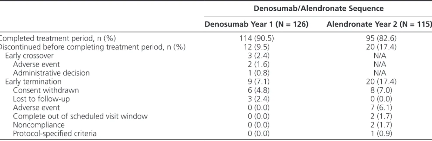
The DAPS study enrolled 250 participants. Of 126 women randomized to the denosumab/alendronate se-quence, 114 (90.5%) completed denosumab treatment in year 1 and 115 (91%) transitioned to alendronate at month 12, including 3 who crossed over early. Alendronate treatment in year 2 was completed by 95 participants (82.6%). Most common reasons for dis-continuation before completion of treatment included withdrawn consent (6 participants [4.8%] in year 1; 8 [7.0%] in year 2), lost to follow-up (3 [2.4%] in year 1; 0 [0.0%] in year 2), and adverse events (0 [0.0%] in year 1; 7 [6.1%] in year 2) (Table 1). Characteristics of the 126 participants initiating denosumab treatment in year 1 and the 115 participants initiating alendronate treatment in year 2 are shown in Table 2. At baseline, participants had a mean age of 65 years and mean BMD T-scores of −2.0, −1.6, and −2.0 at the LS, TH, and FN, respectively. Prior osteoporotic fracture was reported for 37.3% of participants at baseline. Similar charac-teristics were observed for the participants who transi-tioned to alendronate at month 12.
Change in BMD and BTM with denosumab in year 1 and alendronate in year 2
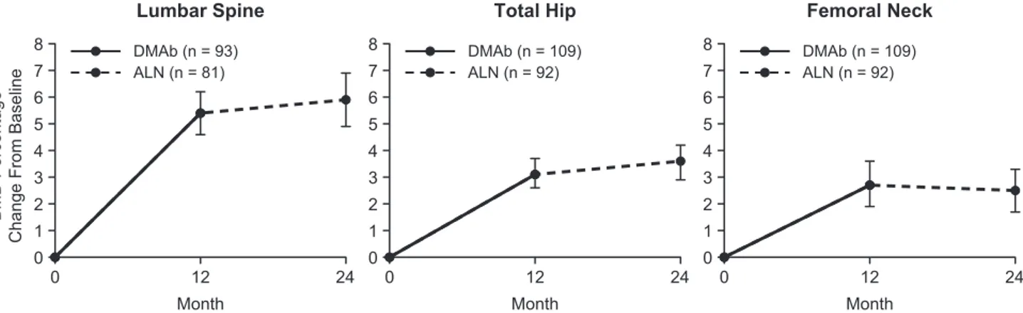
With denosumab treatment in year 1, the mean percent change in BMD from baseline to month 12 was 5.4%, 3.1%, and 2.7% for the LS, TH, and FN,
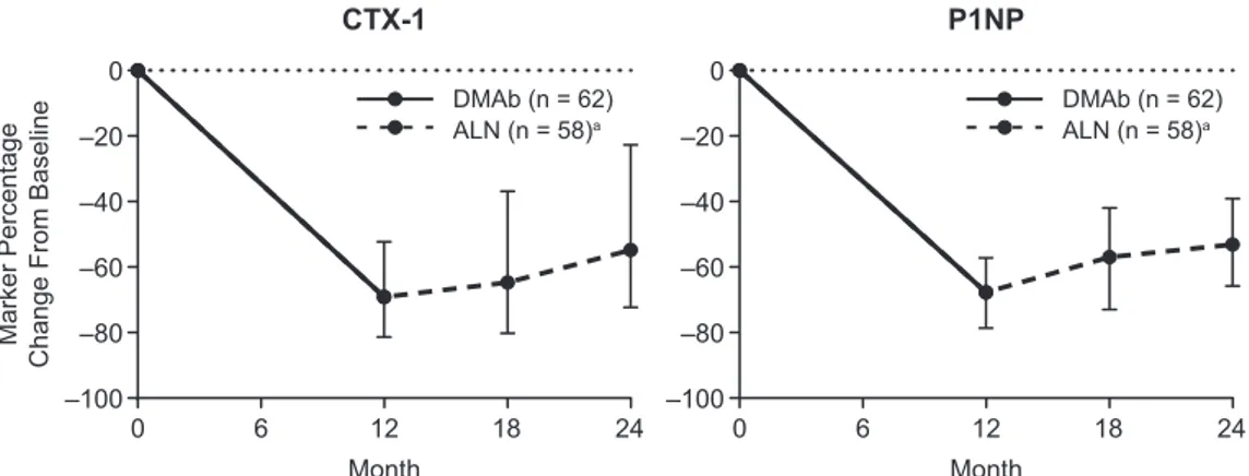
respectively. After transitioning to alendronate in year 2, the mean percent change in BMD from month 12 to month 24 was 0.5%, 0.5%, and −0.2% at the LS, TH, and FN, respectively. Evaluating the entire study period, participants showed an average gain in BMD above baseline of 5.9%, 3.6%, and 2.5% at the LS, TH, and FN, respectively (Fig. 1). The median percent change in CTX-1 from baseline to month 12, month 18, and month 24 was −69.1%, −64.7%, and −54.8%, respect-ively. The median percent change in P1NP from baseline to month 12, month 18, and month 24 was −67.7%, −57.0%, and −53.1%, respectively (Fig. 2).
Participants grouped into those who lost, maintained, or gained BMD in year 2
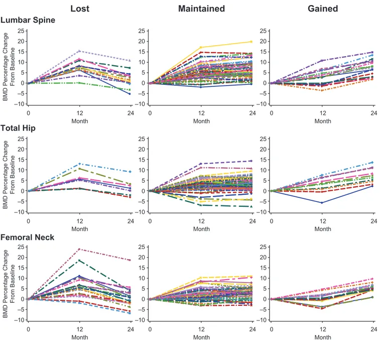
Fig. 3 shows BMD responses for individual partici-pants throughout the study for the groups that lost, main-tained, and gained BMD during year 2 on alendronate. Of the 82 participants with BMD measurements avail-able at the LS, 13 (15.9%) lost BMD, 52 (63.4%) main-tained BMD, and 17 (20.7%) gained BMD at this site. Of the 92 participants with BMD measurements avail-able at the TH and FN, 7 (7.6%) lost BMD, 75 (81.5%) maintained BMD, and 10 (10.9%) gained BMD at the TH, and 20 (21.7%) lost BMD, 56 (60.9%) maintained BMD, and 16 (17.4%) gained BMD at the FN (Table 3). Of the 82 participants with BMD measurements avail-able at the LS, TH, and FN, only 1 participant (1.2%) lost BMD at all 3 sites.
Participant characteristics and BMD change in year 2
The BMD change in year 2 was similar regardless of baseline characteristics such as age, BMD T-score, baseline levels of BTMs, and history of fracture (Table 3). For all skeletal sites, subjects who lost BMD
Table 1. Numbers of Participants Completing Each Treatment Period and Reasons for Study Discontinuation
Denosumab/Alendronate Sequence
Denosumab Year 1 (N = 126) Alendronate Year 2 (N = 115)
Completed treatment period, n (%) 114 (90.5) 95 (82.6)
Discontinued before completing treatment period, n (%) 12 (9.5) 20 (17.4)
Early crossover 3 (2.4) N/A
Adverse event 2 (1.6) N/A
Administrative decision 1 (0.8) N/A
Early termination 9 (7.1) 20 (17.4)
Consent withdrawn 6 (4.8) 8 (7.0)
Lost to follow-up 3 (2.4) 0 (0.0)
Adverse event 0 (0.0) 7 (6.1)
Complete out of scheduled visit window 0 (0.0) 2 (1.7)
Noncompliance 0 (0.0) 2 (1.7)
Protocol-specified criteria 0 (0.0) 1 (0.9)
N = number of participants randomized (year 1) or crossed over (year 2); n = number of participants with the characteristic of interest; N/A = not applicable.
with alendronate in year 2 had shown a greater per-cent change in BMD with denosumab in year 1 (Fig. 3,
Table 3). At the LS, participants who lost BMD in year 2 had gained an average of 7.1% BMD in year 1, while those who gained BMD in year 2 had gained an average of 3.1% BMD in year 1, and a similar differ-ence was observed at the TH (6.2% vs 2.8%). At the FN, participants who lost BMD in year 2 had gained an average of 7.0% BMD in year 1, while those who gained BMD in year 2 had gained an average of 0.6% BMD in year 1. BMD did not fall below pretreatment baseline in the majority of participants and was ob-served most often in participants who lost BMD with alendronate. Among participants who lost BMD in year 2 at a given skeletal site, 23.1% fell below their baseline BMD value at the LS, 28.6% fell below their baseline value at the TH, and 50.0% fell below their baseline value at the FN.
While adherence to oral alendronate in year 2 was lower than adherence to denosumab in year 1 (16), there was no numeric trend between alendronate adherence and the BMD response in year 2 (Table 3). Compliance,
defined as the percentage of provided alendronate tab-lets taken, was also investigated in terms of the BMD change in year 2 using quartiles of compliance, and there were no differences in the percent change in BMD in year 2 among the 4 compliance subgroups (data not shown).
Adverse event summary
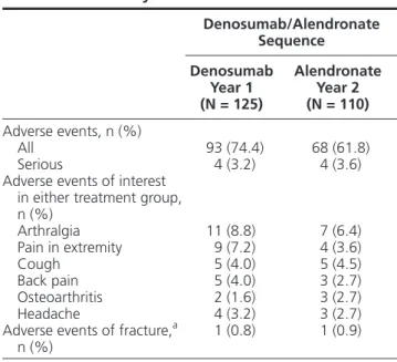
Safety analysis was performed for 125 participants who received at least 1 dose of denosumab in year 1 and 110 participants who received at least 1 dose of
alendronate in year 2 (Table 4). The adverse event
profiles were similar between treatment periods, with 74.4% and 61.8% of participants experiencing ad-verse events during denosumab and alendronate treat-ment, respectively. The most frequent adverse events (denosumab year 1, alendronate year 2) included arth-ralgia (8.8%, 6.4%), pain in extremity (7.2%, 3.6%), back pain (4.0%, 2.7%), and cough (4.0%, 4.5%). Adverse events of fracture were experienced by 1 par-ticipant during year 1 and 1 parpar-ticipant during year 2; both fracture events were classified as osteoporotic and
Table 2. Participant Characteristics at Study Baseline
Denosumab/Alendronate Sequence
Denosumab Year 1 (N = 126) Alendronate Year 2 (N = 115)
Race/ethnicity—white, n (%) 115 (91) 107 (93)
Age, mean (SD), years 65.1 (7.6) 65.1 (7.4)
Years since menopause, mean (SD) 18.2 (11.4) 17.9 (10.9)
BMD T-score at the start of each year, mean (SD)
Lumbar spine –2.04 (1.16) –1.61 (1.29)
Total hip –1.60 (0.74) –1.38 (0.74)
Femoral neck –2.01 (0.55) –1.84 (0.6)
Prior osteoporotic fracture, n (%) 47 (37.3) 41 (35.7)
N = number of participants randomized (year 1) or crossed over (year 2); n = number of participants with the characteristic of interest. Abbreviations: BMD, bone mineral density SD, standard deviation.
0 12 24 0 1 2 3 4 5 6 7 8 Lumbar Spine Month BMD Percentage
Change From Baseline
ALN (n = 81) DMAb (n = 93) 0 12 24 0 1 2 3 4 5 6 7 8 Total Hip Month ALN (n = 92) DMAb (n = 109) 0 12 24 0 1 2 3 4 5 6 7 8 Femoral Neck Month ALN (n = 92) DMAb (n = 109)
Figure 1. BMD percentage change from baseline with denosumab in year 1 and alendronate in year 2. Data show mean and 95% confidence
interval. n = number of participants with measurements at baseline and month 12 (DMAb) or month 24 (ALN). Abbreviations: ALN, alendronate; BMD, bone mineral density; DMAb, denosumab.
nonvertebral. No deaths, osteonecrosis of the jaw, or atypical femoral fractures were reported.
Discussion
Transitioning to alendronate was generally effective at preserving the gain in BMD and suppression of BTMs achieved with 1 year of denosumab treatment. Among the minority of participants who lost BMD at the LS, TH, or FN after transitioning to alendronate, few fell below their pretreatment baseline BMD value, and only 1 participant lost BMD at all skeletal sites. Larger BMD increases in year 1 on denosumab were observed for par-ticipants who lost BMD in year 2 on alendronate, while other participant characteristics showed no numeric trend with the BMD response in year 2 on alendronate. These findings demonstrate that most women receive benefit from oral bisphosphonate therapy following denosumab cessation.
In the pivotal phase 3, randomized FREEDOM trial and open-label extension, treatment with denosumab for up to 10 years was associated with a continued in-crease in BMD, sustained reduction in BTMs, and low incidence of fractures, and was generally well tolerated (9). In head-to-head studies, denosumab treatment led to larger increases in BMD and greater reductions in BTMs compared with alendronate (22, 23), which is in agreement with the results presented here. However, be-cause denosumab is a reversible inhibitor of RANKL, denosumab’s effects on bone turnover are reversible with discontinuation, and cessation of denosumab has been associated with rapid loss of vertebral fracture protec-tion, including multiple vertebral fracture (24). Thus, al-though denosumab treatment can produce large gains in BMD and significant suppression of BTMs, these effects do not protect patients when therapy is discontinued.
For this reason, the use of a “drug holiday” in patients receiving denosumab is not recommended (25). The de-cision to discontinue denosumab treatment should be ac-companied by careful monitoring and use of a follow-on antiremodeling agent.
Limited data are available regarding the optimal
post-denosumab bisphosphonate treatment regimen (26). In
a small case series evaluating women followed for up to 2 years after the FRAME trial, zoledronic acid (n = 11) infusion after denosumab discontinuation showed 73% to 87% preservation of the gains in BMD at the TH or LS after 1 year, with minimal further BMD loss at any skel-etal site in year 2; participants given risedronate (n = 5) showed only partial preservation of BMD (41%–64%) (13, 27). These findings are somewhat inconsistent with a previous, smaller case series showing only minimal ef-ficacy to preserve BMD after denosumab treatment ces-sation (28). This difference in outcome may be related to the inclusion of participants with different ages, previous osteoporosis treatments, and durations of denosumab treatment, and it should be noted that intravenous zoledronic acid was administered 6 or 8 months after the last denosumab injection. In the current analysis, which included a larger number of participants , 1 year of alendronate treatment following 1 year of denosumab led to maintained or increased BMD at the TH or LS in 84% to 92% of participants. These findings demonstrate the potential for bisphosphonate treatment to prevent re-versible bone loss in patients who discontinue denosumab treatment and may suggest that oral alendronate helps maintain BMD in the period immediately following denosumab discontinuation when the effects of treat-ment on bone turnover have dissipated. Ongoing ran-domized clinical trials evaluating bisphosphonate use after denosumab discontinuation should provide clarity on the optimal treatment regimen (29, 30).
0 6 12 18 24 –100 –80 –60 –40 –20 0 –100 –80 –60 –40 –20 0 CTX-1 Month Marker Percentag e
Change From Baseline
ALN (n = 58)a DMAb (n = 62) ALN (n = 58)a DMAb (n = 62) 0 6 12 18 24 P1NP Month
Figure 2. BTM percentage change from baseline with denosumab in year 1 and alendronate in year 2. Data show mean and interquartile range. n
= number of participants with measurements at baseline and month 12 (DMAb) or month 24 (ALN). Abbreviations: ALN, alendronate; BTM, bone turnover marker; CTX-1, serum C-telopeptide; DMAb, denosumab; P1NP, N-terminal propeptide type I procollagen. aAt month 18, n = 60.
Although the majority of participants maintained or gained BMD after transitioning from denosumab to alendronate in the current analysis, 15.9%, 7.6%, and 21.7% lost BMD at the LS, TH, and FN, respect-ively, with only 1 participant losing BMD at all sites. These differences between sites in the response to treat-ment may be due to differences in the proportions of cortical versus trabecular bone as well as load-bearing parameters. Also, degenerative artifact is likely to have a greater impact on BMD at the LS compared with TH. In general, cortical bone may respond less rap-idly to antiresorptive therapies compared with tra-becular bone, as cortical bone has less surface area per unit volume of mineralized bone matrix upon which
bisphosphonates can be adsorbed (31). Specifically,
denosumab is likely to have superior effects on cortical bone as compared with alendronate. In a head-to-head study using high-resolution peripheral computed tom-ography (HR-pQCT), there was greater reduction in cortical porosity at the radius and tibia in patients
re-ceiving denosumab compared with alendronate (32).
Supportive data using another technology to measure cortical porosity indicate the efficacy of denosumab in reducing cortical porosity at the hip, a load-bearing site (33). Although the FN is included in the TH region of interest, the FN comprises a much smaller region and provides a less precise measurement than the TH, which is the preferred region of interest for following Lumbar Spine Total Hip Femoral Neck –10 –5 0 5 10 15 20 25 –10 –5 0 5 10 15 20 25 –10 –5 0 5 10 15 20 25 –10 –5 0 5 10 15 20 25 –10 –5 0 5 10 15 20 25 –10 –5 0 5 10 15 20 25 –10 –5 0 5 10 15 20 25 –10 –5 0 5 10 15 20 25 –10 –5 0 5 10 15 20 25 0 12 24
Month 0 Month12 24 0 Month12 24
0 12 24
Month 0 Month12 24 0 Month12 24
0 12 24 Month BMD Percentage Change From Baseline BMD Percentage Change From Baseline BMD Percentage Change From Baseline 0 12 24 Month 0 Month12 24
Lost
Maintained
Gained
Figure 3. Individual participant BMD changes for groups that lost, maintained, or gained BMD in year 2 on alendronate. Abbreviation: BMD, bone
mineral density.
Table 3.
Participant Characteristics and BMD Change in Y
ear 2 After T ransitioning to Alendr onate Lumbar Spine (N = 82) Total Hip (N = 92) Femoral Neck (N = 92)
Participants stratified by BMD change category fr
om M12 to M24 a Lost Maintained Gained Lost Maintained Gained Lost Maintained Gained n (%) 13 (15.9) 52 (63.4) 17 (20.7) 7 (7.6) 75 (81.5) 10 (10.9) 20 (21.7) 56 (60.9) 16 (17.4)
Baseline characteristics Age (years), mean (SD)
63.8 (5.5) 65.5 (7.7) 64.3 (8.2) 66.4 (8.5) 64.5 (7.1) 68.5 (7.5) 67.1 (8.8) 64.2 (6.7) 65.8 (7.2) BMD T -scor e, mean (SD) −1.8 (2.0) −2.1 (1.1) −1.9 (0.8) b −2.0 (0.5) −1.4 (0.7) −2.2 (0.7) −1.9 (0.4) −2.0 (0.5) −2.2 (0.4) CTX-1 (ng/mL), mean (SD) 0.49 (0.19) 0.53 (0.29) 0.53 (0.25) 0.63 (0.38) 0.49 (0.25) 0.68 (0.17) 0.52 (0.26) 0.53 (0.27) 0.51 (0.26) P1NP ( µg/mL), mean (SD) 52.8 (9.5) 55.7 (25.1) 58.8 (25.1) 52.4 (20.6) 55.3 (22.8) 63.7 (22.7) 54.4 (16.4) 56.3 (25.2) 56.6 (22.9) History of fractur e (yes), n (%) 7 (53.9) 23 (44.2) 10 (58.8) 4 (57.1) 37 (49.3) 3 (30.0) 7 (35.0) 26 (46.4) 11 (68.8) M12 and M24 characteristics n = 12 n = 51 n = 14 n = 7 n = 71 n = 10 n = 19 n = 55 n = 14 % change in BMD fr om M0 to M12, mean (SD) 7.1 (3.1) 5.9 (3.8) 3.1 (3.9) 6.2 (4.5) 3.0 (3.1) 2.8 (4.0) 7.0 (6.3) 2.7 (2.9) 0.6 (2.6) n = 12 n = 51 n = 15 n = 7 n = 71 n = 10 n = 19 n = 55 n = 14 BMD (g/cm 2 ) at M12, mean (SD) 1.0 (0.3) 0.9 (0.2) 0.9 (0.1) 0.8 (0.1) 0.8 (0.1) 0.7 (0.1) 0.7 (0.1) 0.7 (0.1) 0.6 (0.1) n = 12 n = 51 n = 15 n = 7 n = 71 n = 10 n = 19 n = 55 n = 14 BMD T -scor e at M12, mean (SD) −1.2 (2.4) −1.7 (1.2) −1.7 (0.8) −1.7 (0.5) −1.2 (0.7) −2.1 (0.6) −1.5 (0.6 −1.9 (0.6) −2.2 (0.5) n = 8 n = 32 n = 11 n = 6 n = 43 n = 5 n = 15 ) n = 32 n = 7 % change in CTX-1 fr om M0 to M12, mean (SD) −61.3 (18.9) −62.4 (24.7) −52.0 (34.9) −71.9 (20.2) −58.6 (26.9) −67.1 (21.2) −56.7 (27.3) −66.4 (22.8) −44.4 (30.3) n = 9 n = 33 n = 13 n = 6 n = 47 n = 5 n = 16 n = 33 n = 9 % change in P1NP fr om M0 to M12, mean (SD) −31.2 (32.4) −47.3 (28.1) −50.5 (20.9) −23.2 (36.2) −48.0 (25.9) −62.2 (12.2) −34.1 (33.3) −55.4 (22.0) −36.9 (24.2) n = 13 n = 52 n = 17 n = 7 n = 75 n = 10 n = 20 n = 56 n = 16 % change in BMD fr om M0 to M24, mean (SD) 2.1 (4.2) 6.7 (4.2) 7.7 (4.0) b 1.7 (4.1) 3.3 (3.2) 7.4 (3.8) 0.8 (5.4) 3.0 (2.8) 5.4 (2.3) BMD at M24 below M0 value, n (%) 3 (23.1) 1 (1.9) 0 (0.0) b 2 (28.6) 5 (6.7) 0 (0.0) 10 (50.0) 8 (14.3) 0 (0.0) ALN adher ence at M24, n (%) 11 (84.6) 38 (73.1) 14 (82.4) 5 (71.4) 56 (74.7) 10 (100) 15 (75.0) 45 (80.4) 11 (68.8)
N = number of participants with BMD values at M12 and M24; n = number of participants in each BMD gr
oup with available data.
ALN, alendr
onate; BMD, bone mineral density; CTX-1, serum C-telopeptide; P1NP
, N-terminal pr
opeptide type I pr
ocollagen; M, month.
a Based on a 3% BMD thr
eshold. A BMD change ≤ −3% indicated lost BMD; > −3% and < 3% indicated maintained BMD; and ≥ 3% indicated gained BMD;
b n = 16.
individual patients. Together, these differences can lead to variability in the clinical response to osteoporosis therapies.
The current analysis sought to better understand patient characteristics linked to loss of BMD after transitioning from denosumab to alendronate treat-ment, and our results identified the change in BMD with denosumab treatment from baseline to month 12. Specifically, the BMD gain in year 1 on denosumab was numerically higher in individuals who lost BMD in year 2 on alendronate than in those who maintained or gained BMD in year 2. This result may be attrib-utable to “regression to the mean,” a characteristic of imprecision of measurement. Bone remodeling differs between individuals and is influenced by a variety of factors (34, 35). Closure of the remodeling space with antiresorptive treatment in high remodelers might produce greater gains in BMD with treatment. In these individuals, subsequent discontinuation of reversible treatment may result in resumption of the same level of remodeling, which may not be fully inhibited with a bisphosphonate. However, alendronate treatment was able to maintain BMD above the pretreatment base-line level in the majority of women who lost BMD in year 2.
No other baseline or year 1 participant character-istics could consistently identify participants who lost BMD after transitioning to alendronate treatment
in this analysis. This finding suggests that all pa-tients, regardless of baseline characteristics and frac-ture history, may benefit from follow-on therapy with bisphosphonates after discontinuation of denosumab. We did not observe a meaningful effect of adherence to oral alendronate treatment on the change in BMD in year 2 after denosumab discontinuation. However, this may be the result of the overall good adherence in our clinical trial patient population, as compared to clinical practice where adherence can be low. Thus, it is likely that the low rate of nonadherence did not allow us to detect an effect of nonadherence on BMD decline in our patient population. Our findings also stress the import-ance of BMD monitoring while on treatment to identify those at greatest risk for fracture, as those patients with the largest increase in BMD with denosumab might also be the most vulnerable to BMD loss while receiving alendronate. Unfortunately, in clinical practice, most patients who discontinue denosumab treatment do not receive any osteoporosis treatment in the year following discontinuation; moreover, approximately half of those who begin a prescription medication for osteoporosis after denosumab cessation stop the therapy in the sub-sequent year (11).
Several limitations of this analysis should be con-sidered. First, denosumab was only administered for 1 year before transition to alendronate. With longer-term denosumab treatment, there will be con-tinued gains in BMD and the potential for greater bone loss after treatment is stopped, and it could be more difficult to preserve bone mass when transitioning to bisphosphonates. Determining the optimal timing, dose, and bisphosphonate medication to administer after denosumab cessation warrants further study. Second, this post hoc analysis had a modest sample size, particularly for the analysis of participants divided into BMD response groups (ie, lost, maintained, and gained). However, the current sample size is larger than that reported in other ad-hoc case series investigating denosumab discontinuation and bisphosphonate follow-on therapy. Third, the analysis was not powered to detect statistical relationships between participant characteristics and their BMD response in year 2, and all analyses were descriptive in nature. Fourth, the effect-iveness of alendronate to maintain BMD in this study may have been unrealistically high, given the difficulties with compliance and dosing faced in clinical practice (36). Finally, DXA assessment for this study was not centralized; therefore, BMD results may vary by center.
Denosumab treatment increased BMD at all skel-etal sites examined, and the gains in BMD achieved with denosumab were maintained in the majority of participants after transitioning to alendronate. Among
Table 4. Summary of Adverse Events
Denosumab/Alendronate Sequence Denosumab Year 1 (N = 125) Alendronate Year 2 (N = 110) Adverse events, n (%) All 93 (74.4) 68 (61.8) Serious 4 (3.2) 4 (3.6)
Adverse events of interest in either treatment group, n (%) Arthralgia 11 (8.8) 7 (6.4) Pain in extremity 9 (7.2) 4 (3.6) Cough 5 (4.0) 5 (4.5) Back pain 5 (4.0) 3 (2.7) Osteoarthritis 2 (1.6) 3 (2.7) Headache 4 (3.2) 3 (2.7)
Adverse events of fracture,a
n (%) 1 (0.8) 1 (0.9)
N = number of participants who received at least 1 dose of investi-gational product during the corresponding treatment period (safety analysis population); n = number of participants reporting at least 1 adverse event during the corresponding period.
Includes only treatment-emergent adverse events that started on or before the end of the corresponding treatment period.
aReported fractures were classified as osteoporotic and nonvertebral.
participants who lost BMD in year 2 with alendronate, the majority remained above their pretreatment base-line value. Those with larger BMD increases in year 1 often showed greater BMD losses in year 2, with other participant characteristics not related to the re-sponse in year 2. These data highlight the need for oral bisphosphonate therapy following denosumab cessation and BMD monitoring of patients transitioning from denosumab to bisphosphonates.
Acknowledgments
Kate Smigiel, PhD (Amgen Inc.) provided medical writing as-sistance.
Financial Support: This study was sponsored by Amgen Inc. Clinical Trial Information: NCT00518531
Additional Information
Correspondence: David Kendler, MD, 150-943 W. Broadway, Vancouver, BC, Canada V5Z 4E1. E-mail:
davidkendler@gmail.com.
Disclosure Summary: D.K. has grant/research support from Amgen, AstraZeneca, and Eli Lilly and consultant/speakers’ bureau/advisory activities with Amgen, Pfizer, and Eli Lilly. P.C. has grant/research support from CONACYT (Mexico) Fondos Federales (Mexico); consultant/speakers’ bureau/ad-visory activities with Amgen, Eli Lilly, and Pfizer; board mem-bership with IOF and National University of Mexico UNAM; and patent licensing for 613227. P.R.E. has grant/research support from Amgen and Eli Lilly and consultant/speakers’ bureau/advisory activities with Amgen, Alexion, and Eli Lilly. M.M. has grant/research support from Amgen and consultant/ speakers’ bureau/advisory activities with Amgen and Radius Health. Y.R. has grant/research support from the Korean Ministry of Health and Welfare, Korean Ministry of Science and ICT and consultant/speakers’ bureau/advisory activities with Amgen. A.C., S.H., and R.K.S. are company employees and have stock ownership or royalties with Amgen.
Data Availability: Qualified researchers may request data from Amgen clinical studies. Complete details are available
at the following:
https://wwwext.amgen.com/science/clinical-trials/clinical-data-transparency-practices/.
References
1. Roux C, Wyman A, Hooven FH, et al.; GLOW investiga-tors. Burden of non-hip, non-vertebral fractures on quality of life in postmenopausal women: the global longitudinal study of osteoporosis in women (GLOW). Osteoporos Int. 2012;23(12):2863–2871.
2. Adachi JD, Ioannidis G, Olszynski WP, et al. The impact of in-cident vertebral and non-vertebral fractures on health related quality of life in postmenopausal women. BMC Musculoskelet
Disord. 2002;3:11.
3. Burge R, Dawson-Hughes B, Solomon DH, Wong JB, King A, Tosteson A. Incidence and economic burden of osteoporosis-related fractures in the United States, 2005-2025. J Bone Miner
Res. 2007;22(3):465–475.
4. Weycker D, Li X, Barron R, Bornheimer R, Chandler D. Hospitalizations for osteoporosis-related fractures: economic costs and clinical outcomes. Bone Rep. 2016;5:186–191.
5. Black DM, Cummings SR, Karpf DB, et al. Randomised trial of effect of alendronate on risk of fracture in women with existing vertebral fractures. Fracture Intervention Trial Research Group.
Lancet. 1996;348(9041):1535–1541.
6. Black DM, Delmas PD, Eastell R, et al.; HORIZON Pivotal Fracture Trial. Once-yearly zoledronic acid for treatment of postmenopausal osteoporosis. N Engl J Med. 2007;356(18): 1809–1822.
7. Cummings SR, San Martin J, McClung MR, et al.; FREEDOM Trial. Denosumab for prevention of fractures in postmenopausal women with osteoporosis. N Engl J Med. 2009;361(8):756–765. 8. Harris ST, Watts NB, Genant HK, et al. Effects of risedronate
treatment on vertebral and nonvertebral fractures in women with postmenopausal osteoporosis: a randomized controlled trial. Vertebral efficacy with risedronate therapy (VERT) Study Group.
Jama. 1999;282(14):1344–1352.
9. Bone HG, Wagman RB, Brandi ML, et al. 10 years of Denosumab treatment in postmenopausal women with osteoporosis: results from the phase 3 randomised FREEDOM trial and open-label extension. Lancet Diabetes Endocrinol. 2017;5(7):513–523. 10. Bone HG, Bolognese MA, Yuen CK, et al. Effects of Denosumab
treatment and discontinuation on bone mineral density and bone turnover markers in postmenopausal women with low bone mass. J Clin Endocrinol Metab. 2011;96(4):972–980.
11. McClung MR, Wagman RB, Miller PD, Wang A, Lewiecki EM. Observations following discontinuation of long-term Denosumab therapy. Osteoporos Int. 2017;28(5):1723–1732.
12. Popp AW, Zysset PK, Lippuner K. Rebound-associated vertebral fractures after discontinuation of Denosumab-from clinic and biomechanics. Osteoporos Int. 2016;27(5):1917–1921.
13. Horne AM, Mihov B, Reid IR. Bone loss after Romosozumab/ Denosumab: effects of bisphosphonates. Calcif Tissue Int. 2018;103(1):55–61.
14. Tsourdi E, Langdahl B, Cohen-Solal M, et al. Discontinuation of Denosumab therapy for osteoporosis: a systematic review and position statement by ECTS. Bone. 2017;105:11–17.
15. Kendler DL, McClung MR, Freemantle N, et al.; DAPS Investigators. Adherence, preference, and satisfaction of postmenopausal women taking Denosumab or Alendronate.
Osteoporos Int. 2011;22(6):1725–1735.
16. Freemantle N, Satram-Hoang S, Tang ET, et al.; DAPS Investigators. Final results of the DAPS (Denosumab adherence preference satisfaction) study: a 24-month, randomized, cross-over comparison with alendronate in postmenopausal women.
Osteoporos Int. 2012;23(1):317–326.
17. Cosman F, Crittenden DB, Ferrari S, et al. FRAME study: the foun-dation effect of building bone with 1 year of romosozumab leads to continued lower fracture risk after transition to Denosumab. J
Bone Miner Res. 2018;33(7):1219–1226.
18. Miller PD, Hattersley G, Lau E, et al. Bone mineral density response rates are greater in patients treated with abaloparatide compared with those treated with placebo or teriparatide: Results from the ACTIVE phase 3 trial. Bone. 2019;120:137–140.
19. Egerdie RB, Saad F, Smith MR, et al. Responder analysis of the ef-fects of Denosumab on bone mineral density in men receiving an-drogen deprivation therapy for prostate cancer. Prostate Cancer
Prostatic Dis. 2012;15(3):308–312.
20. Nakano T, Yamamoto M, Hashimoto J, Tobinai M, Yoshida S, Nakamura T. Higher response with bone mineral density increase
with monthly injectable ibandronate 1 mg compared with oral risedronate in the MOVER study. J Bone Miner Metab. 2016;34(6):678–684.
21. Sebba AI, Bonnick SL, Kagan R, et al.; Fosamax Actonel Comparison Trial investigators. Response to therapy with once-weekly alendronate 70 mg compared to once-once-weekly risedronate 35 mg in the treatment of postmenopausal osteoporosis. Curr
Med Res Opin. 2004;20(12):2031–2041.
22. Brown JP, Prince RL, Deal C, et al. Comparison of the effect of Denosumab and alendronate on BMD and biochemical markers of bone turnover in postmenopausal women with low bone mass: a randomized, blinded, phase 3 trial. J Bone Miner Res. 2009;24(1):153–161.
23. Kendler DL, Roux C, Benhamou CL, et al. Effects of Denosumab on bone mineral density and bone turnover in postmenopausal women transitioning from alendronate therapy. J Bone Miner
Res. 2010;25(1):72–81.
24. Cummings SR, Ferrari S, Eastell R, et al. Vertebral fractures after discontinuation of Denosumab: a post hoc analysis of the ran-domized placebo-controlled freedom trial and its extension. J
Bone Miner Res. 2018;33(2):190–198.
25. Anagnostis P, Paschou SA, Mintziori G, et al. Drug holidays from bisphosphonates and denosumab in postmenopausal osteopor-osis: EMAS position statement. Maturitas. 2017;101:23–30. 26. Guañabens N, Moro-Álvarez MJ, Casado E, et al.; SEIOMM
Working Group. The next step after anti-osteoporotic drug discon-tinuation: an up-to-date review of sequential treatment. Endocrine. 2019;64(3):441–455.
27. Horne AM, Mihov B, Reid IR. Effect of zoledronate on bone loss after romosozumab/denosumab: 2-year follow-up. Calcif Tissue
Int. 2019;105(1):107–108.
28. Reid IR, Horne AM, Mihov B, Gamble GD. Bone loss after Denosumab: only partial protection with zoledronate. Calcif
Tissue Int. 2017;101(4):371–374.
29. ClinicalTrials.gov. Identifier NCT02499237, Zoledronic Acid
to Maintain Bone Mass After Denosumab Discontinuation (AfterDmab). Bethesda: National Library of Medicine (US);
2000. https://clinicaltrials.gov/ct2/show/NCT02499237.
30. ClinicalTrials.gov. Identifier NCT03087851, Treatment
With Zoledronic Acid Subsequent to Denosumab in Osteoporosis (ZOLARMAB). Bethesda: National Library
of Medicine (US); 2000. https://clinicaltrials.gov/ct2/show/ NCT03087851.
31. Roelofs AJ, Stewart CA, Sun S, et al. Influence of bone affinity on the skeletal distribution of fluorescently labeled bisphosphonates in vivo. J Bone Miner Res. 2012;27(4):835–847.
32. Zebaze RM, Libanati C, Austin M, et al. Differing effects of denosumab and alendronate on cortical and trabecular bone.
Bone. 2014;59:173–179.
33. Zebaze R, Libanati C, McClung MR, et al. Denosumab re-duces cortical porosity of the proximal femoral shaft in postmenopausal women with osteoporosis. J Bone Miner Res. 2016;31(10):1827–1834.
34. Ferretti JL, Cointry GR, Capozza RF, Frost HM. Bone mass, bone strength, muscle-bone interactions, osteopenias and osteoporoses.
Mech Ageing Dev. 2003;124(3):269–279.
35. Frost HM. The mechanostat: a proposed pathogenic mech-anism of osteoporoses and the bone mass effects of mechanical and nonmechanical agents. Bone Miner. 1987;2(2):73–85. 36. Fatoye F, Smith P, Gebrye T, Yeowell G. Real-world persistence
and adherence with oral bisphosphonates for osteoporosis: a sys-tematic review. BMJ Open. 2019;9(4):e027049.