tebroplasty offers a minimally invasive procedure, which can stabilize unstable fractures satisfactorily under local anesthesia, without worsening the co-morbidities. Unfortunately, some complications of vertebroplasty do exist. Acute complications are mostly related to surgical procedures, which include leakage of bone cement, infection, and fracture along the path of the vertebroplasty cannula14,17). Delayed complication is an occur-rence of a new fracture in an adjacent vertebra after plasty. Approximately 20% of the patients treated with vertebro-plasty experience incident fractures within one year and about 50-67% of the incident fractures occur adjacent to the aug-mented vertebra3,11,15,19,20).
Although the biomechanical explanation for the subsequent
INTRODUCTION
Despite the controversy over the role of vertebroplasty, many patients with painful osteoporotic compression fractures have been treated with the placement of bone cement into the frac-tured vertebral bodies10,12). It is well known that vertebroplasty is quite helpful; not only to reduce pain intensity, but also to sta-bilize the unstable parts of fractured vertebral columns2,4). For patients with vertebral fractures, conservative management sometimes fails to prevent the progress of kyphotic deformity, yet surgical treatment using metal implants cannot be per-formed in patients with osteoporotic spines, due to poor bone quality and the presence of co-morbidity. In this situation,
ver-Effect of Bone Cement Volume and Stiffness
on Occurrences of Adjacent Vertebral Fractures
after Vertebroplasty
Jin-Myung Kim, M.S.,1 Dong Ah Shin, M.D., Ph.D.,1,2 Dong-Hak Byun, M.S.,1 Hyung-Sun Kim, M.S.,1 Sohee Kim, Ph.D.,1 Hyoung-Ihl Kim, M.D., Ph.D.1,3
Department of Medical System Engineering and Mechatronics,1 Gwangju Institute of Science and Technology, Gwangju, Korea
Department of Neurosurgery,2 Yonsei University Health System, Seoul, Korea
Department of Neurosurgery,3 Presbyterian Medical Center, Jeonju, Korea
Objective : The purpose of this study is to find the optimal stiffness and volume of bone cement and their biomechanical effects on the adjacent vertebrae to determine a better strategy for conducting vertebroplasty.
Methods : A three-dimensional finite-element model of a functional spinal unit was developed using computed tomography scans of a normal mo-tion segment, comprising the T11, T12 and L1 vertebrae. Volumes of bone cement, with appropriate mechanical properties, were inserted into the trabecular core of the T12 vertebra. Parametric studies were done by varying the volume and stiffness of the bone cement.
Results : When the bone cement filling volume reached 30% of the volume of a vertebral body, the level of stiffness was restored to that of normal bone, and when higher bone cement exceeded 30% of the volume, the result was stiffness in excess of that of normal bone. When the bone ce-ment volume was varied, local stress in the bony structures (cortical shell, trabecular bone and endplate) of each vertebra monotonically increased. Low-modulus bone cement has the effect of reducing strain in the augmented body, but only in cases of relatively high volumes of bone cement (>50%). Furthermore, varying the stiffness of bone cement has a negligible effect on the stress distribution of vertebral bodies.
Conclusion : The volume of cement was considered to be the most important determinant in endplate fracture. Changing the stiffness of bone ce-ment has a negligible effect on the stress distribution of vertebral bodies.
Key Words : Finite element analysis · Bone cements · Vertebroplasty.
Laboratory Investigation
•Received : April 4, 2012 •Revised : July 18, 2012 •Accepted : November 22, 2012 •Address for reprints : Hyoung-Ihl Kim, M.D., Ph.D.
Department of Medical System Engineering and Mechatronics, Gwangju Institute for Science and Technology, 123 Cheondangwagi-ro, Buk-gu, Gwangju 500-712, Korea Tel : +82-62-715-3244, Fax : +82-62-715-3234, E-mail : [email protected]
•This is an Open Access article distributed under the terms of the Creative Commons Attribution Non-Commercial License (http://creativecommons.org/licenses/by-nc/3.0) which permits unrestricted non-commercial use, distribution, and reproduction in any medium, provided the original work is properly cited.
venting adjacent vertebral fractures in relation to the volume and stiffness of the bone cement. In this study, we tried to find the optimal stiffness and volume of bone cement and their biome-chanical effect on adjacent vertebrae, using multilevel vertebral unit modeling to determine a better strategy in vertebroplasty.
MATERIALS AND METHODS
A 3-D linear finite element model was generated to study the biomechanics of three-level spine segments (T11-L1) with bone cement augmentation in the T12 vertebral body. To model a 3-D geometry of the spine, computed tomographic scans of a normal thoracolumbar spinal segment with 1 mm-thick slices in the Digital Imaging and Communications in Medicine file format and a 3D surface model in the STL file format were pro-vided from KISTI (Korea Science and Technology Information, Seoul, Korea). Then, a customized volume mesh was generated by a homemade mesh generation program using MATLAB (Mathworks, MA, USA) (Fig. 1). The finite element model of each spine consists of three vertebral bodies (T11, T12, L1), four cartilage endplates, and posterior elements including the spinous and transverse processes and laminae. Intervertebral discs were inserted between the vertebral bodies. The vertebral bodies were made of outer cortical bone (1 mm thick) and in-ner trabecular bone, and the superior and inferior surfaces were covered by endplates (1 mm thick). The intervertebral discs were made of inner nucleus pulposus and outer annulus fibro-sus. The nucleus pulposus was set to occupy 50% of the total surface area of the disc and its height was 1 cm. The annulus fi-brosus was constructed with 10 folds of fiber layers in a concen-tric circumferential fashion in the outer margin of the interver-tebral discs1).
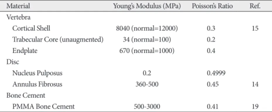
All materials were modeled as linear-elastic solids with their properties assigned as listed in Table 1. Elastic moduli of corti-cal shell, cancellous bone and endplates were reduced from those of normal bone to represent an osteoporotic bone condi-tion16). Decreasing these moduli resulted in a reduction of the overall compression stiffness that made the spinal bodies more susceptible to vertebral compression fractures. A barrel-like bone cement was given the material properties of polymethyl methacrylate (PMMA) (Young’s modulus : 3000 MPa, Poisson’s ratio : 0.41), which is the most common medium used in clinical procedures. To investigate the effect of the stiffness of the bone cement, Young’s modulus of the bone cement was varied from 500 to 3000 MPa. In a clinical setting, Young’s modulus of bone cement can be varied by introducing pores into the cement or by copolymerizing PMMA beads with ethyl-, propyl-, or butyl-methacrylate. The volume of bone cement was varied with respect to the volume of the can-fractures in levels adjacent to the augmented body is not
com-plete, the bone cement and its effect on the non-augmented ver-tebra are agreed to be the determinants of adjacent verver-tebral fractures. To determine the effect of bone cement on the treated and non-augmented vertebrae, simulation studies using finite el-ement modeling are regarded as useful methods to delineate the pathophysiology of adjacent vertebral fractures5,6,8,16). Instead of using human specimen, finite element modeling has the advan-tages of radical avoidance of individual variability and easy ac-quisition of various parameters such as intradiscal pressure, bone strain, and facet joint contact pressure. Finite element modeling has become a valid tool for analyzing spinal biome-chanics with low cost and reduced time. Berlemann et al.8) hy-pothesized that the increased stiffness of the treated vertebral al-ters the load transfer to the adjacent non-augmented level, leading to weakening of the spinal unit. Baroud et al.5,6) and Po-likeit et al.16) demonstrated that augmented vertebra reduced the normal cushioning function of endplates, consequently the pres-sures in the discs and endplates of non-treated vertebra in-creased. The increased stress is likely to place the non-augment-ed vertebra at risk of fracture. However, there is a paucity of biomechanical studies that provide information regarding
pre-Fig. 1. Geometry of multilevel spinal units with customized volume
mesh used in the finite element analysis. The unit consists of three seg-ments (T11, T12, & L1) and bone cement is inserted into T12.
Table 1. Material properties to represent osteoporosis in the study
Material Young’s Modulus (MPa) Poisson’s Ratio Ref.
Vertebra
Cortical Shell 8040 (normal=12000) 0.3 15
Trabecular Core (unaugmented) 34 (normal=100) 0.2
Endplate 670 (normal=1000) 0.4
Disc
Nucleus Pulposus 0.2 0.4999
Annulus Fibrosus 360-500 0.45 14
Bone Cement
the augmented body (T12) underwent a dramatic change with increase in bone cement volume. With the change of 10% to 90% bone cement volume, the stress increase was 0.038 MPa in the cancellous bone of the augmented T12.
The stress level varied in the endplates with changes in bone cement filling volume. With respect to the T12 vertebrae, the magnitude of stress increase was much greater with those end-plates located above the augmented body than those below. With the change of 10% to 90% bone cement volume, the T11 inferior endplate and the T12 superior endplate experienced stress increases of 0.34 MPa and 0.39 MPa, respectively. In con-trast, the T12 inferior endplate and the L1 superior endplate ex-perienced only 0.037 MPa and 0.083 MPa stress increases.
Along with the increase in the magnitude of stress and strain with bone cement volume increase, the overall stress and strain distributions in the structures were greatly altered upon bone cement augmentation, subjecting larger areas to higher stresses and strains with the bone cement volume increase. As the bone cement volume increased, the change in stress distribution and magnitude were apparent in the vertebrae directly above the augmented body. In contrast, the vertebra below the augment-ed level did not show markaugment-ed changes even with 90% filling volume of bone cement (Fig. 3).
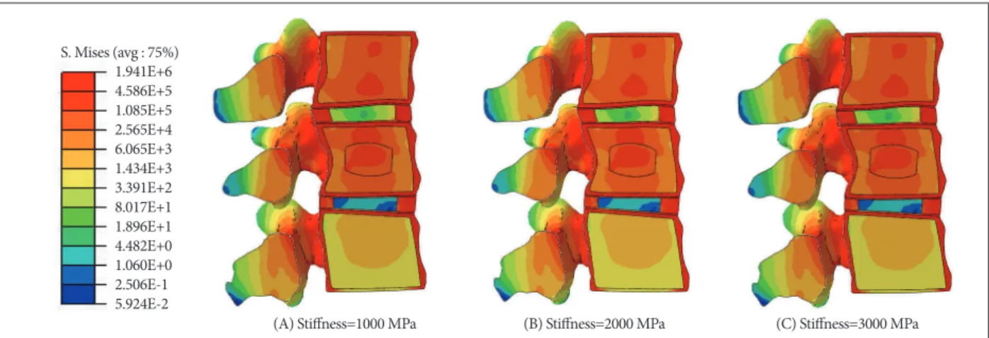
As the stiffness of bone cement was increased from 500 MPa to 3000 MPa, the structures in the augmented body showed a great-er change in strain level compared to the adjacent non-augment-ed vertebrae. Fig. 4 shows the percentile change in maximal prin-cipal strain in the structures of each vertebra, when the elastic modulus of the bone cement was increased from 500 MPa to 3000 MPa. The changes in nucleus pulposus and annulus fibro-sus were not marked.
DISCUSSION
Our study showed that the volume of bone cement is the crit-ical factor with regards to the restoration of biomechancrit-ical stiff-cellous bone of the T12 segment from 10% to 90% filling volume.
The fixed boundary condition restrained the bottom plate of the L1 segment in a translational axis but rotational motion was allowed in order to make the model more realistic. Compres-sive loading with a magnitude of 1000 N was applied to the front face of the top surface of the T11 cortical shell. Both the magnitude and the distribution of local stresses and strains in each material were computed using ABAQUS (SIMULIA, RI, USA) finite element analysis software. The developed finite model was validated by comparing its stiffness to that reported previously.
RESULTS
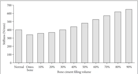
Compressive stiffness was calculated by dividing the axial compressive load of 1000 N by the maximum vertical displace-ment measured at a point on the top of the T11 segdisplace-ment. The stiffness of the vertebral bodies increased monotonically with bone cement volume. When bone cement filling volume reached 30% of the volume of a vertebral body, its stiffness was restored to that of normal bone, and bone cement filling volume of higher than 30% of the volume of a vertebral body resulted in stiffness in excesses of that of normal bone (Fig. 2).
When bone cement volume was varied, the local stress in the bony structures (cortical shell, trabecular bone and endplate) of each vertebra changed. In general, the stress level monotonical-ly increased with bone cement volume in all structures. Under the given compressive loading, the cortical shell of the T11 ver-tebral segment was subject to the greatest stress. Also, it experi-enced the greatest stress increase with increasing bone cement volume compared to cortical shells in other vertebrae. When bone cement volume increased from 10% to 90%, stress in the cortical shell of T11 increased by 0.34 MPa, while the cortical shells of T12 and L1 experienced stress increases of 0.24 MPa and 0.082 MPa, respectively. The stress level change in the corti-cal shell of L1 was negligible over the course of the change of bone cement volume from 10% to 90%,
whereas both cortical shells of T11 and T12 underwent relatively constant stress increases up to 60% bone cement vol-ume and significantly rapidly rapid in-crease beyond that.
Relatively equivalent results of changes in stress level were shown in the trabec-ular bone of each vertebra with changes in bone cement volume. When the bone cement volume was changed from 10% to 90%, the magnitude of stress increase was 0.012 MPa for the cancellous bone of T11 and only 0.0043 MPa for the can-cellous bone of L1. Different from what was seen with stress change in the corti-cal shell, stress in the cancellous bone of
Fig. 2. Stiffness change with different bone cement volumes. When bone cement volume of 30% is
used, stiffness is restored from osteoporotic bone state to the condition of normal bone and further increase in bone cement volume results in stiffness value beyond that of normal bone.
0 100 200 300 400 500 600 700 St iff ne ss (N /m m ) Normal Osteo.
and, as a result, less deformation of the vertebral body, higher than 50% bone cement volume is not currently being suggested in practical procedures, since higher bone cement volumes are accompanied with higher risks of leakage problems.
Our study demonstrated that stresses in all bony structures in adjacent vertebral bodies of the augmented spine segment in-creased with bone cement volume. In particular, stresses in the cranial vertebral body positioned above the augmented seg-ment showed a much greater increase than those in the verte-bral body beneath. Consequently, the level directly above the augmented vertebral body is much more susceptible to subse-ness of osteoporotic vertebral bodies. While Liebschner et al.13)
finite element analysis results showed that 15% bone cement volume was necessary to restore the stiffness of a fractured ver-tebral body, there is a general consensus on biomechanics of augmented vertebral bodies that approximately 30% or lower bone cement is required to restore the mechanical stiffness of a fractured vertebrae with osteoporotic conditions7). Our results also demonstrated that the initial stiffness of the vertebral body was restored when the T12 segment was augmented with ap-proximately 30% bone cement volume.
Even though higher cement volumes led to higher stiffness
Fig. 3. Sagittal plane of the studied spine segments with contours showing von Mises stress distribution for different bone cement filling volumes.
Changes in both magnitude and distribution of stress in each vertebral body can be observed with more apparent changes in upper vertebra (T11) than lower one (L1).
Fig. 4. Percentile difference in maximal principal strains in structures when bone cement stiffness is increased from 1000 MPa to 3000 MPa with
50% bone cement volume. Negligible changes are observed in all structures but in cortical shell and trabecular core of T11 and T12 segment. (A) Normal
(D) 50% Filling volume
(A) Stiffness=1000 MPa
(B) 10% Filling volume
(E) 70% Filling volume
(B) Stiffness=2000 MPa (C) 30% Filling volume (F) 90% Filling volume (C) Stiffness=3000 MPa S. Mises (avg : 75%) 1.941E+6 4.586E+5 1.085E+5 2.565E+4 6.065E+3 1.434E+3 3.391E+2 8.017E+1 1.896E+1 4.482E+0 1.060E+0 2.506E-1 5.924E-2 von Mises Stress (avg. 75%)
2.036E+6 1.000E+5 3.008E+4 9.051E+3 2.723E+3 8.191E+2 2.464E+2 7.414E+2 2.230E+1 6.710E+0 2.019E+0 6.073E-1 1.827E-1 5.496E-2
larger range of patients with patient-specific data, so that an ap-propriate protocol for vertebroplasty can be made for individual patients. Due to morphological variations present in the geo-metrical model of the studied segments, it was very hard to as-sign the center of rotation at which axial compression can be ap-plied regardless of any non-reproducible movements acting on the vertebrae. The influence of disc degeneration on the alterna-tion of load transfer should not be overlooked and should be in-cluded in a complete study. In addition, the results of this study do not provide absolute answers to vertebroplasty but it should be noted that the optimization of bone cement volume is patient specific; the volume of bone cement should be based on the size, body mineral density and stiffness of the vertebra of individual patients.
CONCLUSION
The modeling results suggest that bone cement volume can have significant effect on the occurrences of subsequent verte-bral fractures after vertebroplasty. The compressive stiffness of osteoporotic vertebra can be restored to normal range with only 30% bone cement volume. Stiffness increases further with high-er bone cement volume than 30% but may result in the subse-quent fractures of adjacent vertebral bodies, most likely in cra-nial direction. Low-modulus bone cement does have effect on reducing the strains in the augmented body but only with rela-tively high volumes of bone cement (>50%). Furthermore, vary-ing stiffness of bone cement has a negligible effect on stress dis-tribution of vertebral bodies.
• Acknowledgements
This research was jointly supported by Basic Science Research Program through the National Research foundation of Korea (NRF) funded by the Ministry of Education, Science, and Technology (KRF-2011-0010067) re-search and by a grant from the Institute of Medical System Engineering (iMSE) in the GIST, Korea.
References
1. Adams MA, Bogduk N, Burton K, Dolan P : The Biomechanics of Back
Pain. Edinburgh, London, New York : Churchill Livingstone, 2002, pp100
2. Anselmetti GC, Manca A, Hirsch J, Montemurro F, Isaia G, Osella G, et al. : Percutaneous vertebroplasty in osteoporotic patients : an institu-tional experience of 1,634 patients with long-term follow-up. J Vasc
In-terv Radiol 22 : 1714-1720, 2011
3. Aquarius R, Homminga J, Verdonschot N, Tanck E : The fracture risk of adjacent vertebrae is increased by the changed loading direction after a wedge fracture. Spine (Phila Pa 1976) 36 : E408-E412, 2011
4. Bae H, Hatten HP Jr, Linovitz R, Tahernia AD, Schaufele MK, McCol-lom V, et al. : A prospective randomized FDA-IDE trial comparing Cor-toss with PMMA for vertebroplasty : a comparative effectiveness re-search study with 24-month follow-up. Spine (Phila Pa 1976) 37 : 544-550, 2012
5. Baroud G, Heini P, Nemes J, Bohner M, Ferguson S, Steffen T : Biome-chanical explanation of adjacent fractures following vertebroplasty.
Ra-diology 229 : 606-607; author reply 607-608, 2003
6. Baroud G, Nemes J, Heini P, Steffen T : Load shift of the intervertebral disc after a vertebroplasty : a finite-element study. Eur Spine J 12 : 421-quent fracture than the level below, agreeing with previous
clin-ical findings9,18). It is hypothesized that bone cement volume is responsible for the stiffness of the augmented body but in re-turn, it compromises adverse kinematic effects of the surround-ing motion segments, mostly to the vertebrae directly above.
For low to moderate filling volumes (10-60%), initial fracture is likely to occur at endplates, especially the ones positioned above the augmented vertebrae, since they are under higher stresses and strains than those located beneath the augmented body. Our findings, in terms of distorted cushioning effect by vertebroplasty, agreed with the results of Baroud et al.5,6). They reported that axial cushioning can be achieved by the combined action of outward bowing of annulus fibrosis as well as inward bowing of the verte-bral endplates in normal vertebrae, but rigid cement augmenta-tion underneath the endplates acts as an upright pillar which can diminish the inward bulge of the endplates of the augmented ver-tebra5,6). Similar results were shown in this study that upon bone cement augmentation, the vertebral bodies experienced in-creased stiffness that prevented the disc from bulging in the di-rection of the augmented vertebral body but instead caused asymmetrical endplate deflection. The lower endplate experi-enced relatively lower stress increase, because some of the load was supported by the annulus fibrosus and nucleus pulposus that were positioned between each body. Consequently, higher local strains in the adjacent non-augmented vertebral bodies af-ter bone cement injection signify a higher incidence of endplate or cortical shell fractures of the non-augmented vertebral bod-ies. Since endplates have much lower failure strength than corti-cal shells, the initial fracture is likely to occur in an endplate, followed by a cortical fracture.
When bone cement volume is high (>70%), the stress level in cancellous bone of the augmented segment greatly increases, burgeoning the risk of re-fracture. Strains, especially in struc-tures in the augmented body, are decreased with low-modulus bone cement9). Higher strains in the augmented body can pro-voke the fracture of adjacent vertebral bodies, therefore, the use of low modulus bone cement is considered to be beneficial as it reduces the risk of fracture9). However, with a bone cement vol-ume of less than 30%, a negligible change in the maximum prin-cipal strain was observed. This implies that effect of changes in bone cement stiffness are only present when bone cement vol-ume is relatively high (>70%). Furthermore, no significant dif-ference in stress distribution was observed in the augmented and non-augmented vertebrae with different Young’s modulus of the bone cement.
Our study has several limitations in interpreting the results for clinical applications. Our finite model was based on the verte-bral bodies of a single person and does not cover the whole range of patients treated with vertebroplasty. Furthermore, this model did not consider disruption of mechanical integrity due to fracture. Also, the material properties of each material com-prising the spine can greatly vary with age and sex. In future studies, more quantitative data should be collected to cover a
et al. : 1150 kyphoplasties over 7 years : indications, techniques, and in-traoperative complications. Orthopedics 32 : 90, 2009
15. Pérez-Higueras A, Alvarez L, Rossi RE, Quiñones D, Al-Assir I : Percu-taneous vertebroplasty : long-term clinical and radiological outcome.
Neuroradiology 44 : 950-954, 2002
16. Polikeit A, Nolte LP, Ferguson SJ : The effect of cement augmentation on the load transfer in an osteoporotic functional spinal unit : finite-ele-ment analysis. Spine (Phila Pa 1976) 28 : 991-996, 2003
17. Tanigawa N, Kariya S, Komemushi A, Tokuda T, Nakatani M, Yagi R, et al. : Cement leakage in percutaneous vertebroplasty for osteoporotic compression fractures with or without intravertebral clefts. AJR Am J
Roentgenol 193 : W442-W445, 2009
18. Trout AT, Kallmes DF : Does vertebroplasty cause incident vertebral fractures? A review of available data. AJNR Am J Neuroradiol 27 : 1397-1403, 2006
19. Trout AT, Kallmes DF, Kaufmann TJ : New fractures after vertebroplasty : adjacent fractures occur significantly sooner. AJNR Am J Neuroradiol
27 : 217-223, 2006
20. Voormolen MH, Lohle PN, Lampmann LE, van den Wildenberg W, Juttmann JR, Diekerhof CH, et al. : Prospective clinical follow-up after percutaneous vertebroplasty in patients with painful osteoporotic verte-bral compression fractures. J Vasc Interv Radiol 17 : 1313-1320, 2006 426, 2003
7. Belkoff SM, Mathis JM, Erbe EM, Fenton DC : Biomechanical evalua-tion of a new bone cement for use in vertebroplasty. Spine (Phila Pa
1976) 25 : 1061-1064, 2000
8. Berlemann U, Ferguson SJ, Nolte LP, Heini PF : Adjacent vertebral fail-ure after vertebroplasty. A biomechanical investigation. J Bone Joint
Surg Br 84 : 748-752, 2002
9. Boger A, Heini P, Windolf M, Schneider E : Adjacent vertebral failure after vertebroplasty : a biomechanical study of low-modulus PMMA ce-ment. Eur Spine J 16 : 2118-2125, 2007
10. Boonen S, Wahl DA, Nauroy L, Brandi ML, Bouxsein ML, Goldhahn J, et al. : Balloon kyphoplasty and vertebroplasty in the management of vertebral compression fractures. Osteoporos Int 22 : 2915-2934, 2011 11. Hierholzer J, Fuchs H, Westphalen K, Baumann C, Slotosch C, Schulz R :
Incidence of symptomatic vertebral fractures in patients after percuta-neous vertebroplasty. Cardiovasc Intervent Radiol 31 : 1178-1183, 2008 12. Kallmes DF, Comstock BA, Heagerty PJ, Turner JA, Wilson DJ, Dia-mond TH, et al. : A randomized trial of vertebroplasty for osteoporotic spinal fractures. N Engl J Med 361 : 569-579, 2009
13. Liebschner MA, Rosenberg WS, Keaveny TM : Effects of bone cement volume and distribution on vertebral stiffness after vertebroplasty. Spine
(Phila Pa 1976) 26 : 1547-1554, 2001