RESEARCH ARTICLE
Received: November 30, 2017, Revised: January 2, 2018, Accepted: January 17, 2018 ISSN 2233-7679 (Online)
†
Correspondence to: Ki-Rim Kim
Department of Dental Hygiene, Kyungpook National University, 2559 Gyeongsang-daero, Sangju 37224, Korea Tel: +82-54-530-1422, Fax: +82-54-530-1429, E-mail: [email protected]
Copyright © 2018 by Journal of Dental Hygiene Science
Antimicrobial Activity of Korean Propolis Extracts on Oral Pathogenic Microorganisms
Jiyeon Roh and Ki-Rim Kim 1,†
Department of Dental Hygiene, Yonsei University Wonju College of Medicine, Wonju 26426,
1
Department of Dental Hygiene, Kyungpook National University, Sangju 37224, Korea
Propolis has been used as a natural remedy in folk medicine worldwide. The antibacterial, antiviral, antifungal, and antiprotozoal aspects of its antimicrobial properties have been widely investigated. However, few studies focused on its applications in dentistry. Many dental diseases are related to various microorganisms in the oral cavity. In this study, we assessed the antimicrobial activity of Korean propolis extract, collected from 6 different regions, on oral pathogenic microorganisms. The propolis samples, collected from 6 different regions (P1: Uijeongbu, P2: Ansan, P3:
Hongcheon, P4: Iksan, P5: Gwangju, and P6: Sangju), were dissolved in ethanol at two different concentrations (10 and 50 mg/ml). Three oral bacteria ( Streptococcus mutans , Staphylococcus aureus , and Enterococcus faecalis ) and one fungus ( Candida albicans ) were activated in general broth for 24 hours. Microorganisms were diluted and spread onto agar plates, onto which sterilized 6 mm filter papers with or without each propolis sample were placed. After 24 hours of incubation, clear zones of inhibition were observed. All tests were performed in triplicate. The propolis samples showed significant antibacterial and antifungal activity on oral pathogenic microorganisms; in addition, low-concentration groups showed outstanding antimicrobial efficacy on the 4 different microorganisms. Among the samples, P6 had significantly higher antibacterial activity than that of the others against three different bacteria. In particular, a high concentration of P6 showed a significant antifungal effect. In conclusion, we confirmed that Korean propolis has an inhibitory effect on oral pathogenic bacteria and fungi. Therefore, we suggest the possibility of developing oral medicine and oral care products based on Korean propolis.
Key Words: Antimicrobial activity, Microorganisms, Oral diseases, Propolis
Introduction
Propolis has been known for its antibacterial properties in folk medicine, and recently it found application in medicine and cosmetics 1) . It is generally composed of resin and balsam (around 50%), wax (30%), essential and aromatic oils (10%), pollen (5%), and other substances 2) . There is subtle variability in composition, based on several factors such as botanical sources and geographical zones 3) .
The oral cavity is a suitable habitat for many micro- organisms because of the wet environment and abundance of nutrients. Furthermore, in the oral cavity, soft and hard tissues coexist and form a specific environment that provides both aerobic and anaerobic conditions, in which
various microorganisms interact. If the overall oral bacteria decrease because of excessive intake of antibiotics, this may lead to opportunistic infections by fungi. Thus, various factors such as diseases, medicines, or irradiation can cause different oral diseases due to changes in oral microbial flora.
Among oral bacteria, Streptococcus mutans, a major
dental caries-inducing bacterium, occupies the largest
portion 4) . Enterococcus faecalis, on the other hand, plays a
pivotal role in establishing a microbial community by
connecting various bacteria in the oral cavity 5-7) . Staphy-
lococcus aureus, a Gram-positive staphylococcus typically
found in the respiratory system, on the skin, and in the oral
cavity, is also found in the periodontium where it may
Fig. 1. Collection sites of Korean propolis. P1: Uijeongbu, P2:
Ansan, P3: Hongcheon, P4: Iksan, P5: Gwangju, P6: Sangju.
cause periodontal disease 8,9) . Candida albicans, the most common fungus in the oral cavity and the gastrointestinal tract, causes opportunistic infections by quickly expanding due to host environmental changes induced by various factors 10,11) .
Regarding health, products based on natural components have recently gained focus 12-14) . The biological activity and chemical constituents of propolis have been widely studied, and propolis has been applied using various household items. However, few studies have been con- ducted on Korean propolis, and insufficient studies assessed its antimicrobial effects on microorganisms that inhabit the oral cavity 15-17) .
In this study, we investigated the antimicrobial potential of Korean propolis extracts on standard microorganism strains that cause major oral diseases.
Materials and Methods
1. Microbial samples
S. mutans (KCTC 5365), S. aureus (KCTC 1916), E.
faecalis (KCTC 5290), and C. albicans (KCTC 17712) were purchased from the Biological Resource Center in Korea Research Institute of Bioscience and Biotechnology (KRIBB). Four types of strains were cultured in Brain Heart Infusion (BHI) broth (Difco; BD, Franklin Lakes, NJ, USA) and Bacto Agar (BD) at 37 o C.
2. Propolis extracts
Korean propolis samples, which were collected from various regions of Korea (Fig. 1), were provided by Dong-A University. In brief, crude propolis was extracted with ethanol for 24 hours. The suspensions were separated, concentrated in a rotary evaporator, and redissolved in ethanol. We used two different concentrations of each propolis sample (10 and 50 mg/ml), and dimethyl sulfoxide (DMSO) was used as the negative control.
3. Measurement of microbial growth curve
Each microorganism was serially diluted 10-fold using BHI broth, and 100 μl of all dilutions were seeded into a 96-well microplate. Microbial growth was determined by following changes in optical density (OD 600 ) at 1, 3, 5, 7,
and 18 hours, using a spectrophotometer (Epoch Take3;
BioTek, Winooski, VT, USA). Before analysis, the plates were tapped carefully. BHI broth without bacteria was used as blank.
4. Agar diffusion test
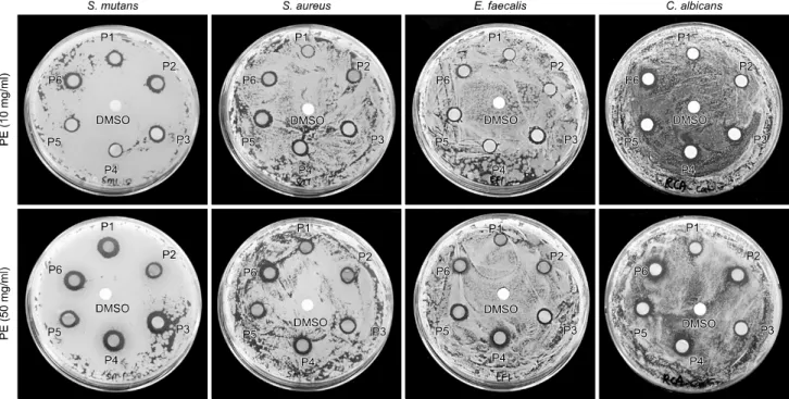
About 15 ml of BHI agar was poured onto sterile 90 mm Petri dishes (SPL, Pocheon, Korea) and allowed to solidify at room temperature. A 0.1 ml aliquot of each microbial suspension was spread on the agar plates separately. Six-millimeter filter papers were immersed in 0.1 ml of various propolis samples, and were placed onto the surface of the agar plates. The plates were incubated for 24 hours at 37 o C. The inhibition zones around each group were measured with Vernier calipers (Mitutoyo, Kawasaki, Japan).
5. Statistical analysis
All experiments were repeated at least 3 times inde- pendently. Data are expressed as the mean value±standard deviation. One-way analysis of variance (ANOVA) was performed to detect the significant effects of variables, followed by Tukey’s test (IBM SPSS Statistics 23.0; IBM Co., Armonk, NY, USA). The differences were considered significant at p<0.05.
Results
1. Growth curves of oral microorganisms
Growth curves of four different oral microorganisms are
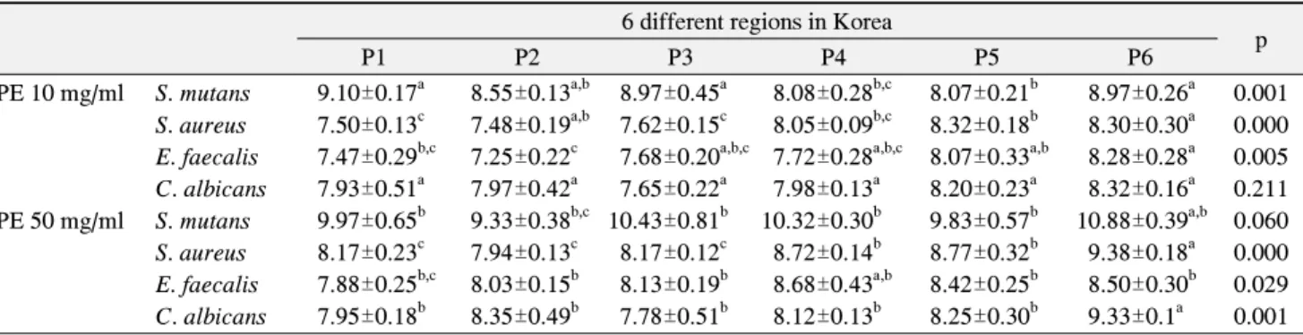
Table 1. Zones of Inhibition (mm)
6 different regions in Korea
P1 P2 P3 P4 P5 P6 p
PE 10 mg/ml S. mutans 9.10±0.17
a8.55±0.13
a,b8.97±0.45
a8.08±0.28
b,c8.07±0.21
b8.97±0.26
a0.001 S. aureus 7.50±0.13
c7.48±0.19
a,b7.62±0.15
c8.05±0.09
b,c8.32±0.18
b8.30±0.30
a0.000 E. faecalis 7.47±0.29
b,c7.25±0.22
c7.68±0.20
a,b,c7.72±0.28
a,b,c8.07±0.33
a,b8.28±0.28
a0.005 C. albicans 7.93±0.51
a7.97±0.42
a7.65±0.22
a7.98±0.13
a8.20±0.23
a8.32±0.16
a0.211 PE 50 mg/ml S. mutans 9.97±0.65
b9.33±0.38
b,c10.43±0.81
b10.32±0.30
b9.83±0.57
b10.88±0.39
a,b0.060 S. aureus 8.17±0.23
c7.94±0.13
c8.17±0.12
c8.72±0.14
b8.77±0.32
b9.38±0.18
a0.000 E. faecalis 7.88±0.25
b,c8.03±0.15
b8.13±0.19
b8.68±0.43
a,b8.42±0.25
b8.50±0.30
b0.029 C. albicans 7.95±0.18
b8.35±0.49
b7.78±0.51
b8.12±0.13
b8.25±0.30
b9.33±0.1
a0.001 Values are presented as mean±standard deviation. All experiments were repeated at least 3 times independently.
P1: Uijeongbu, P2: Ansan, P3: Hongcheon, P4: Iksan, P5: Gwangju, P6: Sangju, PE: propolis extracts, S. mutans: Streptococcus mutans, S. aureus: Staphylococcus aureus, E. faecalis: Enterococcus faecalis, C. albicans: Candida albicans.
a∼c