anterior teeth with severe periodontitis
Chul Eui Hong1, Ju-Youn Lee1,2, Jeomil Choi1,2, Ji-Young Joo1,2,*
1Department of Periodontology, Pusan National University School of Dentistry, Yangsan, Korea
2 Department of Periodontology, Pusan National University Dental Hospital, Dental Research Institute, Yangsan, Korea
Research Article
J Periodontal Implant Sci 2015;45:216-222 http://dx.doi.org/10.5051/jpis.2015.45.6.216
Purpose: After extraction, the alveolar bone tends to undergo atrophy in three-dimensions.
The amount of alveolar bone loss in the horizontal dimension has been reported to be greater than the amount of bone loss in the vertical dimension, and is most pronounced in the buccal aspect. The aim of this study was to monitor the predictive alveolar bone level following the extraction of anterior teeth seriously involved with advanced chronic peri- odontitis.
Methods: This study included 25 patients with advanced chronic periodontitis, whose max- illary anterior teeth had been extracted due to extensive attachment loss more than one year before the study. Periapical radiographs were analyzed to assess the vertical level of alveolar bone surrounding the edentulous area. An imaginary line connecting the mesial and the distal ends of the alveolar crest facing the adjacent tooth was arbitrarily created.
Several representative coordinates were established in the horizontal direction, and the vertical distance from the imaginary line to the alveolar crest was measured at each coor- dinate for each patient using image analysis software. Regression functions predicting the vertical level of the alveolar bone in the maxillary anterior edentulous area were identified for each patient.
Results: The regression functions demonstrated a tendency to converge to parabolic shapes. The predicted maximum distance between the imaginary line and the alveolar bone calculated using the regression function was 1.43±0.65 mm. No significant differences were found between the expected and actual maximum distances. Likewise, the predicted and actual maximum horizontal distances did not show any significant differences. The distance from the alveolar bone crest to the imaginary lines was not influenced by the me- sio-distal spans of the edentulous area.
Conclusions: After extraction, the vertical level of the alveolar ridge increased to become closer to the reference line connecting the mesial and distal alveolar crests.
Keywords: Alveolar bone loss, Periodontitis, Tooth extraction.
Received: Oct. 21, 2015 Accepted: Dec. 1, 2015
*Correspondence:
Ji-Young Joo
Department of Periodontology, Pusan National University Dental Hospital, Dental Research Institute, 20 Geumo-ro, Beomeo-ri, Mulgeum-eup, Yangsan 50612, Korea E-mail: [email protected] Tel:+82-55-360-5203 Fax:+82-55-360-5194
INTRODUCTION
The alveolar bone is formed during tooth eruption, and consists of maxillary and man- dibular components that shape and support the alveolar sockets. When teeth are lost, alve- olar atrophy occurs, ultimately resulting in a short and narrow alveolar ridge [1,2]. Such changes in the alveolar bone have a significant effect on prosthetic restoration planning, and are particularly important for areas with esthetic requirements, such as the maxillary incisors. Since preservation of the alveolar bone after extraction makes implant placement and prosthetic restoration easier in the edentulous jaw [3,4], alveolar ridge preservation
This is an Open Access article distributed under the terms of the Creative Commons Attribution Non-Commercial License (http://creativecommons.org/licenses/by-nc/3.0/).
and reconstruction methods using various bone graft materials to- gether with resorbable and non-resorbable membranes have been introduced in order to minimize the loss of alveolar bone after ex- traction [5-10].
Investigations of changes in the alveolar bone after extraction have frequently been performed through clinical measurements and radiographic analysis [3,5-8]. In particular, after the extraction of a tooth with fracture or severe periodontitis, the alveolar bone shows severe horizontal and vertical atrophy. The reduction in the width of the alveolar ridge is more notable in the horizontal direc- tion than in the vertical direction, and the buccal wall of the alveo- lar bone has been reported to undergo greater vertical atrophy than the lingual wall [5,6,10,11]. Compared to the loss of a premolar or incisor, the loss of a molar has been shown to lead to a greater re- duction in the height of the alveolar bone on the buccal side [12].
Changes in the width of the alveolar ridge after extraction occur in a relatively short time, decreasing most rapidly within the first six months after extraction, followed by continual, slow bone resorp- tion over the course of the rest of the patient’s life [13]. In a study examining histological changes over the course of 12 months after molar extraction in humans, the width of the alveolar ridge de- creased by approximately 50%, with two-thirds of that decrease observed in the first three months [11]. In animal experiments, alve- olar atrophy after tooth extraction has been characterized histolog- ically, with clear changes in width occurring within eight weeks [14].
When a tooth has been lost, the extent of alveolar bone resorp- tion is affected by a range of factors, including the number of ex- tracted teeth and bone walls, the bone density, the extent of alveo- lar bone loss, infection, and the presence or absence of adjacent teeth [5-7]. In addition, alveolar bone loss can also occur in patients with periodontal disease, because the balance between alveolar os- teogenesis and resorption is disrupted, and is known to be affected by a range of factors relating to the alveolar bone [15]. The majority of previous studies were conducted based on observations of teeth with a normal alveolar bone level that suffered either fracture or early-to-moderate periodontitis and were extracted for different reasons. For teeth with severe, chronic periodontitis, the pattern of resorption in the alveolar ridge may differ from those observed in tooth extraction due to dental caries or fracture due to severe alve- olar bone resorption, loss of attachment leading to extraction and the consequent poor prognosis of periodontal treatment with a significant risk of eventual tooth loss. However, this topic has not been sufficiently studied.
The present study assessed vertical changes in the alveolar ridge following the extraction of maxillary anterior teeth in patients with severe, chronic periodontitis accompanied by considerable loss of attachment and alveolar bone. In particular, we investigat- ed the difference between the predicted and actual maximum al- veolar socket depth after anterior teeth extraction. The aim of this study was to monitor the predictive vertical level of alveolar bone following the extraction of anterior teeth seriously involved with advanced chronic periodontitis.
MATERIALS AND METHODS
Study subjects
The subjects were selected from patients who visited the Peri- odontology Department of the Pusan National University Dental Hospital with severe, chronic periodontitis. Patients were included if they had a history of maxillary anterior tooth extraction and prosthetic restoration due to a considerable loss of attachment and alveolar bone with a poor prognosis. Smokers and patients with systemic disease that could affect periodontal treatment were excluded. The patients had been given oral hygiene education, subgingival scaling, and curettage as treatment for severe, chronic periodontitis in the maxillary anterior teeth, and the lost teeth were restored with fixed partial dentures. Only patients with peri- apical radiographs taken before and at least one year after maxil- lary anterior tooth extraction were included. In total, 25 patients were included in the analysis, of whom 10 were male and 15 were female. A total of 25 edentulous maxillary anterior areas were se- lected from these patients, including 10 areas missing a single tooth and 15 areas missing multiple teeth. The patients visited the Department of Periodontology at our institution every three to six months to receive regular follow-up care.
Study methods
The edentulous maxillary anterior areas of the alveolar ridge se- lected from the 25 patients included in the study were subjected to clinical measurements and radiographic analysis using the fol- lowing methods.
Clinical measurements of teeth
Calipers (Digimatic caliper, Mitutoyo Corporation, Kawasaki, Ja- pan) were used to measure the crown length and width of teeth adjacent to the edentulous area. Since these areas had been re- stored with fixed partial dentures, the crown length of the adja- cent teeth was taken as the distance from the gingival margin of the prosthetic restoration to the incisal edge, while the crown width of the adjacent teeth was taken as the width at the incisal edge of the prosthetic restoration.
Tooth measurements from radiological images and determination of the magnification ratio
Periapical radiological images of the maxillary anterior teeth were acquired using a parallel imaging technique and then con- verted into digital images. The crown length and width of adjacent teeth was measured from the digital images in pixels, using Photo- shop version 7.0 ( Adobe, San Jose, CA, USA). The magnification ra- tio of the radiological images was determined by comparison with the earlier clinical measurements.
Width of the edentulous alveolar ridge
After converting the periapical radiological images of the maxil- lary anterior teeth into digital images, a line was drawn to connect
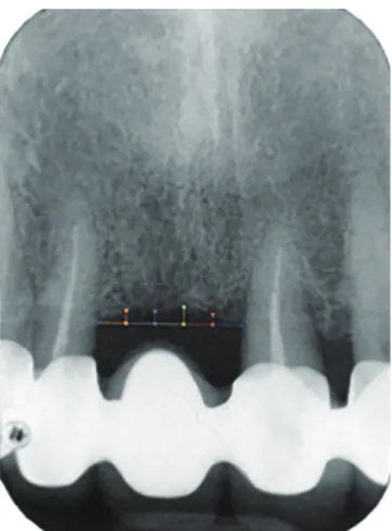
the proximal and distal alveolar crests in the edentulous region us- ing Photoshop version 7.0 (Adobe) (Figure 1). The length of this line was defined as the width of the edentulous alveolar ridge. This was converted into an actual value using the magnification ratio of the periapical radiograph.
Actual maximum socket depth and position of the edentulous alveolar ridge
The maximum distance was found between the line drawn above and the edentulous alveolar ridge. That distance was recorded in pixels, along with its horizontal position. These were defined as the actual maximum socket depth and position, respectively, of the edentulous alveolar ridge, and were converted into actual values using the magnification ratio of the periapical images.
Predicted maximum socket depth and position of the edentulous alveolar ridge
The distance from the line drawn above to the edentulous alveo- lar ridge was measured in pixels at six evenly spaced points along the line, together with the horizontal positions of these points.
These values were converted to actual values using the magnifica- tion ratio of the periapical radiograph. A regression function repre- senting the vertical shape of the alveolar ridge was then obtained using the above six points. The maximum value of this function and the position of the maximum value were obtained and defined as the predicted maximum socket depth and position of the edentu- lous alveolar ridge, respectively.
Maximum socket depth of the alveolar ridge before extraction The periapical radiological image of the maxillary anterior teeth before extraction was converted into a digital image, and a line was drawn connecting the alveolar crests of the adjacent teeth using Photoshop version 7.0 (Adobe). The maximum distance between this line and the alveolar ridge was recorded in pixels, and was de- fined as the maximum socket depth of the alveolar ridge before extraction. This was converted into an actual value using the mag- nification ratio for the radiological image.
Statistical analysis
We used SPSS version 12 for Windows in Korean (SPSS Inc., Chi- cago, IL, USA) for statistical analysis. In order to investigate the dif- ference between the predicted and actual maximum alveolar socket depth, the means and standard deviations were obtained and sta- tistical significance was tested using the Wilcoxon matched-pair signed-rank test. The difference between the predicted maximum socket depth of the edentulous alveolar ridge was compared in cas- es of single tooth loss and multiple tooth loss, using the Mann- Whitney U test to evaluate statistical significance. The same meth- od was used to compare the actual maximum socket depth in cases of single and multiple tooth loss. Pearson correlation coefficients were obtained to evaluate the relationship between the width of the edentulous maxillary anterior alveolar ridge and the actual and predicted maximum socket depths. In order to investigate the dif- ference between the predicted and actual maximum alveolar socket depth, means and standard deviations were obtained, and signifi- cance was tested using the Wilcoxon matched-pair signed-rank test. In order to evaluate the relationship between the predicted and actual maximum alveolar socket depth before and after extrac- tion, means and standard deviations were obtained and Pearson correlation coefficients were calculated. P-values below 0.05 were considered to indicate statistical significance.
RESULTS
The present study investigated edentulous regions in 25 patients with severe, chronic periodontitis who had undergone maxillary anterior tooth extraction due to severe loss of attachment and al- veolar bone. The alveolar socket depth after extraction was evalu- ated retrospectively using periapical radiographs. Twenty-five edentulous sites in the anterior maxilla were selected from 25 pa- tients (10 male, 15 female), including 10 cases of single tooth loss and 15 cases of multiple tooth loss (Table 1).
Figure 1. An imaginary line connecting the mesial and the distal ends of the alveolar crest facing each adjacent tooth was created. The distances between the imaginary line and several representative points were measured for each patient using an image analysis program.
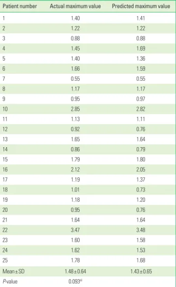
A regression function was obtained to represent the vertical lev- el of the edentulous alveolar ridge, and was expressed as a curve (parabola). The actual maximum socket depth measured from a reference line joining the distal and proximal alveolar crests in the edentulous site was 1.48±0.65 mm. The predicted maximum sock- et depth measured from the reference line was 1.43 ±0.65 mm.
The difference between the actual and predicted maximum socket depth after extraction was not significant (Table 2).
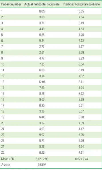
The position of the actual maximum socket depth within the edentulous anterior maxilla was found to be 6.12±2.90 mm distal from the proximal alveolar crest (along the reference line joining the proximal and distal alveolar crests). The position of the pre- dicted maximum socket depth was 6.62±2.74 mm distal to the proximal alveolar crest. The position of the actual maximum socket depth was not significantly different from the position of the pre- dicted maximum socket depth (Table 3).
The relationship of the width of the edentulous alveolar ridge
with the actual and predicted maximum socket depth was evaluat- ed. The alveolar ridge had a width of 13.28±5.62 mm, and showed a positive correlation with both the actual and predicted maximum socket depth. However, these associations were not statistically sig- nificant. The socket depth of the edentulous alveolar bone was compared between cases of single tooth extraction and multiple tooth extraction. The actual maximum socket depths for single and multiple tooth extractions were 1.49 ±0.81 mm and 1.63 ±0.62 mm, respectively, which was not a statistically significant differ- ence. The predicted maximum socket depths for single and multiple tooth extractions were 1.44±0.88 mm and 1.43±0.56 mm, which was not a statistically significant difference (Table 4).
The mean and standard deviation were obtained for the maxi- mum socket depth before extraction, and the relationships with the Table 1. Distribution of the maxillary anterior teeth included in the study.
Patient number Gender Tooth number
1 M 11, 12, 21, 22
2 M 11, 21
3 F 11
4 F 21
5 M 21
6 F 21
7 F 22
8 F 12
9 F 22
10 M 11, 21
11 F 11
12 F 11, 21
13 F 11, 21
14 F 11, 12, 21
15 F 11, 21
16 F 11, 21
17 F 11, 21
18 F 11, 21
19 M 11, 12, 21
20 M 11, 21
21 M 21
22 M 11
23 M 11, 21
24 M 21, 22
25 F 21, 22
M, male; F, female; Tooth numbers reflect the Federation Dentaire Internationale (FDI) numbering system.
Table 2. Actual and predicted maximum values of the vertical alveolar crest level in the edentulous areas (mm).
Patient number Actual maximum value Predicted maximum value
1 1.40 1.41
2 1.22 1.22
3 0.88 0.88
4 1.45 1.69
5 1.40 1.36
6 1.66 1.59
7 0.55 0.55
8 1.17 1.17
9 0.95 0.97
10 2.85 2.82
11 1.13 1.11
12 0.92 0.76
13 1.65 1.64
14 0.86 0.79
15 1.79 1.80
16 2.12 2.05
17 1.19 1.37
18 1.01 0.73
19 1.18 1.20
20 0.95 0.76
21 1.64 1.64
22 3.47 3.48
23 1.60 1.58
24 1.62 1.53
25 1.78 1.68
Mean±SD 1.48±0.64 1.43±0.65
P-value 0.093a)
a)Statistically non-significant difference based on the Wilcoxon matched-pair signed- rank test.
SD, standard deviation.
actual and predicted socket depth after extraction were evaluated.
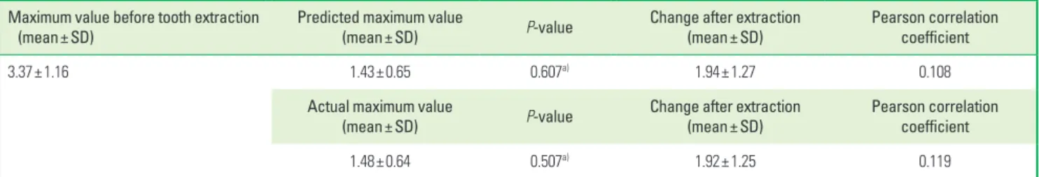
Before extraction, the maximum socket depth was 3.37±1.16 mm, and after extraction, the actual and predicted maximum socket depths were 1.92±1.25 mm and 1.94±1.27 mm, respectively. The maximum socket depth before extraction did not show a statisti- cally significant correlation with the actual or the predicted maxi- mum socket depth after extraction (Table 5).
DISCUSSION
The aim of this study was to investigate changes in the alveolar socket depth after the extraction of maxillary anterior teeth that showed considerable loss of attachment and alveolar bone resorp- tion as the result of severe, chronic periodontitis. Following tooth extraction, changes in the vertical and horizontal width of the al- veolar ridge are known to occur within a relatively short time [11,13,14]. In this study, periapical radiographs taken at least one year after tooth extraction were analyzed with the aim of evaluat- ing alveolar socket depth after the major changes to the tooth ex- traction site had already been completed. In addition, in order to evaluate alveolar socket depth after natural healing, only cases of restoration using fixed partial dentures were selected, without any procedures to preserve or expand the alveolar ridge.
In the present study, in order to evaluate socket depth in the edentulous alveolar bone, an imaginary line between the proximal and distal alveolar crests was used as a reference. Previous studies on changes in the alveolar bone after tooth extraction have shown a small amount of vertical atrophy [8,16,17]. In this study, after extraction, the vertical level of the alveolar ridge followed a para- bolic curve along this reference line, and was positioned 1.48 mm apically from this imaginary line. This distance did not show a sta- tistically significant correlation with the alveolar socket depth be- fore extraction. In other words, regardless of the amount of alveo- lar bone loss before extraction, the alveolar socket depth recovered towards the reference line connecting the proximal and distal al- veolar crests by one year after extraction. The alveolar socket depth before extraction was evaluated based on radiological im- Table 3. Horizontal coordinates corresponding to the actual and predicted
maximum values of the vertical alveolar crest level in the edentulous areas.
Patient number Actual horizontal coordinate Predicted horizontal coordinate
1 10.28 15.05
2 3.80 7.64
3 3.71 3.49
4 4.49 4.53
5 6.88 4.76
6 5.34 5.33
7 2.73 3.37
8 2.61 2.59
9 4.77 3.23
10 7.25 8.54
11 6.08 5.19
12 3.14 7.32
13 12.04 8.11
14 7.80 11.24
15 8.26 8.22
16 9.00 8.29
17 6.95 6.31
18 3.26 6.57
19 14.05 8.98
20 3.32 7.39
21 4.99 4.47
22 5.07 5.05
23 5.71 5.79
24 5.26 6.54
25 6.22 7.61
Mean±SD 6.12±2.90 6.62±2.74
P-value 0.510a)
a)Statistically non-significant difference based on the Wilcoxon matched-pair signed- rank test.
SD, standard deviation.
Table 4. Correlations between the actual and predicted maximum values of the vertical alveolar crest level and the mesio-distal span of the edentulous area (mm), as well as the actual and predicted maximum values of the vertical alveolar crest level in areas of single or multiple tooth loss (mm).
Mesio-distal span (mean±SD) Actual maximum value (mean±SD) Pearson correlation coefficient P-value
13.27±5.62 1.48±0.64 0.105 0.617a)
Predicted maximum value (mean±SD) Pearson correlation coefficient P-value
1.43±0.65 0.074 0.727a)
Single tooth loss Multiple tooth loss P-value
Actual maximum value (mean±SD) 1.49±0.81 1.63±0.62 0.437b)
Predicted maximum value (mean±SD) 1.44±0.88 1.43±0.56 0.782b)
a)Statistically non-significant difference based on the Pearson correlation coefficient.
b)Statistically non-significant difference based on the Mann-Whitney U test.
Table 5. Changes in the vertical alveolar crest level after extraction (mm) and correlation between vertical alveolar crest levels before and after tooth extrac- tion (mm).
Maximum value before tooth extraction
(mean±SD) Predicted maximum value
(mean±SD) P-value Change after extraction
(mean±SD) Pearson correlation
coefficient
3.37±1.16 1.43±0.65 0.607a) 1.94±1.27 0.108
Actual maximum value
(mean±SD) P-value Change after extraction
(mean±SD) Pearson correlation
coefficient
1.48±0.64 0.507a) 1.92±1.25 0.119
a)Statistically non-significant difference based on the Pearson correlation coefficient.
ages, and on average, it was found to be located 3.37 mm apically from the line connecting the adjacent alveolar crests. The vertical level of the alveolar bone was elevated after post-extraction heal- ing. Closer examination of individual cases revealed that the verti- cal level of the alveolar bone consistently converged towards the line connecting the alveolar crests, but the increase in the vertical level of the alveolar bone following extraction varied among cases.
The alveolar socket depth after tooth extraction identified in this study is similar to those reported by previous studies that evaluating changes in the alveolar bone after extraction. Schropp et al. [11] studied alveolar bone healing after tooth extraction us- ing subtraction radiography, and the point on the alveolar ridge closest to the root tip was found to be 1.2 mm lower than the crestal bone level of the adjacent teeth. However, it is interesting to note that the final vertical level of the alveolar ridge after ex- traction decreased in that study, while conversely, in the present study, the final vertical level of the alveolar ridge after extraction increased (Figure 2). Generally, after tooth extraction, the alveolar
bone is known to undergo vertical and horizontal atrophy [5,6,11].
Lekovic et al. [6] made clinical measurements of changes in the al- veolar ridge after extraction, and reported a vertical decrease in the alveolar ridge of 1.50±0.21 mm after six months. Fiorellini et al. [10] examined extraction and healing after four months, find- ing a vertical decrease in the alveolar bone of 1.17±1.23 mm.
The relationship of the distance between the proximal and distal alveolar crests with the maximum alveolar socket depth was eval- uated, but no statistically significant correlation was found. In other words, the maximum alveolar socket depth after extraction was consistent, regardless of the distance between the proximal and distal alveolar crests in the edentulous site. This means that similar recovery of the alveolar socket depth in the anterior maxilla is achievable for both single and multiple tooth extractions. No statistically significant differences were found in the alveolar socket depth depending on whether a single tooth or multiple teeth were lost.
This study analyzed periapical radiographs to evaluate changes in the vertical level of the alveolar ridge before and after the ex- traction of maxillary anterior teeth showing considerable loss of attachment and alveolar bone resorption due to severe, chronic periodontitis. The socket depth after extraction was found to be elevated towards the reference line connecting the proximal and distal alveolar crests. Additional studies are required to determine whether this vertical elevation in the level of the alveolar ridge, as seen in radiological images, reflects an actual elevation in the ver- tical level of the alveolar bone on the buccal and lingual sides.
Moreover, further research will be required to go beyond studying vertical changes to investigate patterns of recovery and factors that affect horizontal bone width.
In conclusion, this study was a retrospective analysis, using ra- diological images, of the vertical level of the alveolar ridge after the extraction of maxillary anterior teeth showing considerable loss of attachment and alveolar bone in patients with severe, chronic periodontitis. After extraction, the vertical level of the al- veolar ridge followed a parabolic shape, and increased to be closer to a reference line connecting the proximal and distal alveolar crests, regardless of the vertical loss of alveolar bone before ex- traction or the proximal-to-distal width of the edentulous area.
Figure 2. Convergence line indicating the vertical level of the alveolar crest after extraction. A: Change in the vertical level of the alveolar crest after ex- traction of teeth with severe bone loss. B: Change in the vertical level of the alveolar crest after extraction of teeth with a normal bone level.
CONFLICT OF INTEREST
No potential conflict of interest relevant to this article was re- ported.
ACKNOWLEDGEMENTS
This work was supported by a 2-Year Research Grant of Pusan National University (2015-2017).
ORCID
Chul Eui Hong http://orcid.org/0000-0002-3996-8911 Ju-Youn Lee http://orcid.org/0000-0002-0772-033X Jeomil Choi http://orcid.org/0000-0002-7491-6711 Ji-Young Joo http://orcid.org/0000-0002-4050-5797
REFERENCES
1. Tallgren A. The continuing reduction of the residual alveolar ridges in complete denture wearers: a mixed-longitudinal study covering 25 years. 1972. J Prosthet Dent 2003;89:427-35.
2. Pinho MN, Roriz VL, Novaes AB Jr, Taba M Jr, Grisi MF, de Souza SL, et al. Titanium membranes in prevention of alveolar collapse after tooth extraction. Implant Dent 2006;15:53-61.
3. Kerr EN, Mealey BL, Noujeim ME, Lasho DJ, Nummikoski PV, Mel- lonig JT. The effect of ultrasound on bone dimensional changes following extraction: a pilot study. J Periodontol 2008;79:283-90.
4. Darby I, Chen ST, Buser D. Ridge preservation techniques for im- plant therapy. Int J Oral Maxillofac Implants 2009;24Suppl:260-71.
5. Lekovic V, Kenney EB, Weinlaender M, Han T, Klokkevold P, Nedic M, et al. A bone regenerative approach to alveolar ridge mainte- nance following tooth extraction. Report of 10 cases. J Periodon- tol 1997;68:563-70.
6. Lekovic V, Camargo PM, Klokkevold PR, Weinlaender M, Kenney EB, Dimitrijevic B, et al. Preservation of alveolar bone in extrac- tion sockets using bioabsorbable membranes. J Periodontol 1998;
69:1044-9.
7. Iasella JM, Greenwell H, Miller RL, Hill M, Drisko C, Bohra AA, et
al. Ridge preservation with freeze-dried bone allograft and a col- lagen membrane compared to extraction alone for implant site development: a clinical and histologic study in humans. J Peri- odontol 2003;74:990-9.
8. Barone A, Aldini NN, Fini M, Giardino R, Calvo Guirado JL, Covani U.
Xenograft versus extraction alone for ridge preservation after tooth removal: a clinical and histomorphometric study. J Peri- odontol 2008;79:1370-7.
9. Camargo PM, Lekovic V, Weinlaender M, Klokkevold PR, Kenney EB, Dimitrijevic B, et al. Influence of bioactive glass on changes in alveolar process dimensions after exodontia. Oral Surg Oral Med Oral Pathol Oral Radiol Endod 2000;90:581-6.
10. Fiorellini JP, Howell TH, Cochran D, Malmquist J, Lilly LC, Spag- noli D, et al. Randomized study evaluating recombinant human bone morphogenetic protein-2 for extraction socket augmenta- tion. J Periodontol 2005;76:605-13.
11. Schropp L, Wenzel A, Kostopoulos L, Karring T. Bone healing and soft tissue contour changes following single-tooth extraction: a clinical and radiographic 12-month prospective study. Int J Peri- odontics Restorative Dent 2003;23:313-23.
12. Pietrokovski J, Massler M. Alveolar ridge resorption following tooth extraction. J Prosthet Dent 1967;17:21-7.
13. Jahangiri L, Devlin H, Ting K, Nishimura I. Current perspectives in residual ridge remodeling and its clinical implications: a review. J Prosthet Dent 1998;80:224-37.
14. Araújo MG, Lindhe J. Dimensional ridge alterations following tooth extraction. An experimental study in the dog. J Clin Peri- odontol 2005;32:212-8.
15. Bartold PM, Cantley MD, Haynes DR. Mechanisms and control of pathologic bone loss in periodontitis. Periodontol 2000 2010;53:
55-69.
16. Serino G, Biancu S, Iezzi G, Piattelli A. Ridge preservation follow- ing tooth extraction using a polylactide and polyglycolide sponge as space filler: a clinical and histological study in humans. Clin Oral Implants Res 2003;14:651-8.
17. Brägger U, Schild U, Lang NP. Effect of chlorhexidine (0.12%) rinses on periodontal tissue healing after tooth extraction. (II).
Radiographic parameters. J Clin Periodontol 1994;21:422-30.