Received:August 25, 2015, Revised:October 26, 2015, Accepted:November 3, 2015
Corresponding to:Seung-Ki Kwok, Division of Rheumatology, Department of Internal Medicine, Seoul St. Mary's Hospital, The Catholic University of Korea College of Medicine, 222 Banpo-daero, Seocho-gu, Seoul 06591, Korea. E-mail:[email protected] pISSN: 2093-940X, eISSN: 2233-4718
Copyright ⓒ 2016 by The Korean College of Rheumatology. All rights reserved.
This is a Free Access article, which permits unrestricted non-commerical use, distribution, and reproduction in any medium, provided the original work is properly cited.
염증성 관절염 마우스 모델에서 Sauchinone의 치료 효과
정승민1,2ㆍ이재선2ㆍ이주현2ㆍ백승예2ㆍ박성환1,2ㆍ곽승기1,2
1가톨릭대학교 의과대학 내과학교실 류마티스내과, 2가톨릭대학교 의과대학 류마티스연구센터
The Therapeutic Effect of Sauchinone on Inflammatory Arthritis in Mice
Seung Min Jung1,2, Jaeseon Lee2, Juhyun Lee2, Seung Ye Baek2, Sung-Hwan Park1,2, Seung-Ki Kwok1,2
1Division of Rheumatology, Department of Internal Medicine, The Catholic University of Korea College of Medicine, 2Rheumatism Research Center, The Catholic University of Korea College of Medicine, Seoul, Korea
Objective. Rheumatoid arthritis (RA) is a chronic inflammatory disease, mainly involving joints and bones. Sauchinone is an anti-inflammatory agent isolated from Saururus chinensis, which was used in oriental medicine. The aim of this study was to evaluate the therapeutic effect of sauchinone on inflammatory arthritis and underlying mechanism of anti-arthritic effect.
Methods. Mice with collagen induced arthritis (CIA) was intraperitoneally injected with sauchinone (20 mg/kg) or vehicle. The clinical and histological evaluations were performed with arthritis scoring and hematoxylin-eosin staining, respectively. CD4+ interleukin (IL) 17+ T cells were determined under Th17 skewing condition treated with sauchinone. To evaluate the effect of sauchinone on osteoclastogenesis, mice bone marrow macrophages (BMMs) and human peripheral blood mononuclear cells (PBMCs) were cultured with macrophage-colony stimulating factor and receptor activator of nuclear factor-κB ligand in the ab- sence or presence of sauchinone. Results. Sauchinone significantly attenuated the inflammatory arthritis in CIA mice both clin- ically and histologically. The proportion of Th17 cells were decreased with treatment with sauchinone in vivo and in vitro. The expressions of Th17 cell markers (IL-17 and retinoic acid receptor-related orphan receptor gamma t) and B cell markers (activation-induced cytidine deaminase) were downregulated in the presence of sauchinone. Sauchinone also suppressed the formation of tartrate-resistant acid phosphatase positive cells from mice BMMs and human PBMCs, and the expression of osteo- clastogenic markers. Conclusion. Sauchinone alleviates inflammatory arthritis in mice through inhibition of Th17 differ- entiation and osteoclastogenesis. Sauchinone, one of traditional herbal medicine, could be a therapeutic candidate for the treat- ment of RA. (J Rheum Dis 2016;23:161-173)
Key Words. Rheumatoid arthritis, Sauchinone, Collage induced arthritis, Experimental arthritis
서 론
류마티스관절염은 전신적인 자가면역질환으로 주로 대 칭적인 관절염과 골미란을 특징으로 하며, 이로 인해 관절 의 변형과 기능의 저하를 가져올 수 있는 질환이다. 류마 티스관절염의 병인은 아직 명확하지 않으나, 다양한 면역
세포와 염증물질의 상호작용이 류마티스관절염의 발생과 악화에 영향을 줄 것으로 생각된다[1]. 류마티스관절염에 서 T 세포의 역할은 특히 광범위하게 연구되어 있으며, 이 중 interleukin (IL)-17을 생산하는 T 세포(type 17 helper T cell, Th17 세포)는 류마티스관절염의 병인에 결정적인 역할을 하는 세포로 알려져 있다. IL-17은 류마티스관절염
활막세포를 자극하여 IL-6의 발현을 증가시키는데, IL-6은 Th17 세포의 중요한 분화 인자로 작용하여 Th17 세포의 분화를 더욱 증폭시키게 된다. 또한 다양한 표적세포에 작 용하여 IL-1, tumor necrosis factor-α (TNF-α), matrix metalloproteinase (MMP) 등 염증성 인자의 생산을 촉진 시킴으로써 활막염을 악화시키고, 세포 외 조직 및 골의 파괴를 일으킬 수 있다. 뿐만 아니라 IL-17은 파골세포 분 화에 가장 중요한 인자 중 하나인 receptor activator of nuclear factor-κB ligand (RANKL)의 발현을 증가시킴으 로써 골미란을 가져온다[2]. 이외에도 류마티스관절염에 서는 조절 T 세포의 기능이 상대적으로 감소하고 B 세포 에 의한 자가항체 생산이 증가하는 등 다양한 면역 기능의 조절 장애가 관찰될 수 있다.
류마티스관절염의 치료는 이러한 비정상적인 면역반응 을 조절하여 활막의 염증과 뼈의 파괴를 감소시키는 것을 목적으로 하고 있다. 현재 류마티스관절염의 치료에는 고 식적인 스테로이드로부터 특정 면역세포나 사이토카인을 표적으로 하는 생물학적 제제에 이르기까지 다양한 약제 가 사용되고 있다. 그러나 일부 환자의 경우에는 여전히 항류마티스 약제에 대한 반응이 부족하며, 약물의 부작용 으로 인하여 약제의 사용이 제한되기도 한다[3,4]. 따라서 보다 안전하고 효과적으로 류마티스관절염을 치료하기 위 해 위해 새로운 약제를 개발하려는 노력이 지속되고 있다.
Sauchinone은 아시아와 북미에 분포하는 삼백초(Saururus chinensis)에서 얻은 활성 성분으로, 삼백초는 예전부터 동 양의학에서 발열이나 염증성 질환에 이용되어 온 약초이 다. 최근 연구에서 sauchinone은 간질환이나 폐질환, 신경 계 질환 등 다양한 질환에서 치료제로서 항염증, 항산화 작용을 한다. 이러한 sauchinone의 효과에 관련해서는 여 러 가지 기전이 연구되어 왔다. Sauchinone은 AMP-acti- vated kinase (AMPK)를 활성화하여 항산화 및 세포보호 효과를 가짐으로써 간독성을 억제하는 역할을 할 뿐 아니 라[5,6], p38 mitogen-activated protein kinase (MAPK)의 인산화를 억제하여 TNF-α 및 macrophage inflammatory protein (MIP)-2의 생산을 감소시킴으로써 급성 폐손상을 완화했다[7]. 또한 성상세포와 미세아교세포 배양 실험에 서 inducible nitric oxide synthase (iNOS)와 cyclo- oxygenase (COX)-2 발현을 감소시키고, signal trans- ducer and activator of transcription 3 (STAT3)의 인산화 를 억제하는 효과를 보여주기도 하였다[8].
따라서 기존의 연구를 바탕으로 sauchinone이 갖는 염증 조절 작용이 염증성 관절염에서도 치료 효과를 나타낼 수 있을 것으로 보고, 자가면역성 관절염 마우스 모델에서 sauchinone의 치료 효과를 확인하며, 이와 관련된 기전에 대해 밝히고자 하였다.
대상 및 방법
실험 동물에서 염증성 관절염 유도 및 약물 처리 7주령의 수컷 DBA/1J 마우스를 Charles River Laborato- ry (Yokohama, Japan)에서 구입하였고, 가톨릭대학교 의 과대학 내의 동물 실험실에서 1주일간 유지한 후에 관절 염을 유도하였다. 동물사육실의 조건은 22.0±2oC, 1일 중 12시간은 200∼300 Lux로 조명하고, 12시간은 모든 빛을 차단하였다. 모든 연구는 가톨릭대학교 동물연구윤리위원 회의 규정에 근거하여 진행되었다(IRB No. 2014-0075- 02).
DBA/1J 마우스에 제 II형 콜라겐(Chondrex, Redmond, WA, USA) 100 μg과 complete Freund’s adjuvant (Chond- rex)를 1:1로 혼합한 뒤 꼬리 기저부에 피하 주사하였다(1차 면역유도). 2주 후, 한쪽 뒷다리 발바닥에 제 II형 콜라겐 100 μg을 incomplete Freund’s adjuvant (Chondrex)와 1:1로 혼합하여 다시 주사하였다(2차 면역유도). 2차 면역유도주 사 후 다음날부터 DBA/1J 마우스에 kg 당 20 mg의 sauchi- none (Sigma Aldrich, St. Louis, MO, USA) 또는 동일한 용 량의 생리식염수를 주 3회 6주간 복강 내 주사하였다.
관절염의 임상적 평가
콜라겐유도관절염 마우스에서 sauchinone의 관절염 억 제 효과를 관찰하기 위하여 1차 콜라겐 투여 시점부터 8주 간 주 3회 관절염의 정도를 점수로 기록하였다. 실험과 관 련되지 않은 세 명의 독립적인 관찰자가 관절의 염증 정도 를 육안으로 평가하였으며, 다음과 같은 기준에 따라 0∼
4점으로 점수를 매겼다.
0=관절에 부기나 발적이 없음.
1=경도의 부기와 발적이 중간발 또는 발목관절에 국한 되어 관찰됨.
2=경도의 부기와 발적이 발목관절부터 중간발에 걸쳐 관찰됨.
3=중등도의 부기와 발적이 발목관절부터 중간발에 걸쳐 관찰됨.
4=발목, 발, 발가락에 걸쳐 중증의 부기와 발적이 관찰됨.
관절염 점수는 2차 콜라겐 주사를 했던 발을 제외하고 세 개의 발에서 매긴 점수를 합쳐서 구하였다. 이에 따라 최고점은 12점으로 계산할 수 있었으며, 8주간 관절염 점 수의 진행상황을 기록하여 sauchinone 투여군과 생리식 염수 투여군에서의 관절염 점수 추이를 비교하였다.
관절염의 조직학적 평가
1차 면역유도 주사 8주 후에 마우스의 앞다리를 제거하 여 피부가죽을 벗겨낸 뒤 10% formalin 용액에 하루 동안 담근 후 Calci-Clear Rapid (National Diagostics, Atlanta, GA, USA)로 7시간 동안 탈회하였다. 수세 및 탈수 후 파 라핀으로 포매하였고, 7 μm의 두께로 절편을 제작하여
hematoxylin-eosin (H&E) 염색을 시행하였다. 관절의 활 막 염증에 대해 3명의 독립된 검사자가 이전에 기술된 기 준에 따라 조직학적 점수를 매기고, sauchinone 투여군과 생리식염수 투여군의 차이를 비교하였다[9].
면역글로불린 G 측정
1차 면역유도 주사 8주 후에 안와 채혈을 통해 얻은 혈청 을 1% bovine serum albumin (BSA)과 0.05% Tween20 (AMRESCO, Cleveland, OH, USA)이 포함된 Tris-buf- fered saline (pH 8.0) 용액에 1:100,000, 1:50,000의 비율 로 희석하였고, 이와 같이 희석된 혈청을 각각 immuno- globulin (Ig)G와 IgG2a의 측정에 사용하였다. 총 IgG의 양과 IgG2a 측정을 위해 mouse IgG and IgG2a enzyme- linked immunosorbent assay (ELISA) Quantitation Kits (Bethyl Laboratories, Montgomery, TX, USA)를 이용하 였고, 모든 과정은 제조사의 방법을 따라 시행하였다.
사이토카인의 측정
마우스 혈청의 상청액 내에 존재하는 사이토카인을 측정하 기 위해 anti-TNF-α 항체, anti-mouse interferon (IFN)-γ 항체, anti-mouse IL-17 항체와 biotin으로 표지 된 각 항 체(R&D systems, Minneapolis, MN, USA)를 이용하여 측정하였다. Biotin이 부착된 각 항체의 반응이 끝난 후, Extravidin-HRP (Sigma Aldrich)를 1% BSA와 0.05%
Tween20이 포함된 phosphate-buffered saline (PBS) 용액 에 1:10,000배의 비율로 희석하여 하나의 well 당 50 μL 씩 넣고 실온에서 2시간 반응시켰다. 다음으로 tetrame- thylbenzidine substrate 용액(eBioscience, San Diego, CA, USA)을 하나의 well 당 50 μL씩 넣고 약 5분간 반응 시킨 뒤 1N H2SO4를 50 μL 넣어 반응을 멈추고 450 nm 의 파장에서 흡광도를 측정하였다. 대조샘플로 재조합 TNF-α, IFN-γ, IL-17 단백질을 이용하여 10 ng/mL부터 156.25 pg/mL의 농도까지 두 배씩 단계적으로 희석하여 정량하였다.
비장세포의 분리 및 자극
마우스 비장세포를 얻기 위해 1차 면역유도 주사 8주 후 에 마우스의 비장을 적출하여 가위로 잘게 자르고 40 μm 의 세포 여과기(BD Falcon, Bedford, MA, USA)로 걸러내 었다. 적혈구를 제거하기 위해 원심분리하여 얻은 비장세 포를 2.06% Tris, 0.83% NH4Cl 용액에 부유시켜 실온에 서 5분간 반응시킨 후, 40 μm의 세포 여과기에 다시 통 과시켰다. 마우스 비장세포는 10% fetal bovine serum (FBS; Gibco, Grand Island, NY, USA)이 포함된 RPMI1640 (Gibco) 배지에 부유시켜 배양하였다. 비장세포를 자극하 기 위하여 24 well plate에 purified anti-mouse CD3e 항 체(BD, Franklin Lakes, NJ, USA)를 1μg/mL의 농도로 분주하여 37oC 배양기에서 2시간 동안 코팅한 뒤 잔여물
을 걷어내고 비장세포를 1×10⁶ cells/mL 농도로 분주하 였고, 상청액을 분리하여 사이토카인 측정에 사용하였다.
세포독성 측정
비장세포를 1×10⁶ cells/mL의 농도로 96 well plate에 200 μL 분주한 후 sauchinone을 5, 10, 15, 20 μM의 농 도로 24시간 동안 자극하였다. Cell counting kit-8 (Dojindo Moleculare Technologies, Rockville, MD, USA) 을 사용하여 시약을 각 well 당 10 μL로 처리하고 2시간 후에 570 nm에서 흡광도를 측정하였다. 대조군으로 사용 된 0.01% dimethylsulfoxide (Sigma Aldrich)로 자극한 세 포의 흡광도를 100%로 환산했을 때 여러 농도의 sauchi- none으로 자극한 세포의 흡광도를 비교하였다.
CD4+T 세포의 분리 및 Th17 세포 분화유도 마우스의 비장세포와 정상인의 말초혈액세포로부터 CD4+T 세포를 분리하였다. 사람의 말초혈액을 이용한 연 구는 가톨릭대학교 임상의학연구센터의 승인을 받아 진행 되었다(KC15TISI0059). PBS로 세척한 마우스의 비장세 포와, PBS로 희석한 후 Ficoll-paque 용액(GE healthcare, Marlborough, MA, USA)을 이용하여 분리한 정상인의 말 초혈액 단핵구 세포를 107개 세포당 10 μL의 CD4 mi- crobead (Miltenyi Biotec, Bergisch Gladbach, Germany) 와 혼합하여 4oC에서 15분간 반응시켰다. 분리한 CD4+ T 세포를 5% BSA와 1 mM ethylenediaminetetraacetic acid (EDTA)가 포함된 PBS 용액으로 세척 후 10% FBS가 포함된 RPMI1640 배지에 재부유시켜, 106개의 세포로 분 주하였고, anti-CD3e 항체(0.5 μg/ mL), anti-CD28 항체 (1 μg/mL) (BD Pharmingen, San Diego, CA, USA), hu- man tumor growth factor (TGF)-β (2 ng/mL), IL-6 (20 ng/mL), anti-IFN-γ 항체(2 μg/mL), anti-IL-4 항체(2 μg/
mL) (R&D Systems)로 자극하여 Th17 세포로 분화를 유도 하였다. 이 때 sauchinone이 Th17 분화에 미치는 영향을 확 인하기 위해, 15 μM 농도의 sauchinone을 30분간 전처리 하였다. Th17 세포로 분화 유도한지 3일째 되는 세포에서 RNA를 추출하였고, 얻어진 상청액으로 IL-17 농도를 측정 하였다.
B 세포의 분리 및 자극
마우스의 비장세포를 PBS 용액으로 세척하고 CD19 mi- crobead (Miltenyi Biotec)를 107개의 세포당 10 μL씩 혼 합하여 4oC에서 15분간 반응시켰다. 5% BSA와 1 mM EDTA가 포함된 PBS 용액으로 세척 후 10% FBS가 포함 된 RPMI1640 배지로 재부유시켜, 106개의 세포로 분주한 뒤, sauchinone을 5 μM 또는 7 μM의 농도로 30분간 전 처리 하고 나서 100 ng/mL의 lipopolysaccharide (LPS;
Sigma Aldrich)로 자극하였다. 4일 후, 세포를 회수하여 RNA를 추출하고 상청액은 따로 보관하여 IgG를 측정하
Table 1. Primer sequence lists for real-time reverse transcription-polymerase chain reaction
Primer Sense (5`- 3`) Anti-sense (5`- 3`)
Mouse IL-17 CCTCAAAGCTCAGCGTGTCC GAGCTCACTTTTGCGCCAAG ROR-γt TGTCCTGGGCTACCCTACTG GTGCAGGAGTAGGCCACATT Blimp-1 CTGTCAGAACGGGATGAACA TGGGGACACTCTTTGGGTAG AID CGTGGTGAAGAGGAGAGATAGTG CAGTCTGAGATGTAGCGTAGGAA TRAP TCCTGGCTCAAAAAGCAGTT ACATAGCCCACACCGTTCTC
OSCAR CCTAGCCTCATACCCCCAG CAAACCGCCAGGCAGATTG
Cathepsin K CAGCAGAGGTGTGTACTATG GCGTTGTTCTTACTTCGAGC
CTR CGGACTTTGACACAGCAGAA AGCAGCAATCGACAAGGAGT
Integrin β3 CCACACGAGGCGTGAACTC CTTCAGGTTACATCGGGGTGA MMP9 CTGTCCAGACCAAGGGTACAGCCT GAGGTATAGTGGGACACATAGTGG β-actin GAAATCGTGCGTGACATCAAAG TGTAGTTTCATGGATGCCACAG
Human MMP9 TGGGGGGCAACTCGGC GGAATGATCTAAGCCCAG
Cathepsin K TGAGGCTTCTCTTGGTGTCCATAC AAAGGGTGTCATTACTGCGGG Integrin β3 GCAATGGGACCTTTGAGTGT GTGGCAGACACATTGACCAC c-fos ATGATGTTCTTCTCGGGCTTCAA GTGGGAATGAAGTTGGCACT NFATc1 GCATCACAGGGAAGACCGTGTC GAAGTTCAATGTCGGAGTTTCTGAG β-actin GGACTTCGAGCAAGAGATGG TGTGTTGGGGTACAGGTCTTTG
AID: activation-induced cytidine deaminase, Blimp-1: B lymphocyte-induced maturation protein-1, CTR: calcitonin receptor, IL-17: interleukin-17, MMP: matrix metalloproteinase, NFATc1: nuclear factor of activated T-cells, cytoplasmic 1, OSCAR:
osteoclast-associated receptor, ROR-γt: retinoic acid receptor-related orphan receptor gamma t, TRAP: tartrate-resistant acid phosphatase.
는 데 사용하였다. 이 때 IgG 농도는 희석하지 않은 상청 액에서 측정하였다.
유세포분석
Th17 세포를 확인하기 위해, 분화 과정이 끝난 세포를 25 ng/mL 농도의 phorbol 12-myristate 13-acetate (PMA) 와 250 ng/mL ionomycin (Sigma Aldrich), 0.67 μL/mL 의 Golgi-Stop (BD)을 처리하고 4시간 후에 peridinin chlorophyll protein (PerCP)으로 표지된 anti-CD4 항체 (Biolegend, San Diego, CA, USA)로 형광염색 하였다. 세 포를 4oC에서 30분간 fixation/permeabilization 용액 (BD)에 처리하고 permeabilization 용액(BD)으로 세척한 후에, fluorescein isothiocyanate (FITC)로 표지된 an- ti-IL-17 항체(eBioscience) 또는 phycoerythrin (PE)-Cy7 으로 표지 된 anti-IFN-γ 항체로 형광염색을 시행하였다.
조절 T 세포를 확인하기 위해서는 분화 과정이 끝난 세포 를 PerCP로 표지된 anti-CD4 항체와 allophycocyanin (APC)로 표지된 anti-CD25 항체(eBioscience)를 이용하 여 형광 염색하였다. 이후 4oC에서 30분간 fixation/per- meabilization 용액(eBioscience)에 반응시키고, per- meabilization (ebioscience) 용액으로 세척한 후에, PE가 표지 된 anti-foxp3 항체(eBioscience)로 형광 염색하였다.
LSRII fortessa (BD) 장비와 FACS DIVA version 10.0 software (BD Biosciences, San Jose, CA, USA)로 CD4+ IL-17+IFN-γ− 세포와 CD4+CD25+FOXP3+ 세포의 분 획을 확인하였다.
파골세포 분화
정상 C57BL/6 마우스의 대퇴골과 경골에서 추출한 골수 세포 또는 사람의 말초혈액 단핵구 세포를 얻어 10% FBS 가 포함된 α-minimal essential medium (Invitrogen, Burlingame, CA, USA)에서 배양하고 다음날 배양접시에 부착된 사람의 단핵구 세포와 부착되지 않은 마우스 골수 세포를 수집하였다. 수집된 세포들을 각각 새로운 배양접 시로 옮겨 10 ng/mL 농도의 재조합 macrophage-colony stimulating factor (M-CSF; R&D Systems)로 3∼4일간 자극하였다. 배양접시에 부착된 세포에 다양한 농도의 sauchinone을 처리하면서 10 ng/mL M-CSF, 25 ng/mL soluble RANKL (PeproTech Inc., Rocky Hill, NJ, USA)로 자극하여 파골세포로 분화를 유도하였다. 이틀마다 동일 한 조성으로 배지를 교환하였으며, 마우스 세포의 경우 배 양 4일에서 6일 사이, 시림세포의 경우는 배양 6일에서 8 일 사이에 세포를 회수하여 RNA를 추출하거나, paraf- ormaldehyde로 고정하였다. 고정한 세포를 tartrate-re- sistant acid phosphatase (TRAP; Sigma Aldrich)로 염색 하였고, TRAP 양성 세포 중 3개 이상의 핵을 가지고 있는 다핵세포의 수를 현미경으로 관찰하였다.
유전자 발현(quantitative reverse transcription-poly- merase chain reaction, qRT-PCR)
위와 같이 분리한 Th17 세포와 B 세포, 파골세포에서 RNAiso Plus (Takara, Otsu, Japan)를 처리하고, phenol- chloroform 추출법으로 총 RNA를 분리하였다. Trans-
criptor First Strand cDNA Synthesis kit (Roche Diagnos- tics, Basel, Switzerland)를 사용하여 추출한 RNA 2 μg으 로부터 cDNA를 합성하였다. 유전자 증폭을 위해 Light- Cycler 480 II (Roche Diagnostics) 장비와 LightCycler480 SYBR Green I Master (Roche Diagnostics)를 사용하였 다. 증폭에 사용된 primer의 염기서열은 Table 1과 같다.
각각의 실험에서 RNA 발현량은 모두 β-actin 발현량으 로 보정하였다.
단백질 발현(Western blot)
마우스의 골수유래 대식세포에 30분간 5 μM, 7 μM 농도 의 sauchinone을 전처리 한 뒤, 100 ng/mL의 RANKL로 4일 간으로 자극하고, Halt protease inhibitor cocktail (Thermo Scientific, Waltham, MA, USA)이 포함된 radio- immunoprecipitation assay (RIPA) 용액으로 단백질을 추 출하였다. 50 μg의 단백질을 12% sodium dodecyl sulfate polyacrylamide gel에서 100 V로 1시간 30분 동안 전기영동 하였고, 전기영동이 끝난 gel 위의 단백질을 polyvynilidene fluoride membrane (Biorad, Hercules, CA, USA)으로 이동 시켰다. Membrane의 비특이적인 반응을 차단하기 위하여 5% skim milk (Dako, Glostrup, Denmark)와 0.1%
Tween20가 포함된 Tris 용액으로 처리한 후, 4oC에서 16시 간 동안 anti-c-fos 항체(1:1,000; Cell Signaling Technology, Inc., Boston, MA, USA), anti-β- actin 항체(1:5,000, Sigma Aldrich)와 반응시키고 0.1% Tween20이 포함된 Tris 용액 으로 세척하였다. Membrane에 horseradish peroxidase로 표지된 goat anti-rabbit IgG (Thermo Scientific) 또는 goat anti-mouse IgG (Santacruz, Dallas, TX, USA)를 2,000배 희 석하여 실온에서 2시간 동안 반응시키고, 0.1% Tween20이 포함된 Tris 용액으로 세척하였고, SuperSignal West Pico Chemiluminescent substrate (Thermo Scientific)로 발광시 켰다. Amersham imager 600 (GE Healthcare Bioscience, Pittsburgh, PA, USA) 장비를 이용하여 membrane에서 단 백질 발현을 확인하였다.
통계 유의성 검증
실험 결과는 평균±평균값의 표준오차로 나타냈다. 통계 적 유의성은 Mann-Whitney U 검정 또는 ANOVA와 Bonferroni 사후 검정을 이용하여 분석하였고, p값이 0.05 보다 미만일 때 유의하다고 기술하였다. 모든 데이터는 SAS software ver. 9.1 (SAS Institute, Cary, NC, USA)을 이용하여 분석하였다.
결 과
Sauchinone의 염증성 관절염 치료 효과
Sauchinone의 항관절염 효과를 관찰하기 위하여 콜라겐 유도관절염 마우스에 주 3회 sauchinone 또는 생리식염수
를 복강내로 주사한 후에 관절염 점수를 조사하였다. 관절 염 점수는 1차 면역유도 주사 후 5주까지는 큰 차이를 보 이지 않았으나, 6주 이후부터는 의미 있는 차이를 보이기 시작하여, 이러한 차이가 이후로도 지속되었다(Figure 1A). 두 군의 혈청을 분리하여 면역글로불린을 측정한 결 과, sauchinone 투여군이 대조군보다 낮게 나타났으나, 통 계적인 유의성은 관찰되지 않았다(Figure 1B). Sauchi- none에 의한 관절염의 조직학적 호전 효과를 관찰하기 위 해 마우스의 발목 관절 조직을 H&E 염색하였을 때 생리 식염수를 투여한 군은 염증세포의 침윤 및 연골의 파괴, 골 미란 등으로 인해 정상적인 관절 구조를 확인하기 어려 운 반면, sauchinone을 투여한 군의 관절 조직은 비교적 잘 보존되어 있음을 확인할 수 있었고(Figure 1C), 활막 염증에 대한 조직학적 점수도 sauchinone 투여군에서 유 의하게 낮았다(Figure 1D).
관절염 치료효과와 관련하여 sauchinone이 체내에서 T 세포의 분화에 미치는 영향을 확인하기 위하여 각군의 마 우스에서 비장세포를 분리하여 세포의 분획을 조사하였 다. CD4+IL-17+ T 세포와 CD4+IFN-γ+ T 세포는 sau- chinone 치료군에서 유의하게 낮은 수치로 관찰되었고, CD4+CD25+Foxp3+ T 세포는 두 군간의 큰 차이를 보이 지 않았다(Figure 1E). 비장세포를 분리하여 CD3 항체로 자극한 후 상청액을 분리하여 TNF-α, IFN-γ 및 IL-17의 농도를 측정하였을 때에는 모두 sauchinone 군에서 더 낮 은 농도를 나타내었다(Figure 1F).
Sauchinone에 의한 Th17 분화의 감소
앞서 실험을 통해 sauchinone을 투여한 마우스에서 Th17 세포의 농도가 감소하는 것을 관찰하였고, Th17 세 포는 류마티스관절염과 같은 자가면역성 관절염에서 매우 중요한 역할을 하는 면역세포임이 잘 알려져 있으므로, sauchinone이 Th17 세포 분화에 미치는 영향을 실험실 검 사로 확인하고자 하였다. 세포실험에서 사용할 sauchi- none의 농도를 결정하기 위해 세포독성 시험을 시행한 결 과 15 μM까지는 세포 생존능이 90% 이상으로 세포독성 을 보이지 않았으며, 20 μM에서도 세포 생존능은 70%
이상으로 관찰되었다(Figure 2A). 세포독성 시험을 바탕 으로 정상 마우스의 비장세포를 분리하여 Th17 세포 분화 조건 하에서 sauchinone을 5∼20 μM의 농도로 처리하였 다. 유세포분석기를 사용하여 분석한 결과 CD4+IL-17+ IFN-γ− T 세포는 sauchinone의 농도에 비례하여 감소함 을 관찰할 수 있었고, 이중 세포 생존능이 90% 이상 보존 되는 농도이면서 억제 효과가 가장 큰 15 μM의 농도로 실험을 지속하였다. CD4+IL17+ T 세포는 sauchinone을 처리하였을 경우 통계적으로 유의하게 감소함을 확인할 수 있었다(Figure 2B). 같은 배양 조건에서 분리한 상청액 에서도 sauchinone을 처리한 군에서 IL-17의 분비가 감소
Figure 1. Sauchinone alleviates the inflammatory arthritis in mice. Mice with collagen induced arthritis were intraperitoneally in- jected with sauchinone (20 mg/kg) or vehicle for six weeks immediately after second immunization. (A) Inflammatory arthritis was assessed with clinical scoring by three independent examiners. (B) The serum level of IgG and IgG2a was measured with ELISA.
(C) Histological analysis was performed with hematoxylin-eosin staining of mice paws. Bar in the left corner of figure indicates 100 μm. (D) Histologic score for synovial inflammation of mice paws was determined by three independent examiners. (E) The compo- sition of mice splenocytes were determined by flowcytometry. The percentages of CD4+IL-17+ T cells, CD4+IFN-γ+ T cells, and CD4+CD25+FOXP3+ T cells were compared between two groups. (F) The production of TNF-α, IFN-γ, and IL-17 from mice sple- nocytes was evaluated with ELISA of culture supernatant after stimulation with anti-CD3 antibodies. Values are presented as three independent experiments±standard error of mean. aCD3: stimulation with anti-CD3 antibodies, ELISA: enzyme-linked im- munosorbent assay, IFN: interferon, Ig: immunoglobulin, IL: interleukin, Nil: no stimulation, NS: not significant, TNF: tumor ne- crosis factor. *p<0.05, ***p<0.001.
Figure 2. Sauchinone inhibits Th17 differentiation. (A) Cytotoxicity of sauchinone was evaluated with cell viability assay using cell count- ing kit-8 after treatment with 0 to 20 μM of sauchinone. The dose of sauchinone with more than 80% of cell viability was considered as a tolerable dose. (B) CD4+ T cells were differentiated into Th17 cells in the presence of sauchinone. The proportions of CD4+IL-17+ cells were determined with flowcytometry. The number of right bottom indicates the proportion of CD4+IL-17+ cells. (C) The production of IL-17 from CD4+ T cells under Th17 skewing condition was measured with ELISA of culture supernatant after pretreatment of sauchinone. (D) The expression of IL-17 and ROR-γt was determined with qRT-PCR of CD4+ T cells under Th17 differentiation condition after pretreatment with sauchinone. Values are presented as three independent experiments±standard error of mean. aCD3: stimulation with anti-CD3 antibodies, FITC: fluorescein isothiocyanate, IFN: interferon, IL: interleukin, ROR-γt: retinoic acid receptor-related or- phan receptor gamma t, PE: phycoerythrin, Th17: Th17 skewing condition, Th17+S: Th17 skewing condition in the presence of sauchinone. *p<0.05.
Figure 3. Sauchinone affects B cell maturation. (A) The expression of Blimp-1 and AID was evaluated with qRT-PCR of CD19+ B cells stimulated with LPS in the presence of sauchinone. (B) The production of IgG from CD19+ B cells stimulated with LPS was measured with ELISA of culture supernatant after pretreatment with sauchinone. Values are presented as three independent experi- ments±standard error of mean. AID: activation-induced cytidine deaminase, Blimp-1: B lymphocyte-induced maturation protein-1, Ig: immunoglobulin, LPS: lipopolysaccharide, Nil: no stimulation, NS: not significant. *p<0.05, **p<0.01.
하였으며(Figure 2C), 세포 내의 IL-17 및 retinoic acid re- ceptor-related orphan receptor gamma t (ROR-γt) mRNA 발현도 역시 sauchinone 처리군에서 감소되어 있 었다(Figure 2D).
Sauchinone의 B 세포 억제 효과
마우스 실험에서 비록 통계학적으로 유의미하지는 않았 으나, sauchinone을 투여한 군에서 면역글로불린이 감소 하는 결과를 관찰하였으므로, sauchinone이 B 세포에 미 치는 영향을 관찰하기 위하여 CD19+ 세포에서 mRNA의 발현과 면역글로불린 생산에 대해 조사하였다. 림프절의 배중심(germinal center) 발달과 B 세포의 항체 형성에 결 정적인 역할을 하는 전사 인자인 B lymphocyte-induced maturation protein-1 (Blimp-1)과 activation-induced cy- tidine deaminase (AID)의 mRNA 발현은 LPS 자극 후 증 가하였으나, sauchinone을 같이 처리할 경우 감소함을 관 찰할 수 있었고, Blimp-1 발현의 경우 sauchinone 7 mg에 서 통계적으로 유의한 감소 효과를 보였다(Figure 3A).
LPS 자극 후의 면역글로불린 생산은 sauchinone의 용량 에 비례하여 감소하는 결과를 나타내었다(Figure 3B).
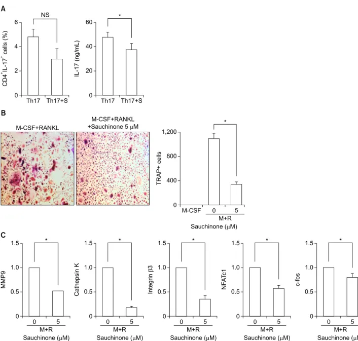
Sauchinone에 의한 파골세포 분화 억제
Sauchinone을 투여한 마우스군의 조직학적 소견에서 골 파괴의 정도가 감소하였고, sauchinone에 의해 Th17 세포 분화가 감소하는 것을 관찰하였으므로, sauchinone이 파 골세포 분화를 억제할 것으로 가정하였다. 이를 확인하기 위해 파골세포 분화 유도 시 sauchinone을 처리하고 TRAP
양성세포의 개수 및 파골세포와 관련된 mRNA 발현을 측 정하였다.
마우스의 골수유래 세포를 M-CSF와 RANKL로 자극하 고 0, 1, 3, 5 μM의 sauchinone을 처리하였을 때 sauchi- none의 농도가 높아질수록 TRAP 양성 세포의 수가 감소 하는 것을 관찰할 수 있었다(Figure 4A). 동일한 분화 조건 에서 5 μM의 sauchinone을 처리하여 배양한 후 mRNA 발현을 분석하였을 때, 파골세포 분화와 관련되는 인자인 TRAP, cathepsin K, calcitonin receptor (CTR), osteoclast- associated receptor (OSCAR), integrin β3 및 MMP9의 발현이 모두 감소하는 것을 관찰할 수 있었다(Figure 4B).
Sauchinone이 파골세포 분화를 억제하는 기전과 관련하 여, 파골세포 형성에 결정적인 역할을 하는 전사 인자인 c-fos의 발현에 미치는 영향을 조사하였다. 파골세포 분화 과정 동안 sauchinone을 5 μM과 7 μM의 농도로 처리할 경우 세포 내 c-fos 단백질 발현이 거의 관찰되지 않아, sauchinone이 c-fos를 억제하여 파골세포 형성을 방해함 을 시사하였다(Figure 4C).
인체세포에서 sauchinone의 효과
동물 실험 결과에서 sauchinone은 Th17 분화를 억제하 고 파골세포 형성을 저해하여 염증성 관절염을 호전시키 는 결과를 보였다. 이러한 결과가 인체세포에서도 같은 경 향을 보이는지 확인하기 위하여 건강인 네 명의 말초혈액 으로 실험을 진행하였다.
건강인의 말초혈액으로부터 단핵구 세포를 분리하여 Th17 세포 분화 조건을 조성한 후 sauchinone을 처리하지
Figure 4. Sauchinone suppresses osteoclastogenesis in a dose-dependent manner. Mice bone marrow macrophages were cultured with M-CSF and RANKL to induced osteoclastogenesis. Sauchinone was added to the culture dishes to evaluate the effect of sauchi- none on osteoclastogenesis. (A) The numbers of TRAP positive cells were counted in culture dishes at various doses of sauchinone.
(B) The expression of osteoclastogenic markers were determined by qRT-PCR in the absence or presence of sauchinone. (C) The ex- pression of c-fos, a transcription factor associated with osteoclastogenesis, was evaluated with Western blot of cells cultured with or without sauchinone. Values are presented as three independent experiments±standard error of mean. M: culture with M-CSF, M+R: culture with M-CSF and RANKL, M-CSF: macrophage-colony stimulating factor, MMP: matrix metalloproteinase, NS: not sig- nificant, OSCAR: osteoclast-associated receptor, qRT-PCR: quantitative reverse transcription-polymerase chain reaction, RANKL:
receptor activator of nuclear factor-κB ligand, TRAP: tartrate-resistant acid phosphatase. *p<0.05, **p<0.01.
Figure 5. Saucinone regulates Th17 differentiation and osteoclastogenesis in human mononuclear cells. To confirm the inhibitory effect of sauchinone on human cells, pripheral blood mononuclear cells isolated from healthy controls were cultured in the ab- sence or presence of sauchinone. (A) The proportion of CD4+IL17+ T cells and the production of IL-17 under Th17 skewing con- dition with or without sauchinone was evaluated by flowcytometry and ELISA, respectively. (B) The number of TRAP positive cells was measured after culture with M-CSF and RANKL in the absence or presence of sauchinone. (C) The expression of osteoclasto- genic markers was compared between cells differentiated to osteoclast with or without sauchinone. Values are presented as three independent experiments±standard error of mean. M: culture with M-CSF, M+R: culture with M-CSF and RANKL, M-CSF: macro- phage-colony stimulating factor, MMP: matrix metalloproteinase, NFATc1: nuclear factor of activated T-cells, cytoplasmic 1, NS:
not significant, RANKL: receptor activator of nuclear factor-κB ligand, Th17: Th17 skewing condition, Th17+S: Th17 skewing condition in the presence of sauchinone, TRAP: tartrate-resistant acid phosphatase. *p<0.05.
않거나, 또는 15 μM의 sauchinone을 처리하여 Th17 세 포로 분화시켰다. Th17 세포의 분획은 suachinone 처리에 의해 감소하였으나, 이는 통계적인 유의성을 보이지는 않 았다. 그러나 IL-17의 경우 sauchinone 처리에 의해 유의 하게 감소함을 확인할 수 있었다(Figure 5A).
다음으로 sauchinone이 파골세포 분화에 미치는 영향을 확인하기 위해 파골세포 분화 과정에서 5 μM의 sauchi- none을 전처리하고 TRAP 양성 세포의 개수와 파골세포 분화 관련 mRNA의 발현을 측정하였다. Sauchinone을 처 리한 군에서 TRAP 양성 세포는 현저하게 감소함을 확인
하였고(Figure 5B), 파골세포 분화와 관련된 mRNA인 MMP9, cathepsin K, intergrin β3, nuclear factor of acti- vated T-cells, cytoplasmic 1 (NFATc1), c-fos도 모두 유의 하게 감소하는 것을 관찰할 수 있었다(Figure 5C).
고 찰
본 연구는 염증성 관절염에서 sauchinone의 치료 효과에 대해 밝힌 첫 번째 연구로, sauchinone은 콜라겐 유도 관 절염 동물 모델에서 Th17 세포의 분화를 억제하고 파골세 포 분화를 감소시킴으로써, 활막의 염증 및 관절의 손상을 완화시키는 효과를 나타내었다. 이러한 sauchinone의 효 과는 사람의 말초혈액 단핵구 세포를 이용한 체외실험에 서도 유사하게 관찰되었다.
삼백초는 동양의학에서는 발열, 황달, 부종 등에 사용해 오던 약제로서, 삼백초의 활성 성분인 sauchinone을 이용 한 여러 동물 모델 연구를 통해 이러한 치료 효과가 과학 적으로 밝혀지고 있다. 현재까지 sauchinone은 주로 항염 증 작용 및 항산화 작용을 통해 치료 효과를 가지는 것으 로 생각되고 있다. 철분 과다 침착으로 인한 독성 간염 및 지방 간염에서 sauchinone은 AMPK를 활성화시킴으로써 산화 스트레스를 막고 세포를 보호하는 작용을 나타내었 다[5,6]. 활성 산소로 인한 세포독성을 보이는 아세트아미 노펜 유발 간손상에서도 sauchinone은 항산화 물질을 활 성화시킴으로써 염증 완화 효과를 보였다[10]. 또한 sau- chinone은 대표적인 염증 물질을 억제하여 치료 효과를 나타내기도 하였다. LPS 유발 급성 폐손상 모델에서 sau- chinone 투여는 기관지폐포세척액 내의 TNF-α와 MIP-2 를 감소시키고 p38의 인산화를 감소시켰으며[7], 패혈증 동물모델에서도 TNF-α를 감소시키고 생존율을 증가시 키는 결과를 보여주었다[11].
본 연구에서도 염증성 관절염 마우스에 sauchinone을 투 여할 경우, T 세포의 TNF-α 분비능이 감소함을 확인할 수 있었다. TNF-α는 류마티스관절염의 병태생리에 필수 적인 사이토카인 중 하나로, TNF-α의 작용을 방해하는 생물학적 제제가 개발되어 류마티스관절염 치료에 중요한 역할을 하고 있으며, 이러한 약제는 질병의 활성도를 전반 적으로 감소시키고 골미란을 억제하는 효과를 보여준다 [12]. Sauchinone은 체내 연구에서뿐만 아니라, 세포주를 이용한 체외 연구에서도 일관되게 TNF-α의 감소를 보여 주고 있어[13-15], 류마티스관절염과 같이 TNF-α의 과 발현이 중요한 기전이 되는 질환에 있어서 치료약제로의 개발가능성을 모색해 볼 수 있다.
본 연구는 sauchinone이 Th17 세포 분화에도 억제 효과 를 가질 수 있음을 처음으로 보여주었다. Th17 세포는 IL-17을 분비하는 주된 세포로서, IL-17A는 TNF-α와 함 께 섬유아세포와 연골세포에서 염증성 사이토카인 분비를 촉진시키고, 조골세포 등의 표적세포에 작용하여 RANKL
의 발현을 유도함으로써, 파골세포 분화를 촉진하여 골감 소를 일으킨다. 따라서 Th17 세포 분화나 IL-17 분비를 촉 진하는 환경은 류마티스관절염을 악화시킬 수 있고, Th17 세포 분화나 IL-17 분비를 억제하는 약제는 류마티스관절 염의 치료제로 사용해 볼 수 있다. 또한 Th17 세포는 류마 티스관절염뿐 아니라, 건선, 건선성 관절염 및 다발성 경 화증 등의 다양한 질환에서 중요한 역할을 담당하고 있기 때문에[2], sauchinone이 이러한 질환 모델에서도 질병 완화 효과를 가질 것으로 기대해 볼 수 있다. 본 실험에서 sauchinone은 Th17 세포의 분획 및 IL-17과 ROR-γt의 발현을 모두 감소시켰고, IL-17의 분비도 감소하는 효과를 보였다. Th17 세포의 독특한 전사인자인 ROR-γt는 STAT3, ROR-α, runt-related transcription factor 1 (Runx1)과 같은 다른 전사인자와 반드시 상호작용해야만 활성화될 수 있으며, 활성화되면 Th17 세포로의 분화를 촉진시키고, IL-23에 대한 수용체 발현을 증가시켜서 Th17 세포를 안정화시키는 역할을 한다[16-18]. Sauchi- none이 Th17 세포 분화를 억제하는 기전에 대해서는 추 가적인 연구가 필요하겠으나, 기존의 연구에서 sauchi- noone은 STAT3를 억제하는 결과를 보여준 바 있으므로 [8,19,20], STAT3의 억제로 인해 ROR-γt의 활성화가 저 해되면서 Th17 세포 분화에 영향을 미칠 가능성이 있을 것으로 생각된다.
본 연구는 sauchinone이 B 세포의 성숙에도 영향을 줄 수 있음을 보여주었다. B 세포의 항체 생산과 관련된 전사 인자인 Blimp-1과 AID의 발현이 sauchinone에 의해 감소 되는 경향을 나타내었고, 체내 및 체외에서 면역글로불린 의 생산도 sauchinone에 의해 억제하는 경향을 보였다. 류 마티스관절염에서 B 세포의 역할은 상대적으로 덜 알려져 있으나, 대다수의 류마티스관절염 환자에서 자가면역항체 의 증가가 관찰되고 B 세포를 표적하는 약제가 류마티스 관절염에서 좋은 치료 효과를 보이고 있어, 자가면역성 B 세포와 이로부터 생산된 자가항체가 류마티스관절염의 병 태생리에 중요한 역할을 할 것으로 보인다[21,22]. 따라서 sauchinone의 항염증 효과에서 B 세포 조절 작용에 대해 보다 심층적인 연구가 필요할 것으로 생각된다.
기존의 여러 연구에서 sauchinone은 염증성 자극을 가한 마우스 대식세포에서 염증 물질의 생산을 감소시키는 결 과를 보였다[13,15,23]. LPS로 자극한 RAW264.7 세포에 sauchinone을 처리할 경우 c-Raf-MEK1/2-ERK1/2 경로 의 인산화를 억제하여 TNF-α의 발현이 감소하였고[13], iNOS, COX 등의 발현도 감소하였을 뿐 아니라, 항산화 물질인 heme oxygenase-1의 발현이 증가하였다[23]. 파 골세포 분화와 관련해서는 시험관에서 골수유래 대식세포 를 LPS로 자극한 후 sauchinone을 처리했을 때 파골세포 분화가 감소하였고, LPS로 자극한 마우스에게 sauchinone 을 투여하자 생체내 골파괴가 감소함을 확인할 수 있었다.
본 연구에서는 염증성 환경이 아니더라도 sauchinone이
파골세포 분화 억제 효과를 갖는 것을 확인할 수 있었고, 이러한 억제 효과는 인체 세포를 이용하여 실험을 진행하 였을 때에도 유사하게 관찰되었다.
금번 연구에서 sauchinone은 T 세포 및 B 세포, 대식세 포 등 다양한 면역세포의 분화를 억제하여 염증 완화 효과 를 나타내었는데, 이러한 분화 억제 효과가 전반적인 세포 에 나타나는 것인지, 주로 염증 세포에 국한된 것인지에 대해서는 추가적인 연구가 필요할 것으로 보인다.
결 론
본 연구는 sauchinone이 Th17 세포 분화를 억제하여 활막 의 염증을 조절하고 파골세포 분화를 감소시켜 염증성 관 절염을 완화시킬 수 있음을 보여 준다. 삼백초는 이전부터 동양의학에서 염증 질환의 치료에 사용되어 오던 약제로서, 삼백초의 활성 대사산물인 sauchinone은 류마티스관절염 에서 치료제로서의 역할을 할 수 있을 것으로 생각된다.
감사의 글
본 연구는 2014년 성의장학 학술연구비의 지원을 받아 이루어진 것임.
CONFLICT OF INTEREST
No potential conflict of interest relevant to this article was reported.
REFERENCES
1. McInnes IB, Schett G. The pathogenesis of rheumatoid arthritis. N Engl J Med 2011;365:2205-19.
2. Miossec P, Korn T, Kuchroo VK. Interleukin-17 and type 17 helper T cells. N Engl J Med 2009;361:888-98.
3. Smolen JS, Landewé R, Breedveld FC, Buch M, Burmester G, Dougados M, et al. EULAR recommendations for the man- agement of rheumatoid arthritis with synthetic and bio- logical disease-modifying antirheumatic drugs: 2013 update.
Ann Rheum Dis 2014;73:492-509.
4. Singh JA, Furst DE, Bharat A, Curtis JR, Kavanaugh AF, Kremer JM, et al. 2012 update of the 2008 American College of Rheumatology recommendations for the use of dis- ease-modifying antirheumatic drugs and biologic agents in the treatment of rheumatoid arthritis. Arthritis Care Res (Hoboken) 2012;64:625-39.
5. Kim YW, Kim YM, Yang YM, Kim TH, Hwang SJ, Lee JR, et al. Inhibition of SREBP-1c-mediated hepatic steatosis and oxidative stress by sauchinone, an AMPK-activating lignan in Saururus chinensis. Free Radic Biol Med 2010;48:567-78.
6. Kim YW, Lee SM, Shin SM, Hwang SJ, Brooks JS, Kang HE, et al. Efficacy of sauchinone as a novel AMPK-activating li- gnan for preventing iron-induced oxidative stress and liver injury. Free Radic Biol Med 2009;47:1082-92.
7. Han HJ, Li M, Son JK, Seo CS, Song SW, Kwak SH, et al.
Sauchinone, a lignan from Saururus chinensis, attenuates neutrophil pro-inflammatory activity and acute lung injury.
Int Immunopharmacol 2013;17:471-7.
8. Song SY, Jung YY, Hwang CJ, Lee HP, Sok CH, Kim JH, et al.
Inhibitory effect of ent-Sauchinone on amyloidogenesis via inhibition of STAT3-mediated NF-κB activation in cul- tured astrocytes and microglial BV-2 cells. J Neuroinflam- mation 2014;11:118.
9. Camps M, Rückle T, Ji H, Ardissone V, Rintelen F, Shaw J, et al. Blockade of PI3Kgamma suppresses joint inflam- mation and damage in mouse models of rheumatoid arthritis. Nat Med 2005;11:936-43.
10. Kay HY, Kim YW, Ryu da H, Sung SH, Hwang SJ, Kim SG.
Nrf2-mediated liver protection by sauchinone, an anti- oxidant lignan, from acetaminophen toxicity through the PKCδ-GSK3β pathway. Br J Pharmacol 2011;163:1653- 65.
11. Seo CS, Lee YK, Kim YJ, Jung JS, Jahng Y, Chang HW, et al.
Protective effect of lignans against sepsis from the roots of Saururus chinensis. Biol Pharm Bull 2008;31:523-6.
12. Scott DL, Kingsley GH. Tumor necrosis factor inhibitors for rheumatoid arthritis. N Engl J Med 2006;355:704-12.
13. Bae HB, Li M, Son JK, Seo CS, Chung SH, Kim SJ, et al.
Sauchinone, a lignan from Saururus chinensis, reduces tu- mor necrosis factor-alpha production through the in- hibition of c-raf/MEK1/2/ERK 1/2 pathway activation. Int Immunopharmacol 2010;10:1022-8.
14. Jang EY, Yang CH, Han MH, Choi YH, Hwang M. Sauchi- none suppresses lipopolysaccharide-induced inflammatory responses through Akt signaling in BV2 cells. Int Immuno- pharmacol 2012;14:188-94.
15. Lee AK, Sung SH, Kim YC, Kim SG. Inhibition of lip- opolysaccharide-inducible nitric oxide synthase, TNF-alpha and COX-2 expression by sauchinone effects on I-kappaBal- pha phosphorylation, C/EBP and AP-1 activation. Br J Pharmacol 2003;139:11-20.
16. Yang XO, Panopoulos AD, Nurieva R, Chang SH, Wang D, Watowich SS, et al. STAT3 regulates cytokine-mediated generation of inflammatory helper T cells. J Biol Chem 2007;282:9358-63.
17. Yang XO, Pappu BP, Nurieva R, Akimzhanov A, Kang HS, Chung Y, et al. T helper 17 lineage differentiation is pro- grammed by orphan nuclear receptors ROR alpha and ROR gamma. Immunity 2008;28:29-39.
18. Zhang F, Meng G, Strober W. Interactions among the tran- scription factors Runx1, RORgammat and Foxp3 regulate the differentiation of interleukin 17-producing T cells. Nat Immunol 2008;9:1297-306.
19. Lee CS, Won C, Yoo H, Yi EH, Cho Y, Maeng JW, et al.
Inhibition of double-stranded RNA-induced inducible nitric oxide synthase expression by fraxinellone and sauchinone in murine microglia. Biol Pharm Bull 2009;32:1870-4.
20. Jeong GS, Lee DS, Park BH, Kwon KB, Kim YC. Sauchinone protects pancreatic β cells against cytokine-mediated toxicity. Toxicol In Vitro 2011;25:505-12.
21. Bax M, Huizinga TW, Toes RE. The pathogenic potential of autoreactive antibodies in rheumatoid arthritis. Semin Immunopathol 2014;36:313-25.
22. Nakken B, Munthe LA, Konttinen YT, Sandberg AK, Szekanecz Z, Alex P, et al. B-cells and their targeting in
rheumatoid arthritis--current concepts and future perspec- tives. Autoimmun Rev 2011;11:28-34.
23. Li B, Lee DS, Choi HG, Kim KS, Kang DG, Lee HS, et al.
Sauchinone suppresses pro-inflammatory mediators by in- ducing heme oxygenase-1 in RAW264.7 macrophages. Biol Pharm Bull 2011;34:1566-71.