Korean J Gastroenterol Vol. 62 No. 3, 165-168 http://dx.doi.org/10.4166/kjg.2013.62.3.165 pISSN 1598-9992 eISSN 2233-6869
CASE REPORT
Korean J Gastroenterol, Vol. 62 No. 3, September 2013 www.kjg.or.kr
불명료한 출혈로 발현된 공장의 이소성 췌장
최우형, 장형진, 승지환, 고봉석, 강상범
가톨릭대학교 의과대학 대전성모병원 소화기내과학교실
A Case of a Jejunal Ectopic Pancreas Presenting as Obscure Gastrointestinal Bleeding
Woo Hyung Choi, Hyoung Jin Chang, Jee Hwan Seung, Bong Suk Ko and Sang Bum Kang
Division of Gastroenterology, Department of Internal Medicine, Daejeon St. Mary’s Hospital, College of Medicine, The Catholic University of Korea, Daejeon, Korea
A jejunal ectopic pancreas, where pancreatic tissue is found outside of the usual anatomical location, is a rare submucosal tumor that may cause obscure gastrointestinal (GI) bleeding. After initial negative endoscopic evaluation of the obscure GI bleeding, including colonoscopy and/or upper endoscopy, it is reasonable to proceed with further evaluation of the small bowel. Diagnostic options for the evaluation of the small bowel may include capsule endoscopy, push enteroscopy, or barium contrast small bowel studies. Here, we report a case of obscure GI bleeding caused by a jejunal ectopic pancreas, diagnosed through capsule endoscopy and barium contrast small bowel studies, which was treated successfully with single incision access laparoscopy. (Korean J Gastroenterol 2013;62:165-168)
Key Words: Pancreas; Gastrointestinal hemorrhage; Capsule endoscopy
Received January 13, 2013. Revised March 7, 2013. Accepted March 25, 2013.
CC This is an open access article distributed under the terms of the Creative Commons Attribution Non-Commercial License (http://creativecommons.org/licenses/
by-nc/3.0) which permits unrestricted non-commercial use, distribution, and reproduction in any medium, provided the original work is properly cited.
교신저자: 강상범, 301-723, 대전시 중구 대흥로 64, 가톨릭대학교 대전성모병원 소화기내과학교실
Correspondence to: Sang Bum Kang, Division of Gastroenterology, Department of Internal Medicine, Daejeon St. Mary’s Hospital, College of Medicine, The Catholic University of Korea, 64 Daeheung-ro, Jung-gu, Daejeon 301-723, Korea. Tel: +82-42-220-9823, Fax: +82-42-221-9038, E-mail: [email protected]
Financial support: None. Conflict of interest: None.
INTRODUCTION
An ectopic pancreas is pancreatic tissue found outside of the usual anatomical location and it may cause inflammation, bleeding, obstruction, and lead to malignant transformation.1 Capsule endoscopy allows for the evaluation of obscure gas- trointestinal (GI) bleeding.
Here, we report a case of a jejunal ectopic pancreas that led to melena. In the present case, a jejunal ectopic pancreas was identified through capsule endoscopy and barium con- trast small bowel studies and was treated with single incision access laparoscopy.
CASE REPORT
A 68-year-old woman with diabetes and essential hyper- tension was admitted to our hospital for evaluation of melena without abdominal pain. Physical examination on admission revealed signs and symptoms of anemia, including dizziness, headache, and pale conjunctiva. She denied any other change in bowel habit or a history of hemorrhoids. The patient had been taking sulfonylurea (glimepiride 2 mg per day) for her diabetes and angiotensin receptor blocker (losartan 50 mg per day) for her hypertension. On presentation, her hemo- globin was 4.7 g/dL (normal, 12-15 g/dL), hematocrit 14.9%
(normal, 36-46%), total bilirubin 0.3 mg/dL (normal, 0.2-1.2 mg/dL), and lactate dehydrogenase 232 IU/L (normal, 218-472 IU/L). The reticulocyte production index was 1.16.
166 최우형 등. 불명료한 출혈로 발현된 공장의 이소성 췌장
The Korean Journal of Gastroenterology
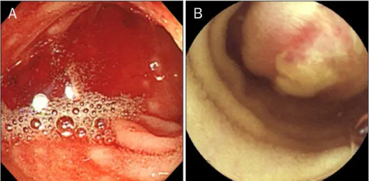
Fig. 1. (A) Fresh blood gushed out from above the terminal ileum. (B) Capsule endoscopy revealed a submucosal lump with erosion located in the jeju- num.
Fig. 2. The small bowel series demon- strated a 2-cm smooth, oval defect (arrow) without irregularity of the over- lying mucosa in the distal jejunum.
Fig. 3. The gross specimen revealed a 1.5×2.0 cm submucosal mass.
The erosion with the black sanguineous crust was considered to be the cause of the obscure gastrointestinal bleeding.
Esophagogastroduodenoscopy and abdominal CT scanning did not reveal any significant abnormalities, such as a mass or obstruction, and colonoscopy revealed small bowel bleeding. In the terminal ileum, up to 20 cm from the ileocecal valve, fresh blood gushed out from above (Fig. 1A). Capsule endoscopy, performed to investigate the source of the ob- scure GI bleeding, revealed a submucosal lump with erosion located in the jejunum (Fig. 1B). A small bowel series was per- formed to verify the presence of the mass and it demon- strated a 2 cm-sized, smooth, oval filling defect without irreg- ularity of the overlying mucosa in the distal jejunum (Fig. 2).
The submucosal lesion in the jejunum was presumed to be the source of the obscure GI bleeding and the cause of the patient’s melena. Surgical wedge resection of the sub- mucosal lesion was carried out using a single incision laparo- scopic approach. A 1.5×2.0 cm submucosal mass with an ul-
Choi WH, et al. Jejunal Ectopic Pancreas with Obscure Hemorrhage 167
Vol. 62 No. 3, September 2013
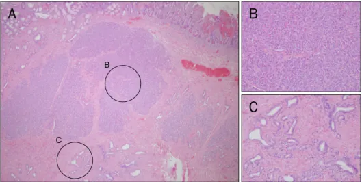
Fig. 4. A portion of the jejunum charac- terized as longer and with more irre- gular villi, no submucosal glands, and no aggregated lymphatic nodules. (A) An ectopic pancreas is located in the submucosa of the jejunum (H&E, ×40).
This lesion demonstrates pancreatic acini (B, ×200) and secretory ducts (C,
×200) in the submucosa of the jeju- num (H&E).
cerated tip was noted in the gross specimen (Fig. 3). Micro- scopic examination revealed the presence of pancreatic ducts and secretory acinar cells without islet cells within the submucosa of the jejunum (Fig. 4). The patient recovered from surgery and had no GI symptoms during the follow-up.
DISCUSSION
An ectopic pancreas, also called a heterotopic or aberrant pancreas, was first described by Jean Schultz in 1729 and re- fers to pancreatic tissue that lacks anatomical and vascular connections with the pancreas.2 It occurs most commonly in the stomach, duodenum, and jejunum and has been re- ported in other locations, including the ileum, Meckel’s diver- ticulum, colon, gall bladder, umbilicus, fallopian tube, media- stinum, spleen, and liver.3 The diameter of the ectopic pan- creatic tissue is generally 1-33 cm and it was 1.5 cm in the current case.4
Gaspar Fuentes and colleagues5 modified von Heinrich’s classification of ectopic pancreas and divided this entity into four types. Type I, a total ectopic pancreas, is composed of all pancreatic cell types, type II contains pancreatic ducts only, type III is composed of acinar cells only, and type IV is com- posed of islet cells only.6 The lesion in the present case was most consistent with a type I lesion, although no islet cells were identified.
An ectopic pancreas is usually discovered incidentally dur- ing radiographic or endoscopic examination of the gut, sur- gery for other abdominal conditions, or upon autopsy.4 A pa- tient with an ectopic pancreas can be normal or present with symptoms such as bleeding, vomiting, or abdominal pain due
to pancreatitis, intestinal obstruction, or intussusception.3 Rarely, an ectopic pancreas can be associated with other pancreatic diseases, including islet cell tumors, pancreatic carcinomas, and pancreatic cysts.1 In the present case, the patient presented with obscure GI bleeding, and an ectopic pancreas was diagnosed during evaluation of this complaint.
The prevalence of GI bleeding due to ectopic pancreas is not well established as the rarity of its morbidity. Dolan et al.7 re- ported that GI bleeding has been reported in 3 out of 73 symptomatic cases among 212 ectopic pancreas patients.
Obscure GI bleeding has been defined as bleeding of un- known origin that persists or recurs after an initial negative endoscopic evaluation, including colonoscopy and/or upper endoscopy.8 The majority of lesions causing obscure GI bleeding are eventually found in the small intestine. Accor- dingly, if conventional evaluation of GI bleeding has failed to reveal a source, it is reasonable to proceed with further evalu- ation of the small bowel.9 Diagnostic options include capsule endoscopy, push enteroscopy, or barium contrast small bow- el studies.9 However, push enteroscopy only evaluates the proximal jejunum, and barium contrast small bowel studies are not particularly sensitive for small lesions. Additionally, push enteroscopy has potential complications, such as bow- el perforation and acute pancreatitis, as well as the rare oc- currence of bleeding and infection.10,11 In this setting, capsu- le endoscopy has a high sensitivity and specificity for detect- ing a bleeding source in patients with obscure GI bleeding.12 For the previously mentioned reasons, capsule endoscopy is well-suited as the first choice for evaluating obscure GI bleeding. In the current patient, a source of obscure GI bleed- ing was detected by capsule endoscopy and confirmed by ba-
168 최우형 등. 불명료한 출혈로 발현된 공장의 이소성 췌장
The Korean Journal of Gastroenterology
rium contrast small bowel studies.
Although endoscopic ultrasound and double balloon en- teroscopy are helpful for diagnosis, an ectopic pancreas is usually buried in the submucosa, which makes it difficult to distinguish from other submucosal tumors.4 Endoscopic di- agnosis is rarely conclusive on endoscopic biopsies because the lesion is usually located too deeply in the submucosa to easily biopsy.3 Surgical resection and histologic evaluation should be undertaken to distinguish an ectopic pancreas from malignant tumors.13
In regard to surgical procedures to address an ectopic pan- creas, single incision laparoscopic surgery constitutes the next step in the development of minimally invasive surgery.14 In the current patient, access to the peritoneal cavity was ach- ieved through a single port in the umbilicus.
In summary, a jejunal ectopic pancreas was revealed to be the source of obscure GI bleeding through capsule endos- copy and barium contrast small bowel studies. The number of reported cases of an ectopic pancreas of the GI tract will likely increase due to the increased use of both capsule en- doscopy and double balloon enteroscopy.15
REFERENCES
1. Rimal D, Thapa SR, Munasinghe N, Chitre VV. Symptomatic gas- tric heterotopic pancreas: clinical presentation and review of the literature. Int J Surg 2008;6:e52-e54.
2. Armstrong CP, King PM, Dixon JM, Macleod IB. The clinical sig- nificance of heterotopic pancreas in the gastrointestinal tract.
Br J Surg 1981;68:384-387.
3. Hirasaki S, Kubo M, Inoue A, Miyake Y, Oshiro H. Jejunal small ectopic pancreas developing into jejunojejunal intussuscep- tion: a rare cause of ileus. World J Gastroenterol 2009;15:3954- 3956.
4. Hsia CY, Wu CW, Lui WY. Heterotopic pancreas: a difficult diag- nosis. J Clin Gastroenterol 1999;28:144-147.
5. Gaspar Fuentes A, Campos Tarrech JM, Fernández Burgui JL, et al. Pancreatic ectopias. Rev Esp Enferm Apar Dig 1973;39:
255-268.
6. Chetty R, Weinreb I. Gastric neuroendocrine carcinoma arising from heterotopic pancreatic tissue. J Clin Pathol 2004;57:314-317.
7. Dolan RV, ReMine WH, Dockerty MB. The fate of heterotopic pan- creatic tissue. A study of 212 cases. Arch Surg 1974;109:762-765.
8. American Gastroenterological Association medical position statement: evaluation and management of occult and obscure gastrointestinal bleeding. Gastroenterology 2000;118:197-201.
9. Leighton JA, Goldstein J, Hirota W, et al. Obscure gastrointestinal bleeding. Gastrointest Endosc 2003;58:650-655.
10. Iddan G, Meron G, Glukhovsky A, Swain P. Wireless capsule endoscopy. Nature 2000;405:417.
11. Mazzarolo S, Brady P. Small bowel capsule endoscopy: a system- atic review. South Med J 2007;100:274-280.
12. Mylonaki M, Fritscher-Ravens A, Swain P. Wireless capsule en- doscopy: a comparison with push enteroscopy in patients with gastroscopy and colonoscopy negative gastrointestinal blee- ding. Gut 2003;52:1122-1126.
13. Yuan Z, Chen J, Zheng Q, Huang XY, Yang Z, Tang J. Heterotopic pancreas in the gastrointestinal tract. World J Gastroenterol 2009;15:3701-3703.
14. Budzyński A, Matłok M, Pędziwiatr M, et al. SILS (single incision laparoscopic surgery)-new surgical approach to peritoneal cavity. Adv Med Sci 2011;56:18-24.
15. Takeda Y, Nakase H, Chiba T. Ectopic pancreas at the jejunum.
Dig Liver Dis 2011;43:e6.