Received:February 5, 2016, Revised:March 11, 2016, Accepted:April 7, 2016
Corresponding to:Seung-Geun Lee, Division of Rheumatology, Department of Internal Medicine, Pusan National University Hospital, Pusan National University School of Medicine, 179 Gudeok-ro, Seo-gu, Busan 49241, Korea. E-mail:[email protected]
pISSN: 2093-940X, eISSN: 2233-4718
Copyright ⓒ 2016 by The Korean College of Rheumatology. All rights reserved.
This is a Free Access article, which permits unrestricted non-commerical use, distribution, and reproduction in any medium, provided the original work is properly cited.
A Case of a Central Retinal Artery Occlusion in a Patient with Rheumatoid Arthritis
Eun-Kyoung Park1, Seung-Geun Lee1, Dong-Wan Koo1, Ji-Heh Park1, Young-Mi Seol2, Shinwon Lee3, Sun-Hack Lee1, In-Su Kim1, Sungwho Park4
Divisions of 1Rheumatology and 2Hemato-oncology, Department of Internal Medicine, Pusan National University School of Medicine,
3Department of Internal Medicine, Pusan National University Hospital, Medical Research Institute, Pusan National University School of Medicine, 4Department of Ophthalmology, Pusan National University School of Medicine, Busan, Korea
A 50-year-old woman, who had been treated for rheumatoid arthritis (RA) over a 10-year period, suddenly presented with mon- ocular vision loss while the RA had a stable course over many years. She was diagnosed with central retinal artery occlusion (CRAO) based on ophthalmologic examinations including optical coherence tomography and fluorescein angiography. There was no evidence of atherosclerosis, infection, and malignancy that can cause CRAO. Considering the association between CRAO and other rheumatic diseases, such as systemic vasculitis and systemic lupus erythematous in previous reports, it was presumed that her RA might have contributed to the development of CRAO. Although cases of CRAO in patients with RA are extremely rare, these findings suggest that physicians need to be aware of the possibility of CRAO in patients with RA who expe- rience decreased visual acuity. (J Rheum Dis 2016;23:326-331)
Key Words. Retinal artery occlusion, Rheumatoid arthritis
INTRODUCTION
Central retinal artery occlusion (CRAO) is a rare con- dition that causes acute, painless, monocular vision loss and results in significant functional morbidity. A CRAO is associated with cardiovascular disease and has several risk factors in common with atherosclerotic disease (e.g., stroke and heart disease) [1,2]. Patients with systemic in- flammatory disorders (e.g., giant cell arteritis, Wegener’s granulomatosis, Churg-Strauss vasculitis, polyarteritis nodosa, systemic lupus erythematosus [SLE], and Behçet’s disease) are at an increased risk for developing a CRAO [3]. In these diseases, an excessive inflammatory burden may contribute to vascular occlusion develop- ment and appropriate immunosuppressive therapy is required. However, the association between CRAO and rheumatoid arthritis (RA) has not been well established.
Here, we report a case of CRAO in a patient with estab-
lished RA.
CASE REPORT
A 50-year-old woman presented to our emergency de- partment with acute, painless vision loss in her left eye.
When she was getting ready for work that morning, she was unable to distinguish objects with her left eye. Ten years earlier, she had been treated for livedo reticularis at a dermatology department and was referred to our rheu- matology clinic because of polyarthralgia and bilateral swelling in the second to fourth metacarpophalangeal, knee, and right third metatarsophalangeal joints. The swelling had persisted for 2 months and was accom- panied by morning stiffness that lasted longer than 1 hour. At the time of referral, the rheumatoid factor (RF) level was 196.2 IU/mL, anti-cyclic citrullinated peptide antibody level was 98.5 U/mL, antinuclear antibodies
Figure 1. Clinical course of the patient during her 10-year follow-up period. CRAO: central retinal artery occlusion, CRP: C-re- active protein, ESR: erythrocyte sedimentation rate.
Figure 2. Fundoscopic findings. (A) A photograph of the right eye’s fundus reveals no specific findings. (B) A photograph of the left eye’s fundus reveals acute central retinal artery occlusion with cherry-red spots that were caused by a retinal infarction and arte- riole cattle trucking. No definite emboli were observed.
were negative, erythrocyte sedimentation rate (ESR) was 120 mm/h, and C-reactive protein (CRP) level was 4.56 mg/dL. The resulting disease activity score with 28-joint assessment (DAS28)-CRP was 5.69. Radiography re- vealed erosions in the right third metatarsophalangeal joint.
The patient was diagnosed with RA, according to the 1987 American College of Rheumatology criteria, and treated using disease-modifying antirheumatic drugs.
She was initially prescribed hydroxychloroquine and sul- fasalazine because no ocular abnormalities were found at a baseline ophthalmologic examination. Hydroxychloro- quine was changed to methotrexate one month later and the patient discontinued hydroxychloroquine at that time (Figure 1). At this time, the patient complained of numb- ness in her left fourth and fifth fingers. Electromyography
was performed and revealed a left ulnar neuropathy, which her physician suspected was caused by vasculitic occlusion. Treatment with high-dose glucocorticoids im- proved the neuropathy. Fortunately, the patient had a sta- ble course of RA over many years and deflazacort had been tapered from 9 to 3 mg per day. The patient did expe- rience intermittent swelling of the right wrist joint during that time.
When the patient presented to the emergency depart- ment, visual acuity in the left eye was counting fingers and an ophthalmologic examination revealed a relative af- ferent pupillary defect. Fundoscopic examination was un- remarkable in the right eye, but revealed cherry-red spots in the left eye that were caused by a retinal infarction and arteriole cattle trucking (Figure 2). We did not observe
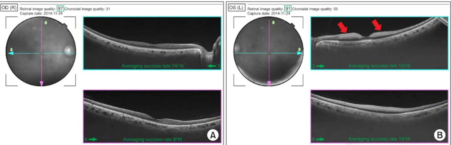
Figure 3. Optical coherence tomography (OCT) shows cross-section images of retina. (A) OCT of the right eye shows normal appearance. (B) OCT of the left eye shows the inner retina layer (nerve fiber layer) infarct presented as thickening and whitening (red arrows).
Figure 4. Fluorescein angiog- raphy at the onset of the central retinal artery occlusion. (A) Arterial filling in the left eye was markedly delayed in the early phase. (B) The arteriovenous transit time was prolonged with no venous flow in the middle phase. (C) Fluorescein pooling is visible in the late phase.
any definite emboli. Optical coherence tomography re- vealed inner retinal layer thickening caused by retinal edema in the left eye (Figure 3). Fluorescein angiography
revealed a markedly delayed arterial filling in the early phase and a prolonged arteriovenous transit time with no venous flow in the middle phase (Figure 4). Therefore,
the patient was diagnosed with a CRAO in the left eye.
The patient did not have fever, headache, temporal ar- tery tenderness, or hip and shoulder girdle stiffness/pain, which was suggestive of giant cell arteritis. The patient did not complain of Raynaud’s phenomenon and did not have hypertension or dyslipidemia, both of which are po- tential causes of CRAO. The patient never smoked, was not taking oral contraceptives, and never had facial cos- metic surgery, a known cause of CRAO. An electrocardio- gram revealed a normal sinus rhythm and an echocardio- graph showed normal left ventricular function with no evidence of heart valve disease. Carotid Doppler ultra- sonography revealed no significant carotid athero- sclerosis or plaque and brain magnetic resonance imaging and angiography were normal. Peripheral angiography was not performed.
Blood lipid profile tests measured a low-density lip- oprotein level of 74 mg/dL, a high-density lipoprotein level of 78 mg/dL, and a triglyceride level of 62 mg/dL.
Laboratory findings to examine the possibility of a hyper- coagulable state showed a prothrombin time of 11.7 s (normal, 10.0∼12.5 s), an activated partial thromboplas- tin time of 25.0 s (normal, 27∼42 s), a D-dimer level of 0.5 μg/mL (normal, 0∼0.5 μg/mL), a fibrinogen level of 346.5 mg/dL (normal, 170∼380 mg/dL), a protein C antigen level of 111% (normal, 72%∼160%), a protein C activity of 99.4% (normal, 73%∼142%), a protein S anti- gen level of 89% (normal, 60%∼150%), a free protein S level of 84.9% (normal, 50%∼150%), a protein S activity of 65% (normal, 65%∼140%), a negative result for factor V Leiden, and a homocysteine level of 7.9 μmol/L (normal, 5∼15 μmol/L). Antinuclear antibody results remained negative and antineutrophil cytoplasmic anti- body, lupus anticoagulant, and anticardiolipin antibody immunoglobulin G and M were all negative. Complement C3 was 93.5 mg/dL (normal, 90∼180 mg/dL) and com- plement C4 was 15.9 mg/dL (normal, 10∼40 mg/dL).
The patient had a DAS28-CRP of 2.55, determined by the presence of swelling and tenderness in her right wrist, an ESR of 25 mm/h, and a CRP level of 0.24 mg/dL. Com- puted tomography of the chest and abdomen was per- formed to rule out other CRAO etiologies, including in- fection or malignancy, but there were no remarkable findings.
The patient was treated for the CRAO with ocular mas- sage, anterior chamber paracentesis, intravenous man- nitol, and an oral antiplatelet agent. After 2 months, visu- al acuity in the left had slightly improved and no retinal
opacification had occurred, although retinal atrophy persisted. After 10 months, visual acuity was 20/1,000, which was still in the counting fingers range. The patient has been followed closely for her RA at our rheumatology clinic. She still has swelling and tenderness in her right wrist, but no RA flare. At the patient’s last visit, CRP level was 0.03 mg/dL and DAS28-CRP was 2.15.
DISCUSSION
Rheumatoid arthritis is a systemic inflammatory disease that is occasionally associated with extra-articular com- plications, including ocular manifestations. The most common ocular complication is secondary Sjögren’s syn- drome, although other ophthalmological conditions (e.g., episcleritis, scleritis, and peripheral ulcerative kera- titis) are known to be associated with systemic in- flammation in patients with RA [4]. Optic neuritis, ante- rior optic neuropathy, and retinal vasculitis associated with RA are rarely reported [5]. A CRAO, the ocular equivalent of a cerebral stroke [6], results in acute, pain- less monocular vision loss. It is considered to be an oph- thalmic emergency because it is associated with sig- nificant functional morbidity. In Korea, the incidence rate of CRAO was 1.80 per 100,000 person-years, with this rate exponentially increasing with age [7]. Embolism is the most common cause of CRAO and is generally caused by the formation of atherosclerotic plaques [6]. Other eti- ologies of CRAO include systemic vasculitis, SLE, and Behçet’s disease, all autoimmune rheumatic diseases [3].
The relationship between CRAO and RA has not been ex- tensively investigated because of its rarity.
Intensive investigation revealed no evidence of in- fection, malignancy, or autoimmune diseases (e.g., SLE or antiphospholipid antibody syndrome), all of which are potential causes of CRAO. Therefore, we cautiously hy- pothesize that RA led to CRAO development. First, RA is a systemic inflammatory rheumatic disease with ex- tra-articular manifestations and, though rare (<1% of pa- tients), rheumatoid vasculitis can accompany long-stand- ing disease. The signs of rheumatoid vasculitis vary and can include petechiae, purpura, digital infarcts, gangrene, livedo reticularis, and lower extremity ulceration. Given that our patient had ulnar neuropathy and livedo retic- ularis, both recognized clinical manifestations of vasculi- tis, rheumatoid vasculitis may have also affected the cen- tral retinal artery and subsequently resulted in CRAO.
Second, RA is associated with cardiovascular diseases
Table 1. Cases of central retinal artery occlusion in rheumatoid arthritis Author
[Reference]
Age (yr)
/sex Cause of CRAO Treatment Nationality Year of
publication Matsuo [8] 67/F Vascular inflammation,
rheumatoid vasculitis
Hyperbaric oxygen, intravenous prostaglandin E1, urokinase
Japan 2001
Kachmaryk et al. [9] 49/F Sickle cell trait,
hypergammaglobulinemia
Not mentioned USA 1995
Park et al.
(present study)
50/F Not clearly defined, may be inflammatory
Conservative, oral antiplatelet agent Korea 2016 CRAO: central retinal artery occlusion, F: female.
and rheumatic inflammation can contribute to athero- sclerotic changes. Significant atherosclerosis was not de- tected via imaging studies in this patient and RA was sta- ble during the follow-up period. However, it is possible that long-term subclinical inflammation might have led to undetected atherosclerotic changes in the micro- vasculature, including the ophthalmic artery, which branches off of the internal carotid artery. Because epi- demiologic data and CRAO case reports in patients with RA are lacking, the pathogenetic association between CRAO and RA could not be thoroughly examined.
However, we speculate that rheumatoid vasculitis and subclinical atherosclerosis associated with long-standing inflammation may have independently or synergistically contributed to CRAO development in our patient.
Further research is needed to investigate pathophysio- logic processes of CRAO in patients with RA.
Our literature review identified two case reports of reti- nal arterial occlusion in patients with RA (Table 1) [8,9].
Matsuo [8] presented a case of branch retinal artery oc- clusion in both eyes. Although the female patient did not have arthralgia, the finger joints in both hands were deformed. This patient had an ESR of 23 mm/h, a CRP level of 0.2 mg/dL, and an RF of 25.2 IU/mL. The author suggested that elevated ESR and RF levels indicated per- sistent RA activity, which led to CRAO development. Our patient had similar ESR and CRP levels, but a much high- er RF level of 196.2 IU/mL. Kachmaryk et al. [9] reported the second case, in which a patient with RA developed a cilioretinal artery occlusion with the sickle cell trait. The patient also had mild hypergammaglobulinemia secon- dary to RA and an elevated RF level. The authors hy- pothesized that increased serum viscosity led to vascular stasis, which resulted in further vascular sludging and cil- ioretinal artery occlusion. We did not check immunoglo- bulin levels in our patient because her albumin/globulin
ratio was normal and she did not have the sickle cell trait.
Together, hyperinflammatory status associated with RA and hypergammaglobulinemia resulting from autoanti- body production could result in occlusion of the retinal vasculature.
No cases of CRAO in an RA patient have been previously reported in South Korea, but 3 cases of CRAO in patients with other rheumatic diseases have been published. Kim et al. [10] reported a case of CRAO that occurred during methylprednisolone pulse therapy treatment for poly- arteritis nodosa. Once CRAO was diagnosed, cyclo- phosphamide pulse therapy was added. The remaining cases occurred in SLE patients. Hwang and Kang [11] re- ported on a combined central retinal vein and artery oc- clusion in a patient with SLE. Their clinical observations led them to believe that CRAO was of a thrombotic, and not of a vasculitic, origin [11]. Song et al. [12] reported that retinal vaso-occlusion was an earlier manifestation of SLE without anticardiolipin antibodies. Evidence from a few other case reports suggested an association between some drugs and CRAO development, including oral con- traceptives [13]. However, an association between gluco- corticoids and CRAO development has not yet been reported.
Further evidence supporting an association between RA disease activity and retinal vasculitis was presented in a report that suggested that suppressing RA-related in- flammation improves vascular health [14]. Another re- port showed that orbital blood flow velocity in RA pa- tients is lower than in healthy controls, which suggests that systemic inflammation may also affect the ocular vessels [15]. Given the increased risk of cerebrovascular disease in RA patients and the influence of systemic in- flammation on blood vessels, CRAO likely has clinical re- lationship with RA rather than simply being an incidental finding in our patient. In the current case, CRAO develop-
ment may reflect long-term subclinical systemic in- flammation. However, more research is needed to verify the association between CRAO and RA.
Conventional treatments for CRAO include vaso- dilators, ocular massage, anterior chamber paracentesis, intravenous mannitol, tissue plasminogen activator, and surgery [2]. Intravenous glucocorticoids can also be used when a CRAO was caused by systemic inflammatory disease. Because our patient showed no direct evidence of high grade active inflammation and RA had been stable, systemic immunosuppressive agents (e.g., high-dose glu- cocorticoids) were not administered and, with the input of an ophthalmologist, the patient was conservatively managed. However, close follow-up was needed in this patient to monitor for CRAO deterioration, RA disease activity, and new rheumatoid vasculitis signs.
SUMMARY
In conclusion, this is the first case of CRAO reported in a Korean patient with RA. Clinical findings in this patient support an association between CRAO and RA, similar to CRAO associations with other systemic inflammatory rheumatic diseases (e.g., SLE and systemic vasculitis).
Thus, clinicians need to consider the possibility of CRAO in patients with RA who complain of decreased visual acuity.
ACKNOWLEDGEMENTS
This work was supported by clinical research grant from Pusan National University Hospital 2016. We specially thank the late Professor Sung-Il Kim who devoted himself to education, research, and patient care in Division of Rheumatology, Department of Internal Medicine, Pusan National University School of Medicine (1963 to 2011).
CONFLICT OF INTEREST
No potential conflict of interest relevant to this article was reported.
REFERENCES
1. Callizo J, Feltgen N, Pantenburg S, Wolf A, Neubauer AS, Jurklies B, et al; European Assessment Group for Lysis in the Eye. Cardiovascular risk factors in central retinal artery occlusion: results of a prospective and standardized medical examination. Ophthalmology 2015;122:1881-8.
2. Cugati S, Varma DD, Chen CS, Lee AW. Treatment options for central retinal artery occlusion. Curr Treat Options Neurol 2013;15:63-77.
3. Perumal B, Black EH, Levin F, Servat JJ. Non-infectious or- bital vasculitides. Eye (Lond) 2012;26:630-9.
4. Artifoni M, Rothschild PR, Brézin A, Guillevin L, Puéchal X.
Ocular inflammatory diseases associated with rheumatoid arthritis. Nat Rev Rheumatol 2014;10:108-16.
5. Tong L, Thumboo J, Tan YK, Wong TY, Albani S. The eye: a window of opportunity in rheumatoid arthritis? Nat Rev Rheumatol 2014;10:552-60.
6. Varma DD, Cugati S, Lee AW, Chen CS. A review of central retinal artery occlusion: clinical presentation and mana- gement. Eye (Lond) 2013;27:688-97.
7. Park SJ, Choi NK, Seo KH, Park KH, Woo SJ. Nationwide in- cidence of clinically diagnosed central retinal artery occlu- sion in Korea, 2008 to 2011. Ophthalmology 2014;121:
1933-8.
8. Matsuo T. Multiple occlusive retinal arteritis in both eyes of a patient with rheumatoid arthritis. Jpn J Ophthalmol 2001;45:662-4.
9. Kachmaryk MM, Trimble SN, Gieser RG. Cilioretinal artery occlusion in sickle cell trait and rheumatoid arthritis. Retina 1995;15:501-4.
10. Kim SS, Lee JH, Nam TS, Kim SI. A case of polyarteritis no- dosa associated with central retinal artery occlusion. J Korean Rheum Assoc 2002;9:236-40.
11. Hwang HS, Kang S. Combined central retinal vein and artery occlusion in systemic lupus erythematosus patient. Retin Cases Brief Rep 2012;6:187-8.
12. Song YH, Kim CG, Kim SD, Kim YY, Choe JY. Systemic lu- pus erythematosus presenting earlier as retinal vaso- occlusion. Korean J Intern Med 2001;16:210-3.
13. Rekhi GS, Dheer S. Oral contraceptive-induced central reti- nal artery occlusion. J Assoc Physicians India 2002;50:1084-5.
14. Moi JH, Hodgson LA, Wicks IP, Wong TY, Van Doornum S.
Suppression of inflammatory disease activity in rheumatoid arthritis is associated with improvements in retinal micro- vascular health. Rheumatology (Oxford) 2016;55:246-51.
15. Erdogmus B, Yazici S, Yazici B, Ataoglu S, Buyukkaya R, Yuksel H, et al. Orbital blood flow velocities in patients with rheumatoid arthritis. J Clin Ultrasound 2007;35:367-71.