J o u r n a l o f R h e u m a t i c D i s e a s e s V o l . 1 9 , N o . 3 , J u n e , 2 0 1 2
http://dx.doi.org/10.4078/jrd.2012.19.3.168 □ Clinical Im age □
168
<Received:April 26, 2012, Revised:June 15, 2012, Accepted:June 18, 2012>
Corresponding to:Jinseok Kim, Department of Internal Medicine, Jeju National University School of Medicine, Jeju, Korea.
E-mail:[email protected] pISSN: 2093-940X, eISSN: 2233-4718
Copyright ⓒ 2012 by The Korean College of Rheumatology
This is a Free Access article, which permits unrestricted non-commerical use, distribution, and reproduction in any medium, provided the original work is properly cited.
Figure 1. Slit lamp examination of the right eye shows a peripheral corneal ulcer (black arrow) and adjacent corneal neovascularization (white arrow).
Figure 2. Deformities due to longstanding rheumatoid arthritis are shown on hand x-ray.
A Case of Peripheral Ulcerative Keratitis in a Patient with Rheumatoid Arthritis
Jung Won Noh
1, Sang Taek Heo
1, Jinho Jeong
2, Jinseok Kim
1Departments of Internal Medicine1, Ophthalmology2, Jeju National University School of Medicine, Jeju, Korea
Case Report
Patient: 85-year-old female
Chief complaint: Blurred vision, pain, and redness of the right eye presenting for 4 weeks
Past medical history: She was diagnosed with rheumatoid ar- thritis (RA) in a public health center 10 years ago. However, she never took medication for RA. She has been receiving regular ophthalmologic follow up because she has a cataract in the right eye. In a test performed two months ago, the cor- nea of the right eye was clear.
Physical examination: Upon admission, she had deformative arthritis with low current disease activity. In the slit lamp ex- amination, a 3 mm deep ulceration of the cornea was ex- hibited in the right eye (Figure 1). Adjacent corneal pannus (neovascularization) was observed and the sclera was not
inflamed. The patient could only count fingers held 30 cm from her face and intraocular tension was 8 mmHg.
Laboratory tests: Laboratory testing revealed white blood cell of 6.4×10³/mm3, hemoglobin of 10.5 g/dL and platelet of 299×103/mm3, erythrocyte sedimentation rate of 118 mm/hr, C-reactive protein of 2.71 mg/dL. Rheumatoid factor was 173.6 IU/mL, anti-cyclic citrullinated peptide antibody was over 1,200 U/mL. Anti-nuclear antibody was weakly positive.
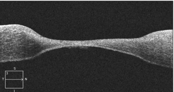
Radiologic findings: Simple hand and knee x-rays showed ad- vanced deformities due to longstanding RA (Figure 2). In the optical coherence tomography test, corneal thinning of the right eye was observed (Figure 3).
Diagnosis and treatment: Following a diagnosis of peripheral ulcerative keratitis (PUK) in this RA patient, she was treated with intravenous steroid pulse therapy (methylprednisolone 1 g per day for 3 days) with scleral patch graft and amniotic mem-
A Case of Peripheral Ulcerative Keratitis in a Patient with Rheumatoid Arthritis 169
Figure 3. Optical coherence tomography exam reveals corneal thinning in the right eye.
brane transplantation. She was discharged with oral pre- dnisolone 30 mg per day.
Discussion
Corneal inflammation as an extraarticular manifestation of RA is a significant complication in patients with RA. In a se- vere case of PUK, corneal deterioration may lead to irrever- sible loss of vision (1). We here present a case of PUK in a patient with long standing RA. In Korea, 2 cases of necrotiz- ing scleritis which developed after pterygium excision in pa- tients with RA were reported (2,3). However, a case of PUK in a patient with RA has never been published in Korea.
Furthermore this patient has never undergone ophthalmic sur- gery and there was no corneal lesion in the right eye upon examination performed 2 months ago.
The severity of the corneal inflammation in RA is often re- lated to the activity of the systemic vasculitis and usually paral- lels the severity of the scleritis, but can occur in eyes with little scleral inflammation (4,5). When the patient visited our hospi- tal, her RA was not active (tender or swollen joint count, 0).
PUK should be treated with a rapid and aggressive approach.
There is no clear consensus of treatment for PUK, but surgical
intervention and systemic immunosuppressive agents such as steroids, cyclophosphamide or biologic agents (infliximab, rit- uximab, daclizumab) have been shown to be effective for ul- cerative keratitis (4,6-9).
References
1. Squirrell DM, Winfield J, Amos RS. Peripheral ulcerative keratitis ‘corneal melt' and rheumatoid arthritis: a case series. Rheumatology (Oxford) 1999;38:1245-8.
2. Kim MJ, Joo CK. A case of enucleation due to extensive necrotiaing scleritis after pterygium excision in a rheuma- toid arthritis patient. J Korean Ophthalmol Soc 1998;39:
777-83.
3. Kim JH, Kim HO, Jeong YG, Yun SU, Lee KJ, Lee CM, et al. A case of scleromalacia perforance that developing after surgery for excision of the pterygium in a patient with rheumatoid arthritis. J Korean Rheum Assoc 2010;17:93-7.
4. Foster CS, Forstot SL, Wilson LA. Mortality rate in rheu- matoid arthritis patients developing necrotizing scleritis or peripheral ulcerative keratitis. Effects of systemic immu- nosuppression. Ophthalmology 1984;91:1253-63.
5. Iliou C, Anthis N, Tsifetaki N, Kitsos G, Voulgari PV.
Clinical images: Corneal melt in a woman with long- standing rheumatoid arthritis. Arthritis Rheum 2012;64:253.
6. Smith JR, Levinson RD, Holland GN, Jabs DA, Robinson MR, Whitcup SM, et al. Differential efficacy of tumor ne- crosis factor inhibition in the management of in- flammatory eye disease and associated rheumatic disease.
Arthritis Rheum 2001;45:252-7.
7. Papaliodis GN, Chu D, Foster CS. Treatment of ocular inflammatory disorders with daclizumab. Ophthalmology 2003;110:786-9.
8. Ahmadi-Simab K, Lamprecht P, Nölle B, Ai M, Gross WL. Successful treatment of refractory anterior scleritis in primary Sjogren's syndrome with rituximab. Ann Rheum Dis 2005;64:1087-8.
9. Smith JR, Mackensen F, Rosenbaum JT. Therapy insight:
scleritis and its relationship to systemic autoimmune disease. Nat Clin Pract Rheumatol 2007;3:219-26.