[Abstract]
Objectives : This study was designed to investigate the neuroprotective effects of Hominis- Placenta (HP)on dopaminergic neurons.
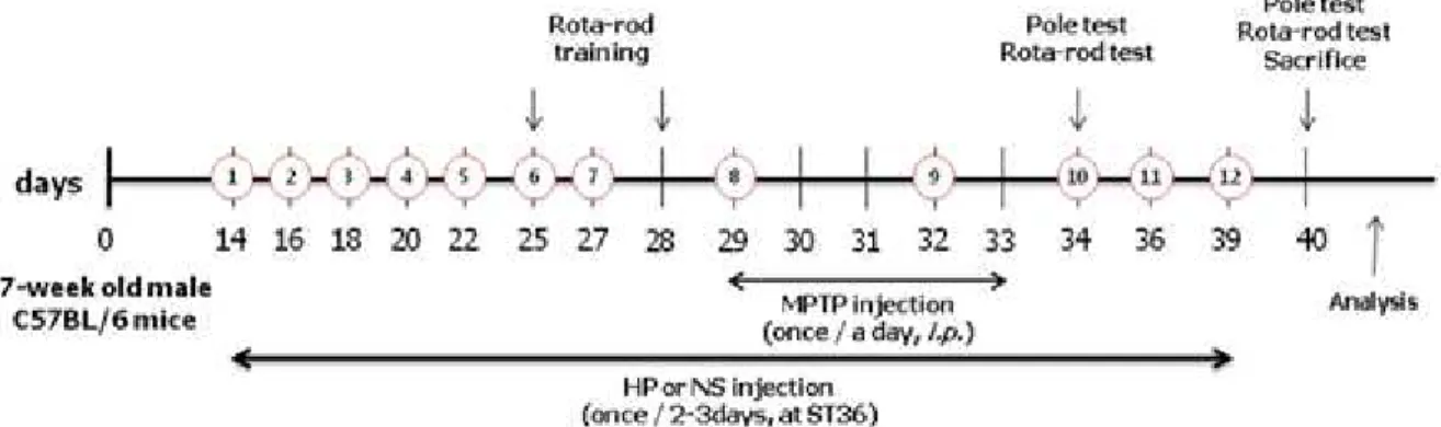
Methods : We examined the effect of i nvitro administration of HP against 1-methyl-4-phenylpyri- dinium(MPP+)-induced dopaminergic cell loss in primary mesencephalic culture and also used behavioral tests and performed analysis in the striatum and the substantia nigra of mouse brain, to confirm the effect of HP on dopaminergic neurons in an i nvivo 1-methyl-4-phenyl- 1,2,3,6-tetrahydropyridine(MPTP)-induced PD mouse model. Animals were assigned to four groups: (1) Group 1(vehicle-treatedgroup), (2) Group 2(MPTPonlytreated group), (3) Group 3(MPTP+ saline-treated/ST
36group), and (4) Group 4(MPTP+HP-treated/ST
36group). HP at 20 μ L of 48 mg/kg dose was injected at ST
36for 4 weeks at 2-day intervals. MPTP in saline was injected intraperitoneally each day for 5 days from the 8
thtreatment of HP. We performed the pole test and rota-rod test on the first and seventh day after the last MPTP injection. To investigate the effect of HP on dopaminergic neurons, we performed analysis in the striatum and the substantia nigra of mouse brain after treatment with HP and/or MPTP.
Results : Treatment with HP had no influence on cell proliferation and caused no cell toxicity in PC
12and HT
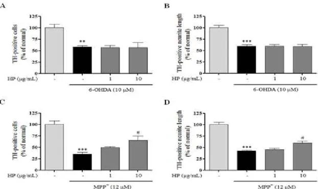
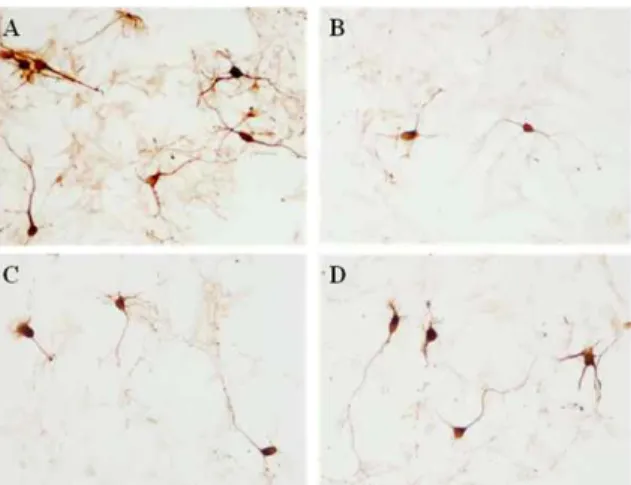
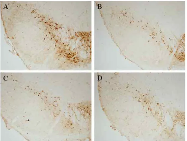
22cells. Our study showed that HP significantly prevented cell loss and protected neurites against MPP+ toxicity. Although the invivo treatment of HP herbal acupuncture at ST 36 showed a tendency to improve movement ability and protected dopaminergic cells and fibers in the substantia nigra and the striatum, it did not show significant changes compared with the MPTP treated group.
Conclusions : These data suggest that HP could be a potential treatment strategy in neuro- degenerative diseases such as Parkinson’s disease.
Key words : Hominis Placenta
pharmacoacupuncture;
Parkinson’s disease;
MPP+;
MPTP;
Behavioral test;
TH-immunohistochemistry
Received :2015. 02. 24.
Revised : 2015. 03. 03.
Accepted : 2015. 03. 04.
On-line : 2015. 03. 20.