Trigeminal neuralgia (TN) is a neurologic disorder characterized by relapsing and lancinating pain in one or more trigeminal nerve distribution unilaterally that lasts for a few seconds to 2 minutes.1 The clinical features and history-taking are very important when diagnosing TN.2 The clinical criteria of the International Headache Society for a TN diagnosis are (1) occurring in one or more divisions of the trigeminal nerve, with no radiation beyond the trigeminal distribution and (2) pain with at least three of the following four characteristics: (a) recurring in paroxysmal attacks lasting from a fraction of a second to 2 minutes; (b) severe intensity; (c) electric-shock-like, shooting, stabbing, or sharp in quality; and (d) precipitated by innocuous stimuli to the affected side of the face.3 However, clinical findings cannot be used to precise- ly distinguish idiopathic TN from symptomatic TN,2 and hence magnetic resonance imaging (MRI) of the brain should be performed to evaluate other causes including multiple sclerosis or tumors.1,2
We report the case of a patient who was diagnosed with recurrent trigeminal neuritis of the maxillary branch confirmed by MRI.
Received: May 24, 2017 Revised: June 9, 2017 Accepted: June 12, 2017 ANNALS OF CLINICAL
NEUROPHYSIOLOGY
CASE REPORT
Ann Clin Neurophysiol 2017;19(2):145-147 https://doi.org/10.14253/acn.2017.19.2.145
Correspondence to Bora Yoon
Department of Neurology, Konyang University Hospital, 158 Gwanjeodong-ro, Seo-gu, Daejeon 35365, Korea
Tel: +82-42-600-9156 Fax: +82-42-545-0050 E-mail: [email protected]
http://www.e-acn.org pISSN 2508-691X eISSN 2508-6960
Copyright © 2017 The Korean Society of Clinical Neurophysiology
This is an Open Access article distributed under the terms of the Creative Commons Attribution Non-Commercial License (http://
creativecommons.org/licenses/by-nc/4.0) which permits unrestricted non-commercial use, distribution, and reproduction in any medium, provided the original work is properly cited.
Chronic recurrent trigeminal neuritis of the maxillary branch confirmed by magnetic resonance imaging
Soon-Ho Hong, Yong-Duk Kim, Sang-Jun Na, Kee Ook Lee, Yun Kyung Park, and Bora Yoon
Department of Neurology, Konyang University College of Medicine, Daejeon, Korea
Trigeminal neuralgia (TN) is generally characterized by lancinating, unilateral, paroxysmal pain occurring in the distribution of the fifth cranial nerve. TN is diagnosed clinically based on the typical patient history, negative findings in a neurologic examination, and the response to medication. Idiopathic TN is the most common type, but TN can result from vascular malfor- mation, compression, trauma, neoplasm, multiple sclerosis, or inflammation. We report a TN case diagnosed as recurrent trigeminal neuritis of the maxillary branch confirmed by magnet- ic resonance imaging.
Key words: Trigeminal neuralgia; Neuritis; Magnetic resonance imaging
146 https://doi.org/10.14253/acn.2017.19.2.145 http://www.e-acn.org
Annals of Clinical Neurophysiology Volume 19, Number 2, July 2017
CASE
A 43-year-old male presented with dysesthesia and recurrent sharp pain in the left nasal and palatal area for 2 days. This was the third time that he had experienced this condition. He had experienced electric-shock-like severe pain in the same area about 5 years previously for the first time. The pain was not aggravated by chewing, tooth brushing, or cold/hot water. At that time he visited a dental clinic and had taken medicines, which eventually relieved the pain. He experienced the same pain in the same area for the second time 8 months previ- ously. Because medication had no effect, three teeth were extracted (first to third upper molars on the left side), and he stated that his pain had disappeared gradually. He subse- quently experienced very brief episodes of intermittent pain, but it was not disabling.
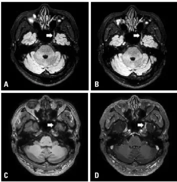
When he went to our clinic he complained of left nasal hypesthesia and pain only, without dizziness, visual distur- bance, hearing impairment, headache, or autonomic symp- toms. There was no history of trauma, infection, or medical problems such as diabetes. A neurologic examination re- vealed facial hypesthesia and allodynia in only the left nasal area (not including the cheek) and without any other neuro- logic deficit. A laboratory study indicated that glycosylated hemoglobin was within the normal range and his thyroid function was normal. The nature of his symptoms and the findings of the neurologic examination suggested left maxil- lary neuritis. We performed cranial nerve MRI with enhance- ment, which revealed diffuse swelling and enhancement in the left maxillary branch of the trigeminal nerve at the cav- ernous portion and foramen rotundum (Fig. 1). The results of a cerebrospinal fluid study including cytology were normal.
We diagnosed left trigeminal neuritis of the maxillary branch, and administered intravenous methylprednisolone for 5 days, which resulted in his symptoms subsiding rapidly.
DISCUSSION
TN is a severe-pain disorder that decreases the quality of life in most patients,4 and an early and accurate diagnosis of TN is important for determining the most appropriate treatment.1 Despite the etiology and pathophysiology of TN are usually uncertain,1,5 Strittmatter et al. reported that the
autonomic nervous system is associated with idiopathic TN, especially in neuroendocrinologic changes such as in corti- sol and adrenocorticotropic hormones.6 Choi et al. studied trigeminal neuropathy induced by T-type Ca2+-channel-me- diated alteration of the thalamocortical rhythm.7
Except for the idiopathic condition, there are many second- ary causes of TN such as vascular compression, neoplasm, de- myelinating diseases, and connective-tissue diseases.1,5 Clini- cal information and history-taking are sufficient for confirming most cases of idiopathic TN.1 However, brain MRI should be performed when the neurologist suspects other causes of TN or there are atypical features. Sakurai et al. reported on a patient with chronic trigeminal neuritis confirmed by MRI, whose symptoms had also occurred 2 months previously and had gradually improved spontaneously without any med- ication, which differs from the present case.8 Although the symptom onset had occurred 2 months previously, inflam- matory changes of the trigeminal nerve could be detected in brain MRI, like in the present case.8 Krafft reported that brain
A B
C D
Fig. 1. Magnetic resonance imaging of the cranial nerve. Diffuse swell- ing and thickening of the maxillary branch of the left trigeminal nerve was noted in a nonenhanced fluid-attenuated inversion recovery with fat saturation axial image (A) and in an enhanced image (B) (arrows).
The enhancement on the left maxillary branch of the trigeminal nerve at the cavernous portion and foramen rotundum was not detected in an enhanced T1-weighted axial image (D) but not in a nonenhanced image (C) (arrows).
147
http://www.e-acn.org https://doi.org/10.14253/acn.2017.19.2.145
Soon-Ho Hong, et al. Recurrent trigeminal neuritis confirmed by MRI
MRI may be considered especially if the atypical features of TN include abnormal findings in neurologic, oral, dental, or ear examinations, bilateral symptoms, dizziness or vertigo, hearing loss or other hearing abnormality, numbness, pain episodes lasting longer than 2 minutes, pain outside of tri- geminal nerve distribution, and visual changes, or age lower than 40 years.1 The guidelines of the American Academy of Neurology and European Federation of Neurological Societies specify that routine imaging may be considered for identify- ing secondary TN in patients with TN without nontrigeminal neurological symptoms.3 Brain MRI is helpful for evaluating the structural lesion in trigeminal nerve.2,9 Especially, struc- tural MRI with special techniques, informing the pathway of trigeminal nerve is useful for confirming inflammation like our case. Besides, the physician can identify other etiology of TN such as petroclival meningioma and abnormalities of root en- try zone by using brain MRI.2,9 Neurologists can use brain MRI to localize and diagnose lesions in the trigeminal pathway.2,9 Like the present case, even if pain due to neuritis is chronic and recurrent, nerve inflammation can be detected by MRI.
Once neuritis has been confirmed in MRI, steroid therapy should be started promptly to ensure a good prognosis even if the etiology of the inflammation has not been identified.
Therefore, brain MRI with enhancement and the fat-suppres- sion technique should be considered if the diagnosis either is uncertain or is incompatible with typical TN.
REFERENCES
1. Krafft RM. Trigeminal neuralgia. Am Fam Physician 2008;77:1291- 1296.
2. Kedarnath NS, Shruthi R. MRI as an essential diagnostic approach for trigeminal neuralgia. J Maxillofac Oral Surg 2015;14(Suppl 1):462-464.
3. Montano N, Conforti G, Di Bonaventura R, Meglio M, Fernandez E, Papacci F. Advances in diagnosis and treatment of trigeminal neu- ralgia. Ther Clin Risk Manag 2015 24;11:289-299.
4. Allsop MJ, Twiddy M, Grant H, Czoski-Murray C, Mon-Williams M, Mushtaq F, et al. Diagnosis, medication, and surgical management for patients with trigeminal neuralgia: a qualitative study. Acta Neurochir (Wien) 2015;157:1925-1933.
5. Cruccu G, Finnerup NB, Jensen TS, Scholz J, Sindou M, Svensson P, et al. Trigeminal neuralgia: New classification and diagnostic grad- ing for practice and research. Neurology 2016;87:220-228.
6. Strittmatter M, Grauer MT, Fischer C, Hamann G, Hoffmann KH, Blaes F, et al. Autonomic nervous system and neuroendocrine changes in patients with idiopathic trigeminal neuralgia. Cephalal- gia 1996;16:476-480.
7. Choi S, Yu E, Hwang E, Llinás RR. Pathophysiological implication of CaV3.1 T-type Ca2+ channels in trigeminal neuropathic pain. Proc Natl Acad Sci U S A 2016;113:2270-2275.
8. Sakurai K, Hara M, Okita K, Kawashima S, Yamawaki T, Shibamoto Y.
Idiopathic trigeminal neuropathy with trigeminal mass lesion on MRI: Neoplasm or not? Cephalagia 2010;30:968-974.
9. DeSouza DD, Hodaie M, Davis KD. Structural magnetic resonance imaging can identify trigeminal system abnormalities in classical trigeminal neuralgia. Front Neuroanat 2016;10:95. eCollection 2016.