Identification of Bacterial and Fungal Isolates by Sequence Analysis of 16S rRNA and Internal Transcribed Spacer
Younhee Park1, Hee Bong Shin2,7, Chang Ki Kim3,7, Kyoung Ho Roh4,7, Jong Hwa Yum5,7, Dongeun Yong6,7, Seok Hoon Jeong6,7, Kyungwon Lee6,7
Department of Laboratory Medicine,
1Kwandong University College of Medicine, Goyang,
2Soonchunhyang University College of Medicine,
4Korea University College of Medicine, Seoul,
5Department of Clinical Laboratory Science, Dongeui University, Busan,
6Department of Laboratory Medicine and
7Research Institute of Bacterial Resistance,
Yonsei University College of Medicine,
3Korean Institute of Tuberculosis, Seoul, Korea
Background: Accurate and rapid identification ofpathogens is one of the most important tasks of the clinical microbiology laboratory, and, in cases of rare pathogens, the identification is difficult and time-con- suming upon the use of conventional methods alone.
Herein, we will report our molecular work involving the identification of bacteria and fungi.
Methods: Sixty bacterial isolates had been collected from November 2004 to May 2007, and 15 fungal isolates had been collected from September 2005 to May 2007. Species identifications were performed using sequence analyses of the 16S rRNA region of bacteria and the internal transcribed spacer (ITS) re- gion of fungi. The data were compared with those of GenBank (http://www.ncbi.nlm.nih.gov/) or EMBL (http://
www.ebi.ac.uk/embl/).
Results: Sixty bacterial isolates included: 23 isolates with genus information (group 1), 17 isolates (group 2) that were too fastidious for genus or species iden-
tification, 16 isolates (group 3) with results from iden- tification kits having low confidence, and 4 isolates (group 4) with odd antibiograms according to the species. In 58 of 60 isolates, identification of the ge- nus or species could be obtained using molecular genetic methods. Thirty-eight isolates (63%) and 20 (33%) of 58 isolates could be identified at the spe- cies and genus levels, repectively. Among the total of 15 fungal isolates, 11 (73%) and 4 (27%) isolates were identified at the species and genus levels, respectively.
Conclusion: 16S rRNA and ITS sequencing analyses are very useful for identifying the species or genus of a pathogenic microorganism in the clinical micro- biology laboratory. (Korean J Clin Microbiol 2010;13:
34-39)
Key Words: 16S rRNA, Internal transcribed spacer, Nu- cleotide sequence, Bacterial identification
Received 31 August, 2009, Revised 7 February, 2010 Accepted 18 February, 2010
Correspondence: Dongeun Yong, Department of Laboratory Medicine, Yonsei University College of Medicine, 250, Seongsanno, Seodae- mun-gu, Seoul 120-752, Korea. (Tel) 82-2-2228-2442, (Fax) 82-2- 364-1583, (E-mail) [email protected]
34 서 론
정확하고 신속한 원인균의 동정은 감염증 진단과 치료에 필 수적인 임상미생물 검사실의 주요 업무이다. 이를 위하여 순수 배양된 세균의 그람 염색성과 형태, 성장 촉진 및 필수 요소, 당발효 혹은 동화 등 생화학적 특성을 이용한 전통적인 방법이 사용되어 왔다. 그러나 표현형에 의존한 전통적인 동정법만으 로는 1) 동일한 세균 종(species) 내의 모든 균주가 동일한 표현 형을 보이지 않을 수 있으며, 2) 참고자료의 부족으로 동정이 어려울 수 있다[1]. 또한 노령인구와 면역억제환자의 증가로 진 균에 의한 감염증이 늘어나고, 과거에는 드물던 세균의 분리가
점차로 증가하고 있어서 세균의 종 동정이 더욱 어려워지고 있 다[2].
최근 감염균 동정에 유전자 염기서열 분석법을 이용할 수 있 음이 보고되고 있다. 그 중 16S rRNA는 세균의 종간 보존 염기 서열과 종 특이 염기서열을 함께 지니고 있어서 세균 분류 및 동정에 널리 이용되고 있고[3-7], internal transcribed spacer (ITS) rRNA 유전자 염기서열 분석법은 진균의 동정에 사용되 고 있다[8-11]. Clinical and Laboratory Standards Institute (CLSI)에서 2008년 염기서열 분석을 통한 미생물 동정의 기준 을 승인하였지만, CLSI 기준 외에도 세균의 종 및 속(genus) 동 정 기준은 다양하게 이용되어 왔다[12-15]. 최근 국내 임상미생 물 검사실에서도 16S rRNA, ITS 염기서열 분석법이 감염균의 동정에 사용되고 있으나, 분리 빈도가 매우 낮은, 제한된 세균 의 동정에서만 일부 보고가 되었다[16,17].
이에 저자들은 최근 20∼30개월간 국내 대학병원 임상미생 물 검사실에서 분자유전학적 방법을 이용한 세균과 진균 동정
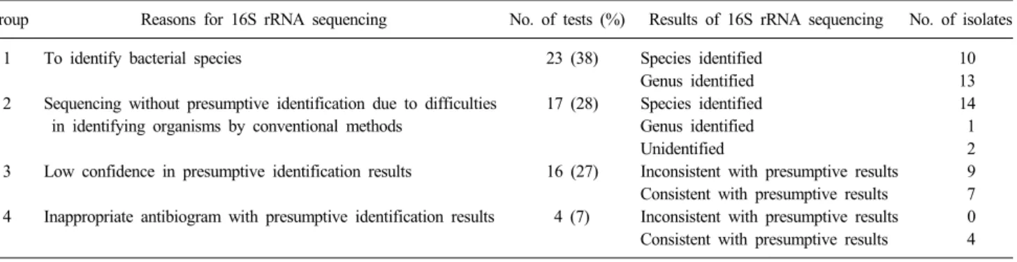
Table 1. Summary of bacterial identification by 16S rRNA sequence analysis
Group Reasons for 16S rRNA sequencing No. of tests (%) Results of 16S rRNA sequencing No. of isolates
1 To identify bacterial species 23 (38) Species identified 10
Genus identified 13
2 Sequencing without presumptive identification due to difficulties 17 (28) Species identified 14
in identifying organisms by conventional methods Genus identified 1
Unidentified 2
3 Low confidence in presumptive identification results 16 (27) Inconsistent with presumptive results 9 Consistent with presumptive results 7 4 Inappropriate antibiogram with presumptive identification results 4 (7) Inconsistent with presumptive results 0 Consistent with presumptive results 4 경험을 정리하여 보고하고, 이를 통하여 분자유전학적 동정법
의 유용성을 평가해 보고하고자 하였다.
재료 및 방법
1. 대상2004년 11월부터 2007년 5월까지 30개월간 배양 의뢰된 임 상 검체에서 증식된 세균 중 전통적인 생화학적 방법 혹은 동 정용 키트(VITEK system, bioMerieux, Marcy l'Etoile, France) 만으로는 동정이 어려웠던 세균 60주를 대상으로 하였다. 또한 2005년 9월부터 2007년 5월까지 20개월간 분리된 진균 중 역 시 전통적인 방법만으로는 동정이 어려웠던 진균 15주에서 분 자유전학적 방법으로 동정을 시도하였다.
2. 세균의 16S rRNA 염기서열 분석
대상 균주를 부유시킨 증류수 100μL를 100oC에서 10분간 중탕 후 4oC에서 13,000 rpm으로 5분간 원심분리하여 불순물 을 침전시켰다. 상청액 10μL를 DNA template로 사용하여 중 합효소연쇄반응을 시행하였다. 한쌍의 시발체(5'-AGA GTT TGA TCC TGG CTC AG-3' 및 5'-AAG GAG GTG ATC CAG CCG CA-3')를 사용하여 Mastercycler gradient (Eppendorf, Hamburg, Germany)로 16S rRNA 유전자를 증폭시켰다[18]. 증 폭조건은 94oC에서 5분간 열 변성 후 94oC 20초, 50oC 40초, 72oC 2분의 과정을 35회 반복 후 72oC에서 5분간 연장반응을 실시하였다. 증폭된 DNA는 전기영동을 통해 band를 확인한 후 QIAquick Gel Extraction kit (QIAGEN GmbH, Hilden, Germany)를 이용하여 증폭산물을 정제하였다. 정제된 DNA는 ABI Prism 3100 Genetic Analyzer (Applied Biosystems, Foster city, CA, USA)를 사용하여 양방향으로 염기서열을 분석하였 다.
3. 진균 ITS 염기서열 분석
DNA 추출 과정은 세균의 16S rRNA 염기서열 분석과 동일 하였다. 한쌍의 시발체(5'- TCC GTA GGT GAA CCT GCG
G-3' 및 5'-TCC TCC GCT TAT TGA TAT GC-3')를 사용하여 Mastercycler gradient (Eppendorf, Hamburg, Germany)로 ITS 부위 유전자를 증폭시켰다[19]. 증폭조건은 95oC에서 5분간 열 변성 후 95oC 30초, 55oC 1분, 72oC 1분의 과정을 35회 반복 후 72oC에서 6분간 연장반응을 실시하였다. 나머지 과정은 세균의 16S rRNA 염기서열 분석과 동일하게 시행하였다.
4. 염기서열 상동성 분석을 통한 세균 및 진균 동정
16S rRNA와 ITS 유전자의 염기서열을 GenBank (http://
www.ncbi.nlm.nih.gov/) 또는 EMBL (http://www.ebi.ac.uk/
embl/)의 자료와 비교하였다. 세균의 종 및 속 동정을 위한 기 준은 자료분석의 일관성을 위하여 CLSI의 기준 대신 연구 시 작부터 적용하였던 기준을 사용하였다[13,15]. 즉, 세균은 참고 염기서열과 99% 및 95% 이상의 일치율을 보일 때 특정 종 및 속으로 동정하였고, 진균은 98% 이상 일치율을 보일 경우 특정 종으로 95% 이상 일치율을 보일 경우 특정 속으로 동정하였다 [13,15].
결 과
1. 16S rRNA 유전자 염기서열 분석을 통한 세균의 동정 염기서열 분석을 시행하였던 균의 검체는 혈액이 31건(52%) 으로 가장 많았고, 창상 14건(23%), 체액 6건(10%), 소변 4건 (7%), 객담 2건(3%) 순이었다. 그람음성 막대균이 56% (포도당 비발효 그람음성 막대균 포함), 그람양성 막대균이 23%, NTM (non-tuberculous mycobacterium)이 3%, 그리고 동정이 되지 않 은 균이 3%였다. 16S rRNA 유전자 염기서열 분석한 균주는 총 60주였고, 종까지 동정된 경우는 38주(63%)였으며, 속까지 동정된 경우는 20주(33%)였다.
16S rRNA 염기서열 분석을 시행한 경우는 1군) 전통적인 생 화학적 방법과 동정용 키트를 이용하여 세균의 속은 동정할 수 있었으나, 종의 감별이 어려웠던 23주(38%), 2군) 증식에 오랜 시간이 소요되거나 동정이 까다로워 세균의 속명을 밝히기 전 에 염기서열 분석을 시행한 17주(28%), 3군) 동정 키트 결과의
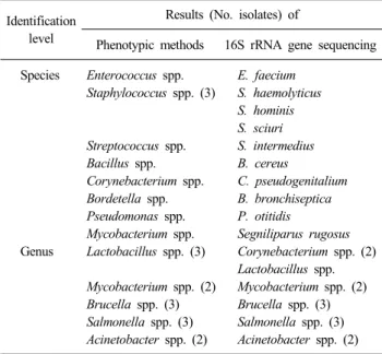
Table 2. Bacterial identifications of group 1 Identification
level
Results (No. isolates) of
Phenotypic methods 16S rRNA gene sequencing Species Enterococcus spp. E. faecium
Staphylococcus spp. (3) S. haemolyticus S. hominis S. sciuri Streptococcus spp. S. intermedius Bacillus spp. B. cereus
Corynebacterium spp. C. pseudogenitalium Bordetella spp. B. bronchiseptica Pseudomonas spp. P. otitidis
Mycobacterium spp. Segniliparus rugosus Genus Lactobacillus spp. (3) Corynebacterium spp. (2)
Lactobacillus spp.
Mycobacterium spp. (2) Mycobacterium spp. (2) Brucella spp. (3) Brucella spp. (3) Salmonella spp. (3) Salmonella spp. (3) Acinetobacter spp. (2) Acinetobacter spp. (2)
Table 3. Summary of fungal identification by ITS sequence analysis Type Specimen
types
Identification by ITS region gene sequencing
Identity (%)
Yeast Blood Pichia ohmeri 100
Pichia guilliermondii 100 Body fluid Cryptococcus neoformans 100
Tissue C. neoformans 100
Wound C. neoformans 100
Mould Body fluid Cladosporium spp. 100
Wound Phaeoacremonium aleophilum 100
Pseudozyma spp. 99
Trichosporon debeurumannianum 100
Eye Alternaria spp. 99
Alternaria spp. 98
Aspergillus fumigatus 99 Pseudallescheria boydii 99 Environment Paecilomyces variotii 100
Dimorphic Wound Coccidioides immitis 100
Abbreviation: ITS, internal transcribed spacer region.
신뢰도가 낮았던 16주(27%) 및 4군) 동정된 세균이 그 세균의 일반적인 감수성 양상과 달라서 추가 확인하기 위해 검사하였 던 4주(7%)였다(Table 1).
세균 종의 결정을 위해 분자생물학적 방법을 추가 시험한 23 주(1군) 중 종이 동정된 경우는 10주(44%)였다. 동정된 종의 속 은 표현형을 이용한 동정 결과와 동일하였다. 이들은 Bacillus cereus, Bordetella bronchiseptica, Corynebacterium pseudogeni- talium, Enterococcus faecium, Pseudomonas otitidis, Segnilipa- rus rugosus, Staphylococcus haemolyticus, Staphylococcus hom- inis, Staphylococcus sciuri 및 Streptococcus intermedius이었다.
분자유전학적 방법으로도 속까지만 동정된 13주는 Acineto- bacter spp. 2주, Brucella spp. 3주, Corynebacterium spp. 2주, Lactobacillus spp. 1주, NTM 2주 및 Salmonella spp. 3주이었다 (Table 2).
증식에 오랜 시간이 소요되거나 증식 조건이 까다로워 균명 을 추정하기 어려웠던 17주(2군) 중 14주는 16S rRNA 염기서 열 분석으로 세균의 종 동정이 가능하였다. Brevundimonas di- minuta, Clostridium septicum, Kingella kingae, Lactobacillus paracasei, Moraxella atlantae, Moraxella nonliquefaciens, Mo- raxella osloensis, Neisseria elongata 3주, Neisseria subflava, Neisseria weaveri, Rahnella aquatilis, Tsukamurella tyrosinosol- vens와 같이 일반적인 배지 및 배양 조건에서 증식이 어려웠거 나 분리빈도가 낮은 세균이었다. 나머지 1주는 Paenibacillus 속까지 동정되었고 2주는 95% 이상 일치하는 염기 서열을 찾 을 수 없었다.
동정 키트 결과의 신뢰도가 낮았던 16주(3군) 중 11주는 종 까지, 5주는 속까지 동정할 수 있었다. 동정 키트로 Burkholde-
ria mallei로 추정된 1주는 Chromobacterium spp.로, Chryseo- bacterium spp.로 추정된 1주는 Burkholderia cepacia로, Clo- stridium perfringens로 추정된 1주는 Propionibacterium acnes 로, Haemophilus parainfluenzae로 추정된 3주는 Pasteurella multocida로, Microbacterium spp.로 추정된 1주는 Arthrobacter woluwensis로, Pseudomonas spp.로 추정된 1주는 Acinetobacter spp.로, Sphingomonas paucimobilis로 추정된 1주는 Leptotri- chia spp.로 각각 동정되었다. 나머지 7주는 각각 Bacillus cer- eus, Moraxella osloensis, Pseudomonas aeruginosa, Pseudo- monas spp., Stenotrophomonas maltophilia, Vibrio fluvialis와 Vibrio vulnificus로 동정되어 키트의 결과와 동일하였다.
동정된 세균의 항균제 감수성이 그 세균의 일반적인 양상과 달랐던 4균주(4군) 모두 추정한 세균명과 염기서열 분석 결과 가 일치하였다. 4군은 Enterococcus faecium 1주, Enterococcus spp. 1주 및 Staphylococcus lugdunensis 2주였다. 문제가 되었 던 감수성 양상을 살펴보면, E. faecium와 Enterococcus spp.는 cephalosporin계 항균제에 대해 감수성을 보였고, S. lugdu- nensis는 oxacillin에 감수성, cefoxitin에 내성을 보였던 균주였 다.
2. ITS 유전자 염기서열 분석을 통한 진균의 동정
효모형 진균은 동정 키트 결과의 신뢰도가 낮은 경우, 곰팡 이형 진균은 전통적 방법으로 동정이 어려웠던 경우 염기서열 분석을 시행하였다. 전체 검체는 창상검체 5건, 안구 검체가 4 건으로 가장 많았고 혈액 검체 2건, 체액 검체 2건 및 기타 검 체 1건이었다. 진균별로는 효모형 진균 5주(33%), 곰팡이형 진 균 9주(60%), 그리고 이상성 진균 1주(7%)였다(Table 3). 그 중 11주(73%)는 종까지, 4주(27%)는 속까지 동정되었다. 효모형
진균과 이상성 진균은 모두 종까지 동정할 수 있었으나 곰팡이 형 진균 9주 중 4주는 속까지 동정할 수 있었다.
고 찰
유전자 염기서열 분석과 같은 분자유전학적 방법은 흔히 분 리되지 않거나 표현형에 대한 자료가 부족한 미생물, 균주에 따라 다양한 생화학적 성상을 나타낼 수 있는 미생물의 동정에 유용할 뿐 아니라, 그동안 밝혀지지 않았던 새로운 병원균을 찾을 수 있는 장점이 있다[12,20]. 진균의 경우 일반적으로 세 균에 비하여 동정에 장시간이 요구되는데 분자유전학적 방법 을 이용하면 비교적 단시간에 동정이 가능하다.
세균의 분자유전학적 동정에서 초기에는 5S, 16S 및 23S rRNA 유전자와 이들 사이에 위치한 유전자가 대상으로 사용 되었으며, 현재는 16S rRNA 유전자가 세균의 분류에서 가장 흔히 사용되고 있다[3-7]. 16S rRNA 유전자의 염기서열은 약 1,550 bp의 길이를 가지며 보존서열과 다형성을 보이는 서열 부위로 구성되어 있어서 염기서열을 이용한 세균의 동정에 유 용하다. 그 이유는 16S rRNA 유전자가 세포의 구조 형성에 필 수적인 역할을 하지 않으므로 세균마다 차이를 보일 수 있는 변이 발생이 용인되기 때문이다[6,7]. 16S rRNA의 염기서열은 많은 균주에서 밝혀져 있는데, 가장 흔히 이용할 수 있는 GenBank에는 90,000가지 이상의 16S rRNA 유전자 염기서열 정보가 저장되어 있어 비교를 통한 세균의 동정이 가능하다.
또한 16S rRNA는 모든 세균에 존재하므로, 이 유전자를 이용 한 동정법은 거의 모든 세균에 적용할 수 있다.
1군에 속하는 균주 중에서 16s rRNA 염기서열 분석으로 종 까지 동정된 9균주와 속까지 동정된 11균주는 표현형을 이용하 여 동정되었던 세균의 결과와 일치하였다. 나머지 3균주 중 2 균주는 표현형 검사에서는 Lactobacillus spp.로 추정하였으나 염기서열 분석에서는 Corynebacterium spp.로, 1균주는 Myco- bacterium spp.로 추정하였으나 염기서열 분석에서 Segnilipa- rus rugosus로 동정되었다. 전통적 표현형 검사법이 대부분의 세균의 동정에 있어 다른 추가적인 검사를 필요로 하는 경우는 높지 않음을 알 수 있었고, 임상미생물 담당 전문의가 검사실 의 인력 상황과 환자 중증도를 고려하여 염기서열 분석을 추가 로 시행할 수 있을 것으로 판단되었다.
Acinetobacter spp., Brucella spp., NTM 및 Salmonella spp.는 16S rRNA 염기서열 분석만을 통해서는 세균의 종까지 동정이 어려운 것으로 보고된 바 있다[12,21-23]. 예를 들어, Brucella spp., Acinetobacter spp., Salmonella spp.는 속 수준까지만 동정 되고 종의 구분은 되지 않는 경우가 흔하다. 이 연구에서도 세 균의 종을 알기 위해 염기서열 분석을 시행한 1군 23주 중 속 까지만 동정된 13주의 대다수(10주)가 이들에 속하였다. 각 세 균은 염기서열 분석을 시행한 유전자에 따라서 동정 신뢰성이
상이할 수 있으므로 rpoB, gyrB, dnaJ 등 세균의 다른 유전자도 이용할 수 있다. Acinetobacter spp.의 경우는 16S-23S riboso- mal ITS 유전자 염기서열이 종의 동정에 더 유용하다는 보고도 있다[12,14,21,24]. 대부분 전향적 시발체에 의해 증폭되는 부 위가 종 간 다형성을 보이는 데 비해 Vibrio spp.는 후향적 시발 체에 의해 증폭되는 염기서열에서 다형성을 보인다. Vibrio spp.의 동정을 위해서 toxR 유전자가 유용하다는 보고도 있다 [23]. 이와 같이 시발체, 균주, 유전자 부위에 따라 염기서열 분 석을 통해 얻을 수 있는 동정의 수준이 상이할 수 있으므로 이 에 대한 충분한 이해와 적절한 적용이 필요할 것이다.
동정 키트 결과의 신뢰도가 낮았던 16검체 중 9검체가 키트 의 결과와 다르게 동정되었다. 드문 세균이나 생화학적 표현형 의 특성이 약한 세균이 분리 동정된 경우 검체를 고려하여 분 자유전학적 확인이 필요할 것으로 판단되었다.
진균의 분자유전학적 동정에서는 주로 리보솜의 ITS 부위와 RNA 오페론(operon)의 D1-D2 부위의 유전자를 대상으로 한다 [8-11]. 진균의 D1-D2 부위의 염기서열은 주로 비병원성 진균 에서 밝혀져 있으며, 병원성 진균에 대해서는 주로 ITS 염기서 열 분석이 이용된다. 또한 ITS 유전자 염기서열 분석을 이용하 면 중합효소연쇄반응 후 증폭산물의 크기가 달라 길이의 차이 에 따라서도 균종을 동정할 수 있다[8,11].
분자유전학적 방법으로 진균 동정을 시행한 검체 중 안구 검 체가 상대적으로 많았던 이유는 배양에 충분한 양의 검체를 얻 기 어렵고 조기진단과 치료가 예후에 중요하므로 안구에서 진 균감염이 의심된 경우 배양과 함께 분자유전학적 검사를 의뢰 하였기 때문이다. 곰팡이형 진균의 경우 대부분 드물게 분리되 는 균이어서 동정이 어려웠기 때문에 분자유전학적 방법이 매 우 유용한 것으로 판단하였다.
본 연구에서 세균에 대해서는 GenBank 등의 염기서열과 각 각 99% 및 95% 이상 일치할 경우, 진균에 대해서는 98% 및 95% 이상 일치할 경우 균종 혹은 균속으로 동정하였다[13,15].
최근 제시된 CLSI 기준 외에도 종 및 속의 동정 기준은 다양하 였고[12-15], 본 연구에서는 연구 시작부터 적용하였던 기준 [13,15]을 자료분석의 일관성을 위하여 채택하였다. CLSI[14]
에도 반영된 바와 같이 미생물 각 세균의 속마다 같은 속도로 진화가 이루어지는 것은 아니므로, 동일한 기준을 적용하는 것 보다는 각각의 세균 속에 알맞은 기준이 필요할 것으로 생각된 다. 염기서열의 비교를 위해 이용되는 GenBank, EMBL 등의 자료는 관련 전문가들에 의해 검토가 되지 않은 자료가 포함되 어 있거나, 미생물의 이름, 분류 및 염기서열에 오류가 있기도 하다. 특히 오래된 자료의 경우 이런 오류를 포함하고 있을 가 능성이 높은 것으로 알려져 있다[12]. 그러므로 임상미생물 검 사실에서 이러한 자료를 이용하여 미생물을 동정하기 위해서 는 자료들의 적절성에 대한 검토가 필요한데, 염기서열을 비교 할 때 자료의 등록 시점 및 자료에 대한 주의 깊은 검토가 요구
된다. 한편 미생물간 염기서열이 거의 동일한 경우 분자유전학 적 방법만을 통해서는 종을 동정하기 어려울 수 있다. 따라서, 생화학적인 활성을 이용한 전통적인 동정방법이 앞으로도 중 요할 것으로 생각된다.
결론적으로 임상미생물 담당 전문의의 판단에 따라서 균 집 락의 육안적 소견 및 현미경 관찰 소견, 전통적인 생화학적 시 험에 추가하여 분자유전학적 미생물 동정법을 적절히 이용한 다면 임상미생물 검사실에서의 병원성 세균 및 진균 동정에 매 우 유용할 것으로 판단되었다.
감사의 글
본 연구는 연세대학교 의과대학 2008년 교내연구비지원 (6-2008-0281)을 받아서 수행하였습니다.
참 고 문 헌
1. Bosshard PP, Abels S, Altwegg M, Böttger EC, Zbinden R.
Comparison of conventional and molecular methods for identi- fication of aerobic catalase-negative gram-positive cocci in the clinical laboratory. J Clin Microbiol 2004;42:2065-73.
2. Walsh TJ, Groll A, Hiemenz J, Fleming R, Roilides E, Anaissie E.
Infections due to emerging and uncommon medically important fungal pathogens. Clin Microbiol Infect 2004;10(Suppl 1):48-66.
3. Patel JB. 16S rRNA gene sequencing for bacterial pathogen identification in the clinical laboratory. Mol Diagn 2001;6:313-21.
4. Chong Y, Lee K, et al. eds. Diagnostic Microbiology. 3rd ed.
Seoul: Seoheung Publishing; 2000:105-15.
5. Tortoli E. Impact of genotypic studies on mycobacterial taxonomy:
the new mycobacteria of the 1990s. Clin Microbiol Rev 2003;
16:319-54.
6. Pace NR. A molecular view of microbial diversity and the biosphere. Science 1997;276:734-40.
7. Thorne JL, Kishino H, Painter IS. Estimating the rate of evolution of the rate of molecular evolution. Mol Biol Evol 1998;15:1647-57.
8. Chen YC, Eisner JD, Kattar MM, Rassoulian-Barrett SL, LaFe K, Yarfitz SL, et al. Identification of medically important yeasts using PCR-based detection of DNA sequence polymorphisms in the internal transcribed spacer 2 region of the rRNA genes. J Clin Microbiol 2000;38:2302-10.
9. Hinrikson HP, Hurst SF, Lott TJ, Warnock DW, Morrison CJ.
Assessment of ribosomal large-subunit D1-D2, internal transcribed spacer 1, and internal transcribed spacer 2 regions as targets for molecular identification of medically important Aspergillus species.
J Clin Microbiol 2005;43:2092-103.
10. Li YL, Leaw SN, Chen JH, Chang HC, Chang TC. Rapid identification of yeasts commonly found in positive blood cultures by amplification of the internal transcribed spacer regions 1 and 2.
Eur J Clin Microbiol Infect Dis 2003;22:693-6.
11. Massonet C, Van Eldere J, Vaneechoutte M, De Baere T, Verhaegen J, Lagrou K. Comparison of VITEK 2 with ITS2-fragment length polymorphism analysis for identification of yeast species. J Clin Microbiol 2004;42:2209-11.
12. Clarridge JE 3rd. Impact of 16S rRNA gene sequence analysis for identification of bacteria on clinical microbiology and infectious diseases. Clin Microbiol Rev 2004;17:840-62.
13. Bosshard PP, Abels S, Zbinden R, Böttger EC, Altwegg M.
Ribosomal DNA sequencing for identification of aerobic gram- positive rods in the clinical laboratory (an 18-month evaluation). J Clin Microbiol 2003;41:4134-40.
14. Clinical and Laboratory Standards Institute. Interpretitive criteria for identification of bacteria and fungi by DNA target sequeincing;
Approved guideline. CLSI document MM18-A. Wayne. PA:
Clinical and Laboratory Standard Institute: 2008.
15. Ciardo DE, Schär G, Altwegg M, Böttger EC, Bosshard PP.
Identification of moulds in the diagnostic laboratory--an algorithm implementing molecular and phenotypic methods. Diagn Microbiol Infect Dis 2007;59:49-60.
16. Sohn KM, Ko KS, Kim J, Rhee JY, Oh WS, Peck KR, et al.
Identification of Gemella species by 16S ribosomal RNA gene sequencing from two patients with infective endocarditis. Korean J Int Med 2006;70:591-6.
17. Yoon S, Kim S, Lee KA, Kim H. A case of Scedosporium apiospermum keratitis confirmed by a molecular genetic method.
Korean J Lab Med 2008;28:307-11.
18. Loffler FE, Sun Q, Li J, Tiedje JM. 16S rRNA gene-based detection of tetrachloroethene-dechlorinating Desulfuromonas and Dehalococcoides species. Appl Environ Microbiol 2000;66:1369- 74.
19. Ferrer C, Colom F, Frasés S, Mulet E, Abad JL, Alió JL. Detection and identification of fungal pathogens by PCR and by ITS2 and 5.8S ribosomal DNA typing in ocular infections. J Clin Microbiol 2001;39:2873-9.
20. Bosshard PP, Zbinden R, Abels S, Böddinghaus B, Altwegg M, Böttger EC. 16S rRNA gene sequencing versus the API 20 NE system and the VITEK 2 ID-GNB card for identification of nonfermenting Gram-negative bacteria in the clinical laboratory. J Clin Microbiol 2006;44:1359-66.
21. La Scola B, Gundi VA, Khamis A, Raoult D. Sequencing of the rpoB gene and flanking spacers for molecular identification of Acinetobacter species. J Clin Microbiol 2006;44:827-32.
22. Shin S, Kim EC, Yoon JH. Identification of nontuberculous mycobacteria by sequence analysis of the 16S ribosomal RNA, the heat-shock protein 65 and the RNA polymerase beta-subunit genes.
Korean J Lab Med 2006;26:153-60.
23. Franco PF and Hedreyda CT. Amplification and sequence analysis of the full length toxR gene in Vibrio harveyi. J Gen Appl Microbiol 2006;52:281-7.
24. Ferroni A, Sermet-Gaudelus I, Abachin E, Quesne G, Lenoir G, Berche P, et al. Use of 16S rRNA gene sequencing for identi- fication of nonfermenting gram-negative bacilli recovered from patients attending a single cystic fibrosis center. J Clin Microbiol 2002;40:3793-7.
=국문초록=
16S rRNA 및 Internal Transcribed Spacer 염기서열 분석법을 이용한 세균 및 진균 동정
1관동대학교 의과대학 진단검사의학교실, 2순천향대학교 의과대학 진단검사의학교실, 3결핵연구원, 4고려대학교 의과대학 진단검사의학교실, 5동의대학교 임상병리과, 6연세대학교 의과대학 진단검사의학교실, 7세균내성 연구소
박윤희1, 신희봉2,7, 김창기3,7, 노경호4,7, 염종화5,7, 용동은6,7, 정석훈6,7, 이경원6,7
배경: 정확하고 신속한 원인균의 동정은 임상미생물 검사실의 주요 업무이다. 표현형을 이용하는 전통적인 동정법만으 로는 드물게 분리되는 균의 동정에 실패하거나 동정에 오랜 시간이 소요되기도 한다. 세균 및 진균의 동정에 분자유전학 적 방법을 추가하였던 경험을 보고하고자 한다.
방법: 2004년 11월부터 2007년 5월까지 30개월간 동정 결과의 추가 확인이 필요하였던 60주의 세균과 2005년 9월부터 2007년 5월까지 20개월간 동일한 목적으로 수집된 15주의 진균을 대상으로 하였다. 세균은 16S rRNA 부위, 진균은 in- ternal transcribed spacer (ITS) 부위의 유전자 염기서열을 분석하였으며, Genbank (http://www.ncbi.nlm.nih.gov/) 및 EMBL (http:// www.ebi.ac.uk/embl/)의 자료와 비교하였다.
결과: 대상 세균은 1군) 전통적인 생화학적 방법과 동정용 키트를 이용하여 세균의 속(genus)은 동정할 수 있었으나, 세균 의 종(species) 감별이 어려웠던 23주(38%), 2군) 증식에 오랜 시간이 소요되거나 동정이 까다로워 세균의 속을 밝히기 전에 염기서열 분석을 시행한 17주(28%), 3군) 동정 키트 결과의 신뢰도가 낮았던 16주(27%) 및 4군) 동정된 세균명과 그 종의 일반적인 감수성 양상이 달랐던 4주(7%)였다. 총 60주 중 58주에서 세균의 종 혹은 속의 동정이 가능하였다.
이 중 38주(63%)는 종까지, 20주(33%)는 속까지 동정할 수 있었다. 총 15주의 진균 중 11주(73%)는 종, 4주(27%)는 속까 지 동정할 수 있었다.
결론: 16S rRNA와 ITS 부위를 이용한 분자유전학적 방법은 임상미생물 검사실에서의 세균의 종 및 속 동정에 많은 도움 을 줄 수 있을 것으로 판단된다. [대한임상미생물학회지 2010;13:34-39]
교신저자 : 용동은, 120-752, 서울시 서대문구 성산로 250 연세대학교 의과대학 진단검사의학교실 Tel: 02-2228-2442, Fax: 02-364-1583 E-mail: [email protected]