Renoprotective Effects of a Highly Selective A3 Adenosine
Receptor Antagonist in a Mouse Model of Adriamycin-induced Nephropathy
The concentration of adenosine in the normal kidney increases markedly during renal hypoxia, ischemia, and inflammation. A recent study reported that an A3 adenosine receptor (A3AR) antagonist attenuated the progression of renal fibrosis. The adriamycin (ADX)-induced nephropathy model induces podocyte injury, which results in severe proteinuria and progressive glomerulosclerosis. In this study, we investigated the preventive effect of a highly selective A3AR antagonist (LJ1888) in ADX-induced nephropathy. Three groups of six-week-old Balb/c mice were treated with ADX (11 mg/kg) for four weeks and LJ1888 (10 mg/kg) for two weeks as following: 1) control; 2) ADX; and 3) ADX + LJ1888.
ADX treatment decreased body weight without a change in water and food intake, but this was ameliorated by LJ1888 treatment. Interestingly, LJ1888 lowered plasma creatinine level, proteinuria, and albuminuria, which had increased during ADX treatment.
Furthermore, LJ1888 inhibited urinary nephrin excretion as a podocyte injury marker, and urine 8-isoprostane and kidney lipid peroxide concentration, which are markers of oxidative stress, increased after injection of ADX. ADX also induced the activation of proinflammatory and profibrotic molecules such as TGF-β1, MCP-1, PAI-1, type IV collagen, NF-κB, NOX4, TLR4, TNFα, IL-1β, and IFN-γ, but they were remarkably suppressed after LJ1888 treatment. In conclusion, our results suggest that LJ1888 has a renoprotective effect in ADX-induced nephropathy, which might be associated with podocyte injury through oxidative stress. Therefore, LJ1888, a selective A3AR antagonist, could be considered as a potential therapeutic agent in renal glomerular diseases which include podocyte injury and proteinuria.
Keywords: Adriamycin; Nephropathy; Adenosine Receptor Antagonist; Mouse Model Hye Sook Min,1* Jin Joo Cha,1*
Kitae Kim,1 Jung Eun Kim,1 Jung Yeon Ghee,1 Hyunwook Kim,2 Ji Eun Lee,2 Jee-Young Han,3 Lak Shin Jeong,4 Dae Ryong Cha,1 and Young Sun Kang1
1Department of Nephrology, Korea University Ansan Hospital, Ansan, Korea; 2Department of Nephrology, Wonkwang University Sanbon Hospital, Gunpo, Korea; 3Department of Pathology, Inha University Medical College, Incheon, Korea; 4Department of Pharmacy, College of Pharmacy, Seoul National University, Seoul, Korea
* Hye Sook Min and Jin Joo Cha contributed equally to this work.
Received: 30 January 2016 Accepted: 13 May 2016 Address for Correspondence:
Young Sun Kang, MD
Department of Nephrology, Korea University Ansan Hospital, 123 Jeokgeum-ro, Danwon-gu, Ansan 15355, Korea E-mail: [email protected]
Funding: This research was supported by Korea University Grant in 2015 (Q1508671).
http://dx.doi.org/10.3346/jkms.2016.31.9.1403 • J Korean Med Sci 2016; 31: 1403-1412
INTRODUCTION
Adenosine is well known to have a cell protective effect in stress- ful situations, such as ischemia, hypoxia, and inflammation (1).
It is released from metabolically active cells into the extracellu- lar space and is produced by the breakdown of released ade- nosine triphosphate (ATP). While the concentration of extracel- lular adenosine is about 300 nM in normal cells (2), its concen- trations rapidly exceed 1 μM in cells undergoing damage (3).
Adenosine’s regulation of tissue function is mediated through activation of a family of 4 G-protein coupled receptors, which includes the A1, A2a, A2b, and A3 adenosine receptors (AR).
Concentrations of extracellular adenosine vary widely depend- ing on external factors and the degree of stress, tissue types, and activation levels of the 4 adenosine receptors (4). Previous stud- ies investigating the role of adenosine have focused on the ago- nists and antagonists of adenosine receptors instead of adenos- ine itself, because of the limitation of its very short half-life (5).
The A3 adenosine receptor (A3AR) has been detected in a wide variety of tissues. A3AR was highly expressed in the testis, but was less likely to be expressed in the lungs, kidney, heart, and central nervous system. Interestingly, inflammatory and cancer cells possess higher A3AR content than other adenosine receptor subtypes. A3AR is able to play a different roles under ischemic conditions, where it shows pro/anti-inflammatory, cellular proliferation/death, and pro/antitumoral effects de- pending on different pathophysiological environments (6). Pre- vious studies demonstrated that inhibition of A3AR leads to kid- ney protection in ischemia-reperfusion injury (7) and myoglo- binuria-induced injury (8). A recent study reported that A3AR antagonist ameliorates the progression of tubulointerstitial fi- brosis in a unilateral ureteral obstruction (UUO) mice model (9).
Few reports have described the expression and distribution of adenosine receptor subtypes in the kidney and the role of A3AR and A3AR antagonists in kidney injury. A new A3AR an- tagonist (LJ1888) may be a powerful, highly selective, species- Nephrology
independent, and orally available agent (10). However, there is rare data for the role of A3AR in glomerular kidney injury. The aim of this study was to investigate the mechanism and reno- protective effects of the highly selective A3AR antagonist (LJ1888) in adriamycin (ADX)-induced nephropathy.
MATERIALS AND METHODS Experimental animals
All 6-week-old Balb/c male mice weighing 22-24 g were purcha- sed from RaonBio Co. (Yongin, Korea). They were randomly as- signed to three groups: control (n = 5); ADX injection (n = 10);
or administration of LJ1888 after ADX injection (n = 10). Mice received a single tail vein injection of ADX diluted with 0.9% sa- line to a final dose of 11 mg/kg. Control mice received the same volume of isotonic saline. Four weeks after ADX injection, mice drank water mixed with 10 mg/kg per day of LJ1888 (Lee et al.
[9] College of Pharmacy, Ewha Womans University, Seoul, Ko- rea) for two weeks as previously described. All mice were pro- vided with a standard diet and water, and were maintained at constant temperature (23°C ± 2°C) and humidity (55% ± 5%) with a 12-hour light/dark cycle. Daily food intake was monitored at regular intervals to confirm drug administration. Plasma cre- atinine level was determined using high-performance liquid chromatography. To determine urinary protein, albumin, and nephrin excretion, mice were caged individually and a 24-hour urine sample was collected at baseline, two, four and six weeks.
Urinary albumin concentrations were determined with a com- petitive enzyme linked immunosorbent assay (ELISA) kit (ALP- CO, Westlake, OH, USA). Urinary protein concentrations were determined by colorimetric detection using bicinchoninic acid (Pierce BCA protein Assay Kit, Pierce Biotech. Inc., Rockford, IL, USA). Urinary nephrin measurement was performed with the commercially available ELISA kit (Exocell, Philadelphia, PA, USA). The urinary 8-isoprostane level was measured using an ELISA kit (Cayman Chemical, Ann Arbor, MI, USA). The extent of peroxidative reaction in the kidney tissue was determined by direct measurement of lipid hydroperoxides (LPO) using an LPO assay kit (Cayman Chemical), as described previously (11).
Mice were sacrificed under anesthesia with intraperitoneal in- jections of sodium pentobarbital (50 mg/kg). Their heart, epi- didymal fat, liver, and kidney tissues were extracted and subse- quently snap-frozen in liquid nitrogen.
Analysis of gene expression by real-time quantitative polymerase chain reaction
We extracted total RNA from experimental cells using the Trizol reagent. Primers were designed for their respective gene sequen- ces using the Primer 3 software, and the secondary structures of the templates were examined and excluded using the mfold program (Supplementary Table 1). Quantitative gene expres-
sion was performed using a Light Cycler 1.5 system (Roche Di- agnostics Corporation, Indianapolis, IN, USA) using SYBR Green technology. In 96-well real-time polymerase chain reaction (PCR) plates, 10 μL SYBR Green master mix was added to 1 μL of RNA (corresponding to 50 ng of total RNA) and 900 nM of forward and reverse primers for a total reaction volume of 20 μL. Real-time reverse transcriptase PCR was performed for 10 minutes at 50°C and for 5 minutes at 95°C. Thirty PCR cycles of denaturation for 10 seconds at 95°C and annealing with exten- sion for 30 seconds at 60°C were then conducted. Gene level ra- tios relative to β-actin (relative gene expression number) were calculated by subtracting the threshold cycle number of the tar- get gene from that of β-actin and squaring this difference. We evaluated the specificity of each PCR product using melting curve analysis, followed by agarose gel electrophoresis.
Protein extraction and western blot analysis
Nuclear and cytoplasmic proteins were extracted from renal cortical tissues and cells using a commercial nuclear extraction kit according to the manufacturer’s instructions (Active Motif, Carlsbad, CA, USA). Protein concentration was determined us- ing the bicinchoninic acid method (Pierce Pharmaceuticals).
For western blotting, 40 μg protein was electrophoresed on a 10% SDS-PAGE mini-gel. Proteins were transferred onto a poly- vinylidene difluoride membrane, and the membrane was hy- bridized in blocking buffer overnight at 4°C with mouse mono- clonal anti-nuclear factor (NF)-κB p65 antibody (1:1,000; Cell Signaling Technology, Danvers, MA, USA), rabbit polyclonal anti-transforming growth factor (TGF)-β1 antibody (1:200; San- ta Cruz Biotechnology, Santa Cruz, CA, USA), rabbit polyclonal anti-nicotinamide adenine dinucleotide phosphate (NADPH) oxidase 4 (NOX4) antibody (1:500; Novus Biologicals, Littleton, CO, USA) and goat polyclonal anti-toll-like receptor 4 (TLR4) antibody (1:500; Santa Cruz Biotechnology). The membrane was subsequently incubated with horseradish peroxidase-con- jugated secondary antibody (1:1,000 dilution) for 60 minutes at room temperature. We detected the specific signals using an enhanced chemiluminescence method (Amersham, Bucking- hamshire, UK).
Histological examination and immunohistochemistry Kidney samples were fixed in 10% buffered formalin and em- bedded in paraffin. Kidney tissue was cut into 4-µm-thick slices and stained with periodic acid Schiff (PAS), Masson’s trichrome, and sirius red staining. Macrophage infiltration was detected by rat anti-mouse F4/80 antibody (1:2,000; Serotec, Raleigh, NC, USA) and incubated at room temperature for 1 hour, followed by use of the Envision kit (Dako, Carpinteria, CA, USA). To per- form immunohistochemical staining for type I collagen, TGFβ1, and α-smooth muscle actin (SMA), kidney sections were trans- ferred to a 10 mM/L citrate buffer solution adjusted to a pH of
6.0. Thereafter, sections were microwaved for 10–20 minutes to retrieve antigens for TGFβ1 and α-SMA, or treated with trypsin (Sigma, St. Louis, MO, USA) for 30 minutes at 37°C for type I collagen and type IV collagen. To block endogenous peroxidase activity, 3.0% H2O2 in methanol was applied for 20 minutes, fol- lowed by incubation at room temperature for 60 minutes with 3% BSA/3% normal goat serum. Slides were incubated over- night at 4°C with rabbit polyclonal anti-TGF-β1 antibody and rabbit polyclonal anti-type I collagen antibody (1:200; Santa Cruz Biotechnology), rabbit polyclonal anti-type IV collagen antibody (1:200; Santa Cruz Biotechnology), and rabbit poly- clonal anti-α-SMA antibody (1:100; Santa Cruz Biotechnology).
For coloration, slides were incubated at room temperature with a mixture of 0.05% 3,3´-diaminobenzidine containing 0.01%
H2O2 and then counterstained with Mayer’s hematoxylin. Nega- tive control sections were stained under identical conditions with a buffer solution that was substituted for the primary anti- body. A pathologist carried out the histological examinations in a blinded manner.
Statistical analysis
Nonparametric analysis was used because of the small sample size. Results are expressed as mean ± s.e.m. Multiple compari- sons were carried out using Wilcoxon’s rank-sum tests and the Bonferroni correction. A Kruskal–Wallis test was used to com- pare more than two groups, followed by a Mann–Whitney U- test, using a microcomputer-assisted program called SPSS for Windows 20.0 (IBM SPSS statistics, Chicago, IL, USA). P value
< 0.05 was considered statistically significant.
Ethics statement
All experiments were conducted in accordance with the Na- tional Institute of Health and guideline after the approval of the Korea University institutional animal care and use committee.
RESULTS
Physical and biochemical parameters of experimental animals
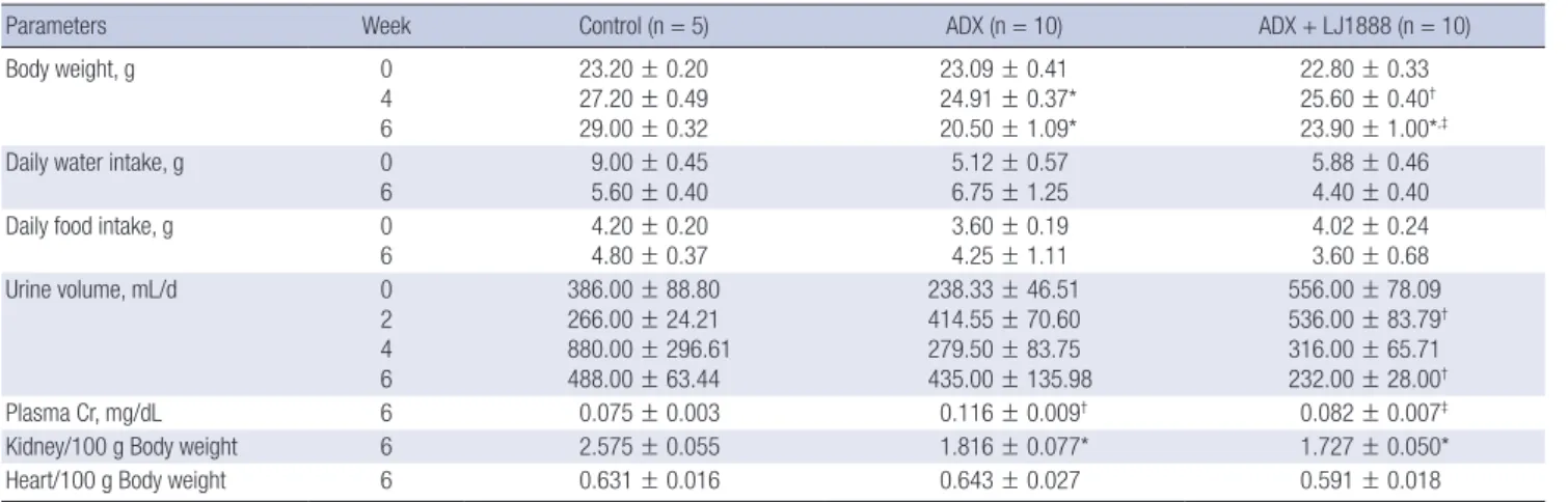
The physical and biochemical parameters for each group of the experimental animals are shown in Table 1. Injection of high dose ADX induced dehydration and cachexia in a previous ani- mal study (12). In our study, ADX administration inhibited wei- ght gain. Mice with ADX-induced nephropathy displayed a sig- nificant decrease in body weight compared to the control mice four weeks after an 11 mg/kg injection of ADX. At the time of sacrifice, mice in the ADX injection group had significantly low- er body weight than mice in the control group, and LJ1888 ad- ministration led to recovery of body weight lost during ADX treatment. The body weight of LJ1888-treated mice was signifi- cantly increased compared to that of ADX-injected mice at six weeks, despite no significant changes in daily water and food intake. Although urine volume tended to increase at two weeks and decrease at four weeks after ADX injection compared with control mice, there was no significant difference based on LJ1888 treatment. Kidney weight was significantly lower in ADX-induc- ed nephropathy than in the control group. However, LJ1888 treat- ment did not induce any significant change in kidney weight.
ADX increased the plasma creatinine level significantly, which was improved by LJ1888 treatment.
Effects of LJ1888 administration on urinary excretion of protein and albumin in ADX-induced nephropathy ADX leads to direct toxic injury of the glomerular filtration bar- rier including podocytes, the glomerular basement membrane and glomerular endothelial cells. Podocyte damage induces loss of protein and nephrin in the urine (13). In order to investi- gate the effects of LJ1888 on podocyte injury in ADX-induced nephropathy, we checked the urinary excretion of protein and
Table 1. Physical and biochemical parameters of experimental animals
Parameters Week Control (n = 5) ADX (n = 10) ADX + LJ1888 (n = 10)
Body weight, g 0
4 6
23.20 ± 0.20 27.20 ± 0.49 29.00 ± 0.32
23.09 ± 0.41 24.91 ± 0.37*
20.50 ± 1.09*
22.80 ± 0.33 25.60 ± 0.40† 23.90 ± 1.00*,‡
Daily water intake, g 0
6 9.00 ± 0.45
5.60 ± 0.40 5.12 ± 0.57
6.75 ± 1.25 5.88 ± 0.46
4.40 ± 0.40
Daily food intake, g 0
6
4.20 ± 0.20 4.80 ± 0.37
3.60 ± 0.19 4.25 ± 1.11
4.02 ± 0.24 3.60 ± 0.68
Urine volume, mL/d 0
2 4 6
386.00 ± 88.80 266.00 ± 24.21 880.00 ± 296.61 488.00 ± 63.44
238.33 ± 46.51 414.55 ± 70.60 279.50 ± 83.75 435.00 ± 135.98
556.00 ± 78.09 536.00 ± 83.79† 316.00 ± 65.71 232.00 ± 28.00†
Plasma Cr, mg/dL 6 0.075 ± 0.003 0.116 ± 0.009† 0.082 ± 0.007‡
Kidney/100 g Body weight 6 2.575 ± 0.055 1.816 ± 0.077* 1.727 ± 0.050*
Heart/100 g Body weight 6 0.631 ± 0.016 0.643 ± 0.027 0.591 ± 0.018
Values are expressed as (means ± SEM).
ADX, adriamycin; Cr, creatinine.
*P < 0.01; †P < 0.05 vs. control; ‡P < 0.05 vs. ADX.
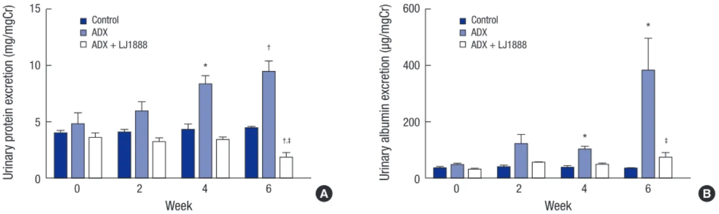
albumin. Urinary protein excretion was significantly increased at four and six weeks after ADX injection compared with the control group. LJ1888 treatment inhibited urinary protein ex- cretion during two weeks of treatment in ADX-induced neph- ropathy (Fig. 1A). In addition, urinary albumin excretion signifi- cantly increased four and six weeks after ADX injection com- pared with the control group, and significantly decreased after two weeks of LJ1888 treatment in ADX-induced nephropathy (Fig. 1B).
Effects of LJ1888 on oxidative stress and urinary nephrin excretion in ADX-induced nephropathy
The mechanism of ADX-induced nephropathy occurs in part through podocyte injury via oxidative stress. ADX exacerbates cellular damage in the renal cortex, inducing renal fibrosis (14).
Examination of urinary 8-isoprostane excretion and kidney lip- id peroxidation (LPO) concentration is an approach for diag- nostic assessment of oxidative stress in the kidney (15). In our study, the urinary level of 8-isoprostane markedly increased six weeks after ADX injection compared with the control group, and significantly decreased after two weeks of LJ1888 treatment
Fig. 1. Effects of LJ1888 on urinary excretion of protein and albumin in experimental ADX-induced nephropathy. (A) Twenty-four hour urinary protein excretion level. (B) Urinary excretion of albumin level. Urinary protein, albumin, and nephrin levels were corrected for urine creatinine levels.
Values are expressed as (means ± SEM).
ADX, adriamycin.
*P < 0.01; †P < 0.05 vs. control; ‡P < 0.001 vs. ADX.
Urinary protein excretion (mg/mgCr)
Week
0 2 4 6
15
10
5
0
*
†
†,‡
Control ADX ADX + LJ1888
Urinary albumin excretion (µg/mgCr)
Week
0 2 4 6
600
400
200
0
*
*
‡
Control ADX ADX + LJ1888
A B
Fig. 2. Effects of LJ1888 on oxidative stress in ADX-induced nephropathy. (A) Twen- ty-four hour urinary level of 8-isoprostane. Urinary 8-isoprostane levels were correct- ed for urine creatinine levels. (B) LPO concentration in the kidney tissue. LPO con- centration levels were corrected for kidney weight. (C) Urinary excretion of nephrin.
Values are expressed as (means ± SEM).
ADX, adriamycin; LPO, lipoperoxidase.
*P < 0.01 vs. control; †P < 0.001; ‡P < 0.01 vs. ADX.
Urinary 8-isoprostane excretion (pg/mgCr)
Control ADX ADX+LJ1888
4,000 3,000 2,000 1,000 0
*
*,†
Kidney LPO concentration (µM/mg tissue)
Control ADX ADX+LJ1888
8 6 4 2 0
*
‡
Urinary excretion of nephrin (µg/mgCr)
0 2 4 6
Week 10,000
8,000 6,000 4,000 2,000 0
†
* Control
ADX ADX + LJ1888
A B
C
(Fig. 2A). Likewise, LPO concentration in the kidney was signifi- cantly increased six weeks after ADX injection compared with the control group, but significantly decreased after LJ1888 treat- ment (Fig. 2B). In addition, we measured urinary nephrin ex- cretion to determine podocyte injury. As shown in Fig. 2C, each group showed no significant difference in urinary nephrin ex- cretion during four weeks after ADX administration. However, nephrin was markedly increased at six weeks after ADX injec- tion compared with the control group. Interestingly, LJ1888 treat- ment yielded the greatest inhibitory effects on urinary nephrin excretion.
Effects of LJ1888 on proinflammatory and profibrotic mechanisms in ADX-induced nephropathy
To determine whether LJ1888 has a protective effect in ADX ne- phropathy, we next investigated the mRNA expression of proin- flammatory and profibrotic molecules in renal cortical tissue.
Gene expression of profibrotic cytokines including type IV col- lagen, plasminogen activator inhibitor (PAI)-1, macrophage/
monocyte chemoattractant protein (MCP)-1, and NOX4 were remarkably increased in ADX-injected mice. On the other hand,
LJ1888 treatment significantly suppressed the expression of PAI-1, MCP-1, and NOX4. TGF-β1 mRNA levels in the renal cor- tex (Fig. 3A). Similarly, proinflammatory cytokines such as TLR4, tumor necrosis factor (TNFα), interleukin (IL)-1β, and interfer- on (IFN)-γ were highly expressed after ADX administration com- pared to the control group, and their mRNA expression was sig- nificantly suppressed after LJ1888 treatment. As expected, A3AR mRNA levels significantly increased after ADX injection, but did not change significantly after LJ1888 administration (Fig.
3B). Moreover, western blot analysis confirmed that the renal cortical expression of NF-κB, TGF-β1, and NOX4 were all mark- edly induced in ADX-injected mice. On the other hand, LJ1888 significantly reduced the expression of all these proteins. TLR4 did not differ among the three groups in the kidney (Fig. 3C).
Effects of LJ1888 on histomorphological changes in ADX- induced nephropathy
Fig. 4 shows representative renal pathology and immunohisto- chemical staining for each group of experimental animals at the time of sacrifice. PAS and Masson’s trichrome staining dem- onstrated that glomerular and tubulointerstitial damage includ-
Fig. 3. Effects of LJ1888 on inflammation and fibrosis in ADX-induced nephropathy. (A and B) Effects of LJ1888 on the expression of inflammatory and fibrotic cytokines. (C) Representative western blots of NF-κB, TGF-β1, NOX4, and TLR4 in the kidney and quantitative analysis scoring of western blot.
Values are expressed as (means ± SEM).
ADX, adriamycin; Col-4, type IV collagen; PAI-1, plasminogen activator inhibitor-1; MCP-1, macrophage/monocyte chemoattractant peptide-1; TGF-β1, transforming growth factor-β1; NOX-4, NADPH oxidase 4; TLR-4,Toll-like receptor 4; A3AR, A3 adenosine receptor; TNFα, tumor necrosis α; IL-1β, interleukine-1β; IFN-γ, interferon-γ; NF-κB, nu- clear factor-κB.
*P < 0.05; †P < 0.01 vs. control; ‡P < 0.05; §P < 0.001; llP < 0.01 vs. ADX.
mRNA expression of target genes (target gene/β-actin)
Col-4 PAI-1 MCP-1 TGF-β1 NOX4
20 15 10 5 0
†,‡
†
†
*
†
†,§
ll §
Control ADX ADX + LJ1888
†
mRNA expression of target genes (target gene/β-actin)
TLR4 A3AR TNFα IL-1β IFN-γ
15
10
5
0
†
†
† †
†
†,§ †,§
†,§ †,§
Control ADX ADX + LJ1888
†
Densitometric analysis of western blot (fold increase of control)
NF-kb TGFβ1 NOX4 TLR4 4
3 2 1 0
‡
*
*
*
*
Control ADX ADX + LJ1888
Control ADX ADX+LJ1888
NF-κB TBP
TGFβ1 β-actin
TLR4 β-actin NOX4 β-actin
A B
C
ing glomerulosclerosis, excessive collagen deposition, intersti- tial fibrosis, and tubular atrophy were dramatically increased in ADX-induced nephropathy. The glomeruli had much more se- vere pathological changes than the tubulointerstitium in ADX- induced nephropathy. ADX led to development of progressive glomerulosclerosis and demonstrated increased accumulation of type I collagen, type IV collagen, TGF-β1, and α-SMA in the glomeruli. In contrast, LJ1888 administration significantly ame-
liorated severe damage lesions of glomerulosclerosis. Infiltra- tion of inflammatory cells such as macrophages and T lympho- cytes in the renal glomeruli is an important pathological finding in the early progression of ADX-induced nephropathy (12). F4/80 is macrophage specific to the inflammatory process. F4/80 stain- ing demonstrated that macrophage infiltration in the glomeruli was markedly increased after ADX injection and dramatically recovered with LJ1888 treatment. Nephrin has important effects Fig. 4. Representative renal histological and immunohistochemical staining in experimental models at the time of sacrifice. Representative sections show that the histological changes and immunohistochemical staining for PAS, F4/80, Masson’s trichrome and α-SMA, nephrin, type I collagen, TGF-β1, and type IV collagen are increased in ADX-in- duced nephropathy and decreased after treatment with LJ1888. Original magnification × 400.
ADX, adriamycin; PAS, periodic acid-Schiff; α-SMA, α-smooth muscle actin; TGF-β1, transforming growth factor-β1.
PAS
ControlADXADX+LJ1888
α-SMA F4/80 Masson-trichrome
Nephrin
ControlADXADX+LJ1888
Type1 collagen TGF-β Type IV collagen
in controlling the integrity and functions of the podocyte slit di- aphragm structure (16). In a previous study, ADX induced di- rect toxic damage to the podocytes and loss of nephrin in urine (13). Immunohistochemical staining revealed that the numbers of nephrin-positive cells were dramatically decreased in the glomeruli of ADX-injected mice and were significantly restored by LJ1888 treatment. As shown in Fig. 5, quantitative analysis with immunohistochemical staining demonstrated those re- sults.
DISCUSSION
In the present study, we first demonstrated that a highly selec- tive A3AR antagonist reduced proteinuria and albuminuria, improved renal function, and recovered histomorphological changes that occurred during ADX-induced nephropathy. We concluded that this selective A3AR antagonist had a notable ef- fect on urinary nephrin excretion and its glomerular expres- sion, which may prevent podocyte injury. Clinically, protein- uria is the most characteristic manifestation of podocyte injury in kidney disease. We used ADX to develop the renal glomeru- lar injury. ADX induces proteinuria and nephrin loss in urine by injuring the glomerular filtration barrier, decreasing glomer- ular endothelial cell thickness and glomerular charge selectivity and increasing podocyte foot process effacement (17,18). The
Fig. 5. Quantitation of immunohistochemical staining. (A) Glomerulosclerosis index, (B) F4/80 positive cell staining score, (C) Tubulointerstitial fibrosis index, (D) Quantitation of immunohistochemical staining of nephrin, type I Collagen, TGF-β1 and Type 4 collagen.
Values are expressed as (means ± SEM).
ADX, adriamycin.
*P < 0.01 vs. control; †P < 0.001; ‡P < 0.01 vs. ADX.
Glomerulosclerosis index
Control ADX ADX + LJ1888
2.0 1.5 1.0 0.5 0
*
*,†
Tubulointerstitial fibrosis index
Control ADX ADX + LJ1888
1.5
1.0
0.5
0
*
*,‡
F4/80 (+) cells/HPF
Control ADX ADX + LJ1888
80 60 40 20 0
*
*,†
Quantitation of immunohistochemical staining
Nephrin Type I Col TGFβ1 Type IV Col 2.5
2.0 1.5 1.0 0.5 0
*
*,†
* *
*,†
*,† *,†
Control ADX ADX + LJ1888
*
A B
C D
renal histological changes due to ADX appear about one week after ADX administration and degenerate into severe injury by four weeks (19).
Adenosine has various effects in the kidneys, including the regulation of renin secretion, glomerular filtration rate, renal vascular tone, tubular glomerular feedback, fluid and electro- lyte transport of the tubular and collecting duct system, and metabolic renal function. The concentration of extracellular adenosine is maintained at approximately 100 nM under nor- mal conditions and rapidly increases during renal cell damage (20). A previous study demonstrated that long-term exposure to high concentrations of adenosine induced secretion of intra- cellular proinflammatory cytokines, activation of fibroblasts, and deposition of collagen in the extracellular matrix (21). A re- cent study showed that an increase in adenosine levels led to ischemic renal fibrosis and finally induced renal interstitial fi- brosis in renal cell damage in the unilateral ureteral obstruction model (22).
Activation of A3AR by adenosine leads to a decrease in ade- nylyl cyclase activity and cAMP production, which induce stim- ulation of phospholipase C (PLC) activity, Ca2+ mobilization, and phosphorylation of phosphatidylinositol 3-kinase (PI3K) and AKT. AKT modulates the mitogen-activated protein kinase (MAPK) family including c-JUN N-terminal kinase (JNK), extra- cellular signal-regulated kinase (ERK), and p38 MAPK (6). There-
fore, A3AR activation stimulates p38 MAPK phosphorylation, leading to decreased hypoxic damage of cardiomyocytes (23).
On the other hand, A3AR activation suppresses the p38 MAPK pathway, leading to down-regulation of the inflammatory sta- tus in human synoviocytes (24). A3AR has different effects in different cell types, such as cell proliferation and cell death, which may be related to PI3K/AKT and ERK1/2 pathway crosstalk (25).
Many studies have shown the biologic functions and therapeu- tic applications of A3AR and A3AR antagonists in the brain, heart, lung, and immune system (6).
LJ1888 dramatically suppressed the urinary excretion of neph- rin. In immunohistochemical staining, the numbers of neph- rin-positive cells were dramatically decreased by ADX and sig- nificantly restored by LJ1888 treatment. Nephrin is one of the molecules that constitute the podocyte slit diaphragm and it is involved the maturation of podocytes during glomerular devel- opment (26). Nephrin induces phosphorylation and activation of the PI3K-dependent AKT downstream signaling pathway.
Several AKT targets bind to the phosphorylation-dependent 14-3-3 proteins, which are involved in the control of cell growth, migration, proliferation, and apoptosis. Nephrin-induced PI3K/
AKT signaling plays a preventative role in podocyte apoptosis (27). A3AR transmits a cellular proliferation signal through the PI3K/AKT signaling pathway. Stimulated AKT is able to phos- phorylate many downstream substrates such as Raf. Therefore, by blocking Raf through AKT activation and inducing cell death, A3AR inhibits the MAPK pathway, including RAS, Raf, MEK1/2, and ERK1/2 (25). A3AR gene expression in renal cortical tissue was dramatically increased in ADX-induced nephropathy. Al- though LJ1888 did not suppress A3AR gene expression, LJ1888 inhibited A3AR-associated PI3K/AKT and RAS/RAF/MEK/ERK downstream pathways, protecting podocytes from apoptosis.
Additional studies are required to demonstrate the interaction of A3AR and nephrin downstream pathways.
There is a considerable body of evidence regarding oxidative stress in various kidney diseases. In ADX-induced nephropa- thy, oxidative stress and inflammation are important mecha- nisms of progressive proteinuric nephropathy leading to renal fibrosis (13). Renal tissue hypoxia induces the elevation of ade- nosine levels, leading to renal fibrosis (22). Oxidative stress in- duces stimulation of TGF-β1 expression and production of ex- tracellular matrix, leading to glomerular and tubulointerstitial fibrosis. The MAPK family signaling is activated by oxidative stress in renal cells. The JNK pathway is associated with pro- gression of renal fibrosis and tubular apoptosis (28). Stimula- tion of the ERK pathway is related to proliferative glomerulone- phritis (29). Activation of the p38 MAPK pathway by IL-1, TNF-α, and TFG-β1 induces extracellular matrix accumulation, leading to tubulointerstitial fibrosis (30). A previous study demonstrat- ed that LJ1888 significantly suppressed JNK and ERK phosphor- ylation during TGF-β1 upregulation in the kidney (9). In this
study, we observed that LJ1888 treatment attenuated the in- creased urinary excretion of 8-isoprostane and the increased LPO concentration in kidney tissue after ADX injection.
In addition, mRNA expression of proinflammatory molecules including TLR4, TNFα, IL-1β, and IFN-γ increased in ADX-in- duced nephropathy, and dramatically recovered during LJ1888 treatment. The TLR4/NK-κB pathway is considered to be an important link between inflammation and oxidative stress in kidney disease (31,32). Gene expression of profibrotic mole- cules including type IV collagen, PAI-1, MCP-1, TGF-β1, and NOX4, as wells as NF-κB protein, were increased by ADX and significantly decreased by LJ1888. Renal histological and im- munohistochemical staining demonstrated similar results con- sistently. Type I collagen, type IV collagen, TGF-β1, and α-SMA accumulation in the fibrotic area of the renal glomeruli were increased in ADX-induced nephropathy, as was macrophage infiltration. Those profibrotic and proinflammatory molecules were genetically suppressed significantly by LJ1888 administra- tion. The extensive glomerular and tubulointerstitial damage, including glomerulosclerosis, excessive collagen deposition, interstitial fibrosis, and tubular atrophy, were dramatically in- creased in ADX-induced nephropathy. In contrast, LJ1888 ad- ministration significantly ameliorated severe damage lesions in both the glomerular and tubular compartments. We suggest that LJ 1888 ameliorated oxidative stress, inflammation, and fi- brosis in ADX-induced nephropathy through antioxidative me- chanisms via A3AR.
In summary, we are the first to show that LJ1888, a highly se- lective A3AR antagonist, exhibits renoprotective effects by ame- liorating podocyte injury, oxidative stress, inflammation, glo- merulosclerosis, and tubulointerstitial fibrosis in ADX-induced nephropathy. Treatment with LJ1888 induced renoprotective effects by attenuating podocyte injury through decreased uri- nary excretion of nephrin. Therefore, we suggest that LJ1888 could be a new therapeutic drug for proteinuric renal disease.
We recommend further study of LJ1888 in progressive renal dis- ease to demonstrate its therapeutic effect more clearly.
DISCLOSURE
The authors have no potential conflicts of interest to disclose.
AUTHOR CONTRIBUTION
Research conception & design: Min HS, Cha JJ, Kang YS. Per- forming the experiments: Min HS, Kim JE, Ghee JY, Jeong LS.
Data acquisition: Kim JE, Min HS, Cha JJ, Cha DR. Data analysis and interpretation: Han JY, Cha DR, Kang YS. Statistical analy- sis: Min HS, Cha JJ, Kang YS. Drafting of the manuscript: Min HS, Cha JJ. Critical revision of the manuscript: Min HS, Cha JJ, Kim HW, Lee JE, Kang YS. Receiving grant: Kang YS, Cha DR.
Approval of final manuscript: all authors.
ORCID
Hye Sook Min http://orcid.org/0000-0002-9576-6054 Jin Joo Cha http://orcid.org/0000-0001-6779-0113 Kitae Kim http://orcid.org/0000-0003-2433-1747 Jung Eun Kim http://orcid.org/0000-0002-5260-7563 Jung Yeon Ghee http://orcid.org/0000-0002-5817-237X Hyunwook Kim http://orcid.org/0000-0002-4274-7562 Ji Eun Lee http://orcid.org/0000-0002-8299-4816 Jee-Young Han http://orcid.org/0000-0003-3224-4029 Lak Shin Jeong http://orcid.org/0000-0002-3441-707X Dae Ryong Cha http://orcid.org/0000-0003-0063-2844 Young Sun Kang http://orcid.org/0000-0002-4061-386X REFERENCES
1. Vallon V, Mühlbauer B, Osswald H. Adenosine and kidney function. Physi
ol Rev 2006; 86: 901-40.
2. Hirschhorn R, Roegner-Maniscalco V, Kuritsky L, Rosen FS. Bone marrow transplantation only partially restores purine metabolites to normal in adenosine deaminase-deficient patients. J Clin Invest 1981; 68: 1387-93.
3. Cronstein BN, Naime D, Firestein G. The antiinflammatory effects of an adenosine kinase inhibitor are mediated by adenosine. Arthritis Rheum 1995; 38: 1040-5.
4. Jacobson KA, Gao ZG. Adenosine receptors as therapeutic targets. Nat Rev Drug Discov 2006; 5: 247-64.
5. Brandon CI, Vandenplas M, Dookwah H, Murray TF. Cloning and phar- macological characterization of the equine adenosine A3 receptor. J Vet Pharmacol Ther 2006; 29: 255-63.
6. Borea PA, Varani K, Vincenzi F, Baraldi PG, Tabrizi MA, Merighi S, Gessi S.
The A3 adenosine receptor: history and perspectives. Pharmacol Rev 2015; 67: 74-102.
7. Lee HT, Emala CW. Protective effects of renal ischemic preconditioning and adenosine pretreatment: role of A(1) and A(3) receptors. Am J Physi
ol Renal Physiol 2000; 278: F380-7.
8. Lee HT, Ota-Setlik A, Xu H, D’Agati VD, Jacobson MA, Emala CW. A3 ad- enosine receptor knockout mice are protected against ischemia- and myo- globinuria-induced renal failure. Am J Physiol Renal Physiol 2003; 284:
F267-73.
9. Lee J, Hwang I, Lee JH, Lee HW, Jeong LS, Ha H. The selective A3AR an- tagonist LJ-1888 ameliorates UUO-induced tubulointerstitial fibrosis. Am J Pathol 2013; 183: 1488-97.
10. Jeong LS, Choe SA, Gunaga P, Kim HO, Lee HW, Lee SK, Tosh DK, Patel A, Palaniappan KK, Gao ZG, et al. Discovery of a new nucleoside template for human A3 adenosine receptor ligands: D-4’-thioadenosine deriva- tives without 4’-hydroxymethyl group as highly potent and selective an- tagonists. J Med Chem 2007; 50: 3159-62.
11. Lee MH, Song HK, Ko GJ, Kang YS, Han SY, Han KH, Kim HK, Han JY, Cha DR. Angiotensin receptor blockers improve insulin resistance in type 2 diabetic rats by modulating adipose tissue. Kidney Int 2008; 74: 890-900.
12. Wang Y, Wang YP, Tay YC, Harris DC. Progressive adriamycin nephropa-
thy in mice: sequence of histologic and immunohistochemical events.
Kidney Int 2000; 58: 1797-804.
13. Szalay CI, Erdélyi K, Kökény G, Lajtár E, Godó M, Révész C, Kaucsár T, Kiss N, Sárközy M, Csont T, et al. Oxidative/nitrative stress and inflammation drive progression of doxorubicin-induced renal fibrosis in rats as revealed by comparing a normal and a fibrosis-resistant rat strain. PLoS One 2015;
10: e0127090.
14. Deman A, Ceyssens B, Pauwels M, Zhang J, Houte KV, Verbeelen D, Van den Branden C. Altered antioxidant defence in a mouse adriamycin mod- el of glomerulosclerosis. Nephrol Dial Transplant 2001; 16: 147-50.
15. Montuschi P, Barnes PJ, Roberts LJ 2nd. Isoprostanes: markers and medi- ators of oxidative stress. FASEB J 2004; 18: 1791-800.
16. Machado JR, Rocha LP, Neves PD, Cobô Ede C, Silva MV, Castellano LR, Corrêa RR, Reis MA. An overview of molecular mechanism of nephrotic syndrome. Int J Nephrol 2012; 2012: 937623.
17. Jeansson M, Björck K, Tenstad O, Haraldsson B. Adriamycin alters glo- merular endothelium to induce proteinuria. J Am Soc Nephrol 2009; 20:
114-22.
18. Fujimura T, Yamagishi S, Ueda S, Fukami K, Shibata R, Matsumoto Y, Kai- da Y, Hayashida A, Koike K, Matsui T, et al. Administration of pigment ep- ithelium-derived factor (PEDF) reduces proteinuria by suppressing de- creased nephrin and increased VEGF expression in the glomeruli of adri- amycin-injected rats. Nephrol Dial Transplant 2009; 24: 1397-406.
19. Lee VW, Harris DC. Adriamycin nephropathy: a model of focal segmen- tal glomerulosclerosis. Nephrology (Carlton) 2011; 16: 30-8.
20. Vallon V, Osswald H. Adenosine receptors and the kidney. In: Wilson CN, Mustafa SJ, editors. Adenosine Receptors in Health and Disease. Dordre- cht: Springer, 2009, p443-70.
21. Dai Y, Zhang W, Wen J, Zhang Y, Kellems RE, Xia Y. A2B adenosine recep- tor-mediated induction of IL-6 promotes CKD. J Am Soc Nephrol 2011;
22: 890-901.
22. Tang J, Jiang X, Zhou Y, Xia B, Dai Y. Increased adenosine levels contrib- ute to ischemic kidney fibrosis in the unilateral ureteral obstruction mod- el. Exp Ther Med 2015; 9: 737-43.
23. Leshem-Lev D, Hochhauser E, Chanyshev B, Isak A, Shainberg A. Ade- nosine A(1) and A (3) receptor agonists reduce hypoxic injury through the involvement of P38 MAPK. Mol Cell Biochem 2010; 345: 153-60.
24. Varani K, Vincenzi F, Tosi A, Targa M, Masieri FF, Ongaro A, De Mattei M, Massari L, Borea PA. Expression and functional role of adenosine recep- tors in regulating inflammatory responses in human synoviocytes. Br J Pharmacol 2010; 160: 101-15.
25. Merighi S, Benini A, Mirandola P, Gessi S, Varani K, Leung E, Maclennan S, Baraldi PG, Borea PA. Modulation of the Akt/Ras/Raf/MEK/ERK path- way by A3 adenosine receptor. Purinergic Signal 2006; 2: 627-32.
26. Doné SC, Takemoto M, He L, Sun Y, Hultenby K, Betsholtz C, Tryggvason K. Nephrin is involved in podocyte maturation but not survival during glomerular development. Kidney Int 2008; 73: 697-704.
27. Huber TB, Hartleben B, Kim J, Schmidts M, Schermer B, Keil A, Egger L, Lecha RL, Borner C, Pavenstädt H, et al. Nephrin and CD2AP associate with phosphoinositide 3-OH kinase and stimulate AKT-dependent sig- naling. Mol Cell Biol 2003; 23: 4917-28.
28. Ma FY, Flanc RS, Tesch GH, Han Y, Atkins RC, Bennett BL, Friedman GC, Fan JH, Nikolic-Paterson DJ. A pathogenic role for c-Jun amino-terminal kinase signaling in renal fibrosis and tubular cell apoptosis. J Am Soc Ne
phrol 2007; 18: 472-84.
29. Pat B, Yang T, Kong C, Watters D, Johnson DW, Gobe G. Activation of ERK in renal fibrosis after unilateral ureteral obstruction: modulation by anti- oxidants. Kidney Int 2005; 67: 931-43.
30. Stambe C, Atkins RC, Tesch GH, Masaki T, Schreiner GF, Nikolic-Paterson DJ. The role of p38α mitogen-activated protein kinase activation in renal fibrosis. J Am Soc Nephrol 2004; 15: 370-9.
31. Min HS, Kim JE, Lee MH, Song HK, Lee MJ, Lee JE, Kim HW, Cha JJ, Hyun
YY, Han JY, et al. Effects of Toll-like receptor antagonist 4,5-dihydro-3-phe- nyl-5-isoxasole acetic acid on the progression of kidney disease in mice on a high-fat diet. Kidney Res Clin Pract 2014; 33: 33-44.
32. Mkaddem SB, Bens M, Vandewalle A. Differential activation of toll-like receptor-mediated apoptosis induced by hypoxia. Oncotarget 2010; 1:
741-50.
Supplementary Table 1. Primer sequences for real-time quantitative PCR
Target genes Primer sequences (´5-´3)
TLR4, forward GGGAACAAACAGCCTGAGAC
TLR4, reverse AGACCCATGAAATTGGCA T
TNF-α, forward CCGATGGGTTGTACCTTGTC
TNF-α, reverse GGCAGAGAGGAGGTTGACTTT
IL-1β, forward CTC ACA AGC AGA GCA CAA GC
IL-1β, reverse ACG GAT TCC ATG GTG AAG TC
MCP-1, forward CTGGATCGGAACCAAATGAG
MCP-1, reverse CGGGTCAACTTCACATTCAA
PAI-1, forward TCCTCATCCTGCCTAAGTTCTC
PAI-1, reverse GTGCCGCTCTCGTTTACCTC
TGF-β1, forward AGCCCGAAGCGGACTACTAT
TGF-β1, reverse CTGTGTGAGATGTCTTTGGTTTTC
Col-IV, forward GCTCTGGCTGTGGAAAATGT
Col-IV, reverse CTTGCATCCCGGGAAATC
A3AR, forward CTCTTCTTGTTTGCGCTGTG
A3AR, reverse GCACATTGCGACATCTGGT
IFN-γ, forward GCGTCATTGAATCACACCTG
IFN-γ, reverse TGAGCTCATTGAATGCTTGG
NOX4, forward TTGGTGAATGCCCTCAACTT
NOX4, reverse TTCTGGGATCCTCATTCTGG
β-actin, forward GGACTCCTATGTGGGTGACG
β-actin, reverse CTTCTCCATGTCGTCCCAGT
In this experiment, each sample was run in triplicate, and the corresponding non-re- verse transcribed mRNA samples were used as negative controls. The mRNA level of each sample was normalized to that of β-actin mRNA.
TLR4, Toll like receptor 4; TNF-α, Tumor necrosis factor-α; IL-1, interleukin-1; MCP- 1, monocyte chemoattractant peptide-1; PAI-1, plasminogen activator inhibitor-1; TGF- β1, transforming growth factor; Col-IV, type IV collagen; A3AR, A3 adenosine recep- tor; IFN-γ, interferon; NOX4, NADPH oxidase 4.