Bone Cement Augmentation Procedures for Spinal Pathologic Fractures by Multiple Myeloma
Efficacy and safety of bone cement augmentations for spinal pathologic fractures related to multiple myeloma, and usefulness of radionuclide studies for surgical decision were retrospectively evaluated. Forty eight vertebrae from 27 patients for bone cement augmentation procedures and 48 vertebrae from 29 patients for conservative treatment were enrolled. Clinical results using visual analogue scale (VAS) and Oswestry disability index (ODI), and radiologic results were assessed. For clinical decisions on treatment of spinal pathologic fracture, bone scan or single photon emission computed tomography was done for 20 patients who underwent surgery. Mean follow-up was 16.8 months. In terms of clinical results, immediate pain relief was superior in the operated group to that in the conservative group. ODI, maintenance of vertebral height and local kyphotic angle at the last follow-up were superior in the operated group in comparison to the conservative group. At one year follow-up, cumulative survival rate were 77.4% and 74.7% in the operated and conservative groups, respectively (log rank test > 0.05). Leakage of bone cement was noted at 10 treated vertebrae. Bone cement augmentations presented short- term pain relief for spinal pathologic fractures by myeloma with relative safety in highly selected patients, and radionuclide imaging studies were useful for the surgical decision on these procedures.
Keywords: Multiple Myeloma; Spine; Fractures, Spontaneous; Bone Cements Kee-Yong Ha,1 Chang-Ki Min,2
Jun-Yeong Seo,3 Young-Hoon Kim,1 Joo-Hyun Ahn,1 Nak-Min Hyun,1 and Yoon-Chung Kim1
Departments of 1Orthopedic Surgery and 2Hematology, Seoul St. Mary’s Hospital, College of Medicine, The Catholic University of Korea, Seoul; 3Department of Orthopedic Surgery, Jeju National University Hospital, Jeju, Korea
Received: 15 May 2014 Accepted: 25 July 2014 Address for Correspondence:
Young-Hoon Kim, MD
Department of Orthopedic Surgery, Seoul St. Mary’s Hospital, College of Medicine, The Catholic University of Korea, 222 Banpo-daero, Seocho-gu, Seoul 137-701, Korea Tel: +82.2-2258-6118, Fax: +82.2-505-9834 E-mail: boscoa@catholic.ac.kr
http://dx.doi.org/10.3346/jkms.2015.30.1.88 • J Korean Med Sci 2015; 30: 88-94
INTRODUCTION
Multiple myeloma, one of hematologic malignancies, is known to have a high chance of skeletal related events (SREs) from its pathologic characteristics (1, 2). Direct and/or indirect activa- tion of osteoclast, and suppression of osteoblast proliferation are related to these SREs (3-5). Among these SREs, involvement of the spinal column predispose to pathologic fracture and neu- rologic complications.
In recent time, minimally invasive stabilization using bone cement has been demonstrated to be a safe and effective sup- portive option for spinal pathologic fractures without neurolog- ic involvement (6, 7). Other treatment modalities such as radio- therapy, bisphosphonates and supportive treatment are also tried as adjuvant therapies (8, 9).
In our institute, each patient with this problem is addressed by a multi-disciplinary team consisting of the hematologist, ra- dio-oncologist and spine surgeon for optimal treatment strate- gy. Our strategy is similar to the suggestion of Kasperk et al. (7).
However, in our clinical pathway, radiotherapy is only consid- ered in the patient with the spinal pathologic fracture and neu- rologic involvements by epidural myeloma. In addition, radio- nuclide studies have been tried for the decision of surgically aug-
mented levels. Because most patients presented with multi-lev- el fractures, the authors tried to select the most mechanically unstable and symptomatic levels using image studies including a radionuclide study and clinical symptoms.
This retrospective study was designed to evaluate clinical and radiologic results of bone cement augmentation procedures as an additional treatment modality for spinal pathologic fractures by multiple myeloma.
MATERIALS AND METHODS Study population
Patients who suffered from pathological spinal fractures in con- junction with multiple myeloma were included in this retrospec- tive study. Diagnosis of multiple myeloma was made according to the guidelines and recommendation of International Myelo- ma Working Group. Treatments including systemic chemother- apy, bone marrow transplantation, and general supportive ther- apy were mainly performed according to the guidelines of the Intenational myeloma foundation in both group patients (10, 11). During 2009-2011, 27 patients who had undergone bone cement augmentation procedures with minimum 6 months follow-up were included as the surgery group (group I). Clinical
and radiological results were compared with 29 patients who had not undergone surgery, and treated conservatively as the non-surgery group (group II).
Evaluation and treatment protocol of pathologic spinal fracture
Mechanical back pain relieved by rest was the main symptom in these enrolled patients. All pathologic fractures were initially evaluated with plain radiographs and magnetic resonance im- ages (MRIs). Involvement of posterior element or epidural ex- tensions of myeloma were also evaluated. As candidates for bone cement augmentation procedure, unremitting back pain with visual analogue scale (VAS) ≥ 5 after supportive treatments were considered. For the patients who were supposed to un- dergo surgery, bone scan or single photon emission computed tomography (SPECT) was evaluated. The findings of these ra- dionuclide studies were taken into account for selection of the level (Fig. 1). The location of fractured vertebrae was also con- sidered. Mechanically demanding levels such as the thoraco- lumbar junction or lumbar level were recommended for bone cement augmentation procedures. Non-surgical treatment is more likely recommended for fractures of the mid-thoracic lev- el. For the group II, patient’s wish was one of the reasons of non- surgical treatment. General pain control and tolerable range ambulation with brace (tailor-made hard corsets) were recom- mended for the group II. Bisphosphonates were prescribed in
all patients. Wearing of the brace was basically recommended for 3 months after initial diagnosis, and as required. For the group I, vertebroplasty or kyphoplasty was performed under local anesthesia with a mild sedation. In all surgery cases, bipe- dicular approach was used, and the bone cement was introduc- ed (mean injected cement amount 4.3 mL/level). For the ky- phoplasty, two balloon catheters (Kyphon®/Medtronic, Mem- phis, TN, USA) were used to make void, and to restore the height of the fractured vertebra. In choosing vertebroplasty or kypho- plasty, in the case of severe height loss (more than 50%) and suspicious destruction of posterior wall of the vertebral body, kyphoplasty was preferred. Postoperatively, leakage of bone ce- ment was evaluated with postoperative CT scans.
Assessment of clinical outcomes
Basic demographics including the level of fractured vertebrae and the stage of myeloma were recorded and compared. Dur- ing the follow-up periods, mortality was also evaluated. Clinical outcomes for back problem caused by pathologic spinal frac- tures were evaluated with VAS (0-10) and Oswestry disability index (ODI). Each parameter was questioned and recorded at baseline, 1 month and the last follow-up periods. Complications related to the surgery were evaluated.
Assessment of radiomorphological results
All radiological parameters were measured by two senior or-
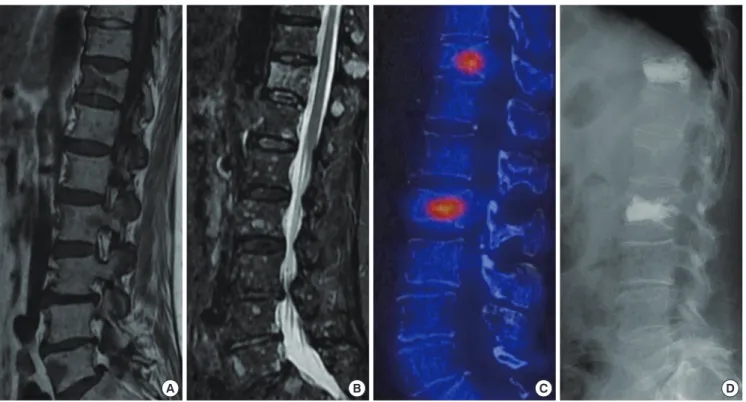
A B C D
Fig. 1. A 70-yr-old woman was diagnosed as multiple myeloma stage III b by laboratory tests and bone marrow biopsy. T1-weighted (A) and fat-suppression MRIs (B) show pathologic fracture at the T12 with multiple nodular signal changes involving multiple vertebrae. Single positron emission CT scan (C) shows abnormal hot uptake at the T12 and L2 vertebrae. Kyphoplasty was performed (D).
thopedic residents who were blinded to the purpose of this study and clinical results. Height loss and local kyphotic angle of the affected vertebral body were measured using initial, postopera- tive 1 month and the last follow-up radiographs. Vertebral height was measured at the point of maximal collapse of the affected vertebral body and height loss (%) was calculated with a formu- la of ([Lower vertebral height +Upper vertebral height]/2 – af- fected vertebral height)/([Lower vertebral height +Upper verte- bral height]/2) × 100. Local kyphotic angle (°) was measured with Cobb’s method between adjacent vertebrae of the affected vertebra (14). Bone cement leakage was evaluated and classi- fied into epidural, paravertebral and discal pattern of leakage using postoperative CT scans. The ratio of the number of posi- tive vertebrae on radionuclide scans to the number of affected vertebrae on plain radiographs and MRIs was calculated.
Statistical analysis
Data with continuance variables which included age, VAS, ODI and radiographic measurements were analyzed by t-test. Intra- and interobserver reliability for radiological parameters were assessed using Cohen’s kappa statistics. Cumulative survival rate and comparison between the groups were analyzed by Ka- plan-Meier analysis and log-rank test. All statistical analyses were conducted by using Statistical Product and Service Solu- tions (SPSS) version 13.0. A value of P < 0.05 was considered to be statistically significant.
Ethics statement
This study was retrospectively done using medical and radio- logical data in a single institute. The study design was approved by the institutional review board (Approval No. KC13RISI0373 of Seoul St. Mary’s Hospital, The Catholic University of Korea).
RESULTS
General demographics of both groups were summarized in Ta- ble 1. There were no significant differences in age, initial VAS
and ODI, and initial height loss or kyphotic angle. Forty eight vertebrae were augmented in 27 patients of the group I. Kypho- plasty was used in 19 patients. The augmented and affected levels of each group were presented in Fig. 2. During a mean of 16.8 months (range 6-33) follow-ups of the enrolled patients, 20 patients (11 in the group I and 9 in the group II) died from com- plications of multiple myeloma (mainly infection including pneumonia, and cerebral vascular accident). One year follow- up cumulative survival rate were 77.4% and 74.7% in the group I and II, respectively. There was no difference in survival rate between two groups (log rank test, P = 0.94). During the follow- up, other SREs besides spinal fracture had occurred in 3 patients.
Subsequent fractures of the spine were noted in 10 patients (6 patients in the group I and 4 in the group II, respectively). Addi- tional cement augmentation procedure has not been perform- ed in all patients.
Clinical outcomes
Even though the baseline VAS and ODI did not show a signifi- cant difference between two groups, 1 month postoperative VAS of the group I (3.2 ± 0.8) was significantly lower than that of the group II (6.1 ± 0.9) (P < 0.05). At the last follow-up, the patients did not report a significant difference in the intensity of back pain (VAS; 3.5 ± 1 in the group I and 4 ± 0.9 in the group II, re- spectively, P = 0.69). In terms of ODI, the scores at 1 month fol- low-up were 54.9% ± 9.8% in the group I and 72.8% ± 6.8% in the group II, which showed a statistical difference (P < 0.01).
Moreover, these difference persisted until the last follow-ups (ODI; 56.1% ± 9.5% and 60.2% ± 5.9% in the group I and II, re- spectively, P < 0.05) (Fig. 3).
Radiomorphological results
Mean Inter- and intra-observer kappas for radiomorphological parameters were 0.59 (SD, 0.07) and 0.55 (SD, 0.08), respective-
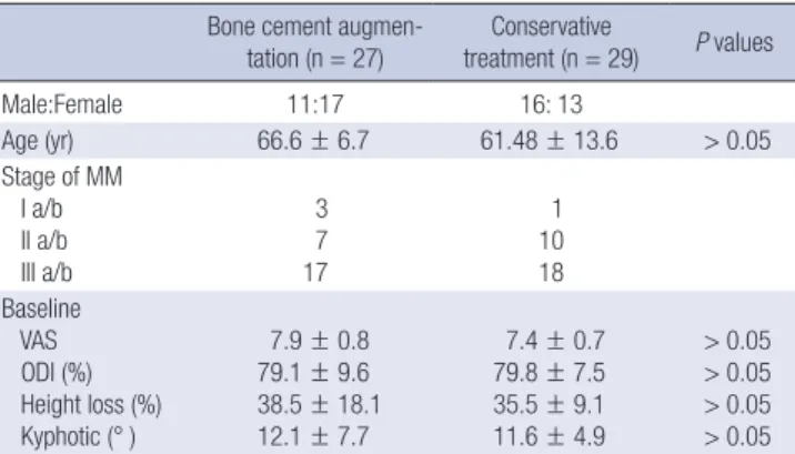
Table 1. Baseline demographics of the enrolled patients Bone cement augmen-
tation (n = 27) Conservative
treatment (n = 29) P values
Male:Female 11:17 16: 13
Age (yr) 66.6 ± 6.7 61.48 ± 13.6 > 0.05
Stage of MM I a/b II a/b III a/b
3 7 17
1 10 18 Baseline
VAS ODI (%) Height loss (%) Kyphotic (° )
7.9 ± 0.8 79.1 ± 9.6 38.5 ± 18.1 12.1 ± 7.7
7.4 ± 0.7 79.8 ± 7.5 35.5 ± 9.1 11.6 ± 4.9
> 0.05
> 0.05
> 0.05
> 0.05 MM, multiple myeloma; VAS, visual analogue scale; ODI, Oswestry disability index.
Fig. 2. Distribution of the affected vertebrae in this study. D indicates the dorsal spine.
L indicates the lumbar spine.
No. of vertebrae
D5 D6 D7 D8 D9 D10 D11 D12 L1 L2 L3 L4 L5 12
10 8 6 4 2 0
Operated group Conservative group
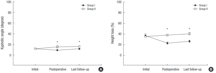
ly. Ten patients suffered from subsequent fractures following the surgery or initial diagnosis for spinal pathologic fractures during the follow-up period. Three patients showed subsequent fractures just adjacent to the previously affected segment. How- ever, subsequent fractures occurred at skipped segments in 7 patients. Even though baseline height loss (group I, 38.5% ± 18.1%; group II, 36% ± 9.1%) and kyphotic angle (group I, 12.2°
± 7.7°; group II, 12° ± 4.9°) of the affected vertebrae did not show significant differences, restoration of the vertebral height and local kyphosis were successfully achieved through augmenta- tion procedures. Moreover, the restoration of height (27.6% ± 12.1%) and local kyphosis (11.7° ± 5.5°) in the group I were su- perior to that (height loss, 40.6% ± 11.8%; kyphotic angle, 16° ± 5.5°) of the group II at the last follow-up (Fig. 4). Among the 48 augmented vertebrae, cement leakage was noted in 10 verte- brae (20.8%) on postoperative CT scans. Leakage into the adja-
cent disc was the most frequent pattern (5 vertebrae). Leakages into the paravertebral (4 vertebrae) and the epidural (1 verte- bra) spaces were also noted. In 20 patients (71%) of the group I, radionuclide scans including bone scan and SPECT were done.
Fifty eight vertebrae were diagnosed as a pathologic fracture with the evidence of morphological change at plain radiographs and signal change at MRIs. Among 58 vertebrae, 50 vertebrae (86.2%) showed abnormal uptake at the radionuclide images.
Complications related to the surgery
Even though bone cement leakage occurred in 10 out of 49 aug- mented vertebrae, there was no leakage leading to clinical symp- toms or revision surgery. No other complications related to the surgery such as infection, embolism, bleeding or neurologic deficit occurred in any patient.
Fig. 3. Clinical results. (A) Pain reduction at 1 month postoperatively is superior to that of the conservative treatment. (B) At the 1 month and last follow-up, the scores of ODI show a significant difference between the two groups (Group I indicates surgery group, group II indicates supportive treatment, *indicates statistically significant differences).
Visual analogue scale
Initial Postoperative Last follow-up 10
8 6 4 2 0
*
Group I Group II
Group I Group II
Oswestry disability index (%)
Initial Postoperative Last follow-up 100
90 80 70 60 50 40 30
*
*
A B
Fig. 4. Radiomorphological results. The maintenance of local kyphosis (A) and restoration of height loss (B) are superior in the surgery group to those in the conservative treat- ment. And these results were also maintained at the last follow-ups (Group I indicates surgery group, group II indicates supportive treatment, *indicates statistically significant differences).
Kyphotic angle (degree)
Initial Postoperative Last follow-up 100
80 60 40 20 0
* *
Group I Group II
Group I Group II
Height loss (%)
Initial Postoperative Last follow-up 100
80 60 40 20 0
* *
A B
DISCUSSION
Multiple myeloma, one of hematologic malignancies is known to have high incidence of skeletal related events such as patho- logic fracture or spinal cord compression by epidural myeloma (2, 13, 14). As myelomatous cells have a predilection for bone and peculiar characteristics on host bone, osteolytic lesion and subsequent mechanical failure of the involved bone are some of the major clinical manifestations of multiple myeloma (3-7).
Among these SREs, spinal pathologic fractures are the most common problem. Decreased mobility, neurologic complica- tions, kyphotic posture and respiratory compromise are subse- quent problems following multiple spinal fractures besides pain due to mechanical failure. Bisphosphonates and radiotherapy have been tried to reduce and treat the clinical manifestations related to SREs in addition to systemic therapy for multiple my- eloma (8, 16).
Surgical interventions also have roles in the spinal involve- ment of multiple myeloma. However, surgical interventions are limited by the general conditions of the patients, osteoporotic conditions of the bone and multiplicity of the lesions. There- fore, open stabilization and/or decompression is reserved and considered only for the patients with neurologic compromises.
Minimally invasive internal stabilization of the affected verte- bra using the bone cement has many advantages and is known to have supportive evidences for painful osteoporotic spinal fractures or other spinal pathologic fractures (6, 7, 17-19). How- ever, there are few studies comparing this with other treatment modalities (7). Moreover, as the bone cement augmentation procedures are not free of complications, repeated procedures at the multiple segments are more likely to result in adverse ef- fects such as complications resulting from bone cement leak- age or systemic complications.
Therefore, surgical intervention including bone cement aug- mentation procedures should be considered in the selected pa- tients. For this clinical decision, level of the affected vertebra may be one of important factors to be considered. The thoraco- lumbar or lumbar spine may need more mechanical support than the thoracic spine. On this background, authors are prone to recommend surgical intervention in this retrospective series.
This would be one of limitations of this study and further stud- ies are required to address this hypothesis.
In terms of clinical results assessed using VAS and ODI, these bone cement augmentation procedures presented immediate pain relief (superior VAS at 1 month in the treated group). And these results are similar to those of other studies (18, 20, 21).
Moreover, clinical results representing back disability (ODI) at the last follow-up showed significant differences between two treatment groups. Even though most patients experienced an early pain relief after the procedures, the patients suffered from kyphotic postural change and decreased pulmonary capacity at
the long-term follow-ups. These clinical manifestations might contribute the differences of VAS and ODI at the last follow-up.
Radiological changes including local kyphosis and height loss of the involved vertebra might be also related to these clinical results. However, since whole spine alignment was not evaluat- ed in the present study, further studies on relationship between clinical results and global sagittal spinal alignment are expected to address this issue. In the surgery group, restoration of radio- morphological changes of the affected vertebrae was well main- tained during the follow-up. However, as mentioned above, gra- dual kyphosis of posture, loss of height and decreased pulmo- nary capacity due to multiple compression fracture are major complaints of the patients at the follow-ups. In general, survival rate of the myeloma patients with pathologic fractures is about 20% lower than that of the patients without pathologic fractures (22). Survival rate in this study did not show a significant differ- ence between two treatment groups. Although this study does not have enough cohort number to evaluate a significant survi- vorship, considering that main complications leading to death are infections including pneumonia, prevention of kyphosis and preservation of pulmonary capacity would be important for the long-term clinical results. Therefore, further studies are needed to explain that intervention of the fractures involving the thoracic spine could influence the maintenance of pulmo- nary capacity, and the long-term clinical results.
Basically, a cold spot or negative finding on radionuclide scan such as bone scan and SPECT is known to be related to the be- haviors of myelomatous cells which decrease osteoblasts activ- ity and increase osteoclasts activity (23, 24). However, in our se- ries, uptake of the affected vertebrae was noted in 86.2% of the morphologically fractured vertebrae at the initial radionuclide studies. False positive vertebrae were not noted in any patient.
The radionuclide studies were initially planned to be helpful for determining the augmented segments. And the findings of these radionuclide studies were considered for selective augmenta- tions for the patients with the myeloma spinal fractures. We could not get firm evidences of clinical efficacies of this clinical path- way with this study. However, on surgical decision for selection of the augmentation lesions, considering the involved levels (such as the thoraco-lumbar or lumbar) and radionuclide stud- ies may be helpful. Further studies including comparative stud- ies could present more reliable evidences.
As the myelomatous cells are known to be highly sensitive to radiotherapy, radiotherapy is preferred modality to manage the clinical manifestations of multiple myeloma. However, the pa- tients who underwent radiotherapy for the spine lesions were excluded in this study. Although several studies have reported the clinical efficacy of radiotherapy for spinal pathologic frac- tures by myeloma, clinical usefulness of radiotherapy is to treat neurologic problems or prevent the pathologic fractures rather than to treat the pathologic fractures. In addition, the purpose
of this study is to investigate the efficiency of treatment for spi- nal pathologic fractures. Therefore, we excluded the patients who received radiotherapy. However, authors also agree that radiotherapy should be considered for lesions with epidural ex- tensions, and recommend this primarily. Interestingly, subse- quent problems such as further collapse or occurrence of epidu- ral myeloma at the initial lesions were not noted in both groups of the study. These clinical courses were quite different from authors’ cases of epidural myeloma (25). These findings made the authors hypothesize that clinical courses of the epidural or extra-skeletal myeloma may be quite different from myeloma presenting pathologic fractures without epidural myeloma.
The retrospective design, relative small cohort and short fol- low-up (16.8 months) are limitations of this study. However, this study presents the clinical efficacy and safety of bone ce- ment augmentation procedures for spinal pathologic fractures of multiple myeloma. Moreover, selective augmentation con- sidering the involved level and findings of radionuclide studies could be useful for surgical decision making. Although these surgical treatments could present immediate pain control and good surgical results, patients have complained of postural chan- ges (kyphosis) and decrease of pulmonary cavity over time. Fur- ther prospective study should include these clinical aspects as assessment items.
In conclusion, during the 16.8 months follow-up, bone ce- ment augmentations for spinal pathologic fractures related to multiple myeloma presented efficient short-term pain relief compared to the conservative treatments. Radionuclide studies were useful in detecting pathologic fracture, and provided some information for surgical decisions. Selective bone cement aug- mentation procedures by considering the involved level and radionuclide studies may provide efficient surgical outcomes and reduce the surgical burdens.
DISCLOSURE
No other funds or benefits in any form have been or will be re- ceived from a commercial party related directly or indirectly to the subject of this study.
AUTHOR’S CONTRIBUTION
Conceived and design: YH Kim, KY Ha, CK Min. Data collection and analysis: YH Kim, NM Hyun, JY Seo. Manuscript writing:
YH Kim. Revision: JH Ahn, YC Kim. Final approval: all authors.
ORCID
Young Hoon Kim http://orcid.org/0000-0003-1237-4600 Kee-Yong Ha http://orcid.org/0000-0002-4059-7943 Jun-Yeong Seo http://orcid.org/0000-0001-8881-7952
REFERENCES
1. Prestwich RJ, Ackroyd S, Gilson D. Is surgery required in the manage- ment of spinal cord compression in myeloma patients? Clin Oncol (R Coll Radiol) 2011; 23: 161.
2. Terpos E, Cibeira MT, Blade J, Ludwig H. Management of complications in multiple myeloma. Semin Hematol 2009; 46: 176-89.
3. Raje N, Roodman GD. Advances in the biology and treatment of bone disease in multiple myeloma. Clin Cancer Res 2011; 17: 1278-86.
4. Vallet S, Anderson KC. CCR1 as a target for multiple myeloma. Expert Opin Ther Targets 2011; 15: 1037-47.
5. Edwards CM, Edwards JR, Lwin ST, Esparza J, Oyajobi BO, McCluskey B, Munoz S, Grubbs B, Mundy GR. Increasing Wnt signaling in the bone marrow microenvironment inhibits the development of myeloma bone disease and reduces tumor burden in bone in vivo. Blood 2008; 111: 2833- 42.
6. Zou J, Mei X, Gan M, Yang H. Kyphoplasty for spinal fractures from mul- tiple myeloma. J Surg Oncol 2010; 102: 43-7.
7. Kasperk C, Haas A, Hillengass J, Weiss C, Neben K, Goldschmidt H, Som- mer U, Nawroth P, Meeder PJ, Wiedenhöfer B, et al. Kyphoplasty in pa- tients with multiple myeloma a retrospective comparative pilot study. J Surg Oncol 2012; 105: 679-86.
8. Morgan GJ, Child JA, Gregory WM, Szubert AJ, Cocks K, Bell SE, Navar- ro-Coy N, Drayson MT, Owen RG, Feyler S, et al.; National Cancer Re- search Institute Haematological Oncology Clinical Studies Group. Effects of zoledronic acid versus clodronic acid on skeletal morbidity in patients with newly diagnosed multiple myeloma (MRC Myeloma IX): second- ary outcomes from a randomised controlled trial. Lancet Oncol 2011;
12: 743-52.
9. Rades D, Huttenlocher S, Dunst J, Bajrovic A, Karstens JH, Rudat V, Schild SE. Matched pair analysis comparing surgery followed by radiotherapy and radiotherapy alone for metastatic spinal cord compression. J Clin Oncol 2010; 28: 3597-604.
10. Wadleigh M, Tefferi A. Classification and diagnosis of myeloprolifera- tive neoplasms according to the 2008 World Health Organization crite- ria. Int J Hematol 2010; 91: 174-9.
11. International Myeloma Working Group. Criteria for the classification of monoclonal gammopathies, multiple myeloma and related disorders: a report of the International Myeloma Working Group. Br J Haematol 2003;
121: 749-57.
12. Ha KY, Kim YH. Risk factors affecting progressive collapse of acute osteo- porotic spinal fractures. Osteoporos Int 2013; 24: 1207-13.
13. Lourbopoulos A, Ioannidis P, Balogiannis I, Stavrinou P, Koletsa T, Ka- racostas D. Cervical epidural plasmacytoma presenting as ascending paraparesis. Spine J 2011; 11: e1-4.
14. Yang Z, Tan J, Xu Y, Sun H, Xie L, Zhao R, Wang J, Jiang H. Treatment of MM-associated spinal fracture with percutaneous vertebroplasty (PVP) and chemotherapy. Eur Spine J 2012; 21: 912-9.
15. Giuliani N, Bataille R, Mancini C, Lazzaretti M, Barillé S. Myeloma cells induce imbalance in the osteoprotegerin/osteoprotegerin ligand system in the human bone marrow environment. Blood 2001; 98: 3527-33.
16. Rosen LS, Gordon D, Kaminski M, Howell A, Belch A, Mackey J, Apffel- staedt J, Hussein MA, Coleman RE, Reitsma DJ, et al. Long-term efficacy and safety of zoledronic acid compared with pamidronate disodium in the treatment of skeletal complications in patients with advanced multi-
ple myeloma or breast carcinoma: a randomized, double-blind, multi- center, comparative trial. Cancer 2003; 98: 1735-44.
17. Pflugmacher R, Kandziora F, Schroeder RJ, Melcher I, Haas NP, Klos- termann CK. Percutaneous balloon kyphoplasty in the treatment of pa- thological vertebral body fracture and deformity in multiple myeloma: a one-year follow-up. Acta Radiol 2006; 47: 369-76.
18. Bartolozzi B, Nozzoli C, Pandolfo C, Antonioli E, Guizzardi G, Morichi R, Bosi A. Percutaneous vertebroplasty and kyphoplasty in patients with multiple myeloma. Eur J Haematol 2006; 76: 180-1.
19. Burton AW, Mendoza T, Gebhardt R, Hamid B, Nouri K, Perez-Toro M, Ting J, Koyyalagunta D. Vertebral compression fracture treatment with vertebroplasty and kyphoplasty: experience in 407 patients with 1,156 fractures in a tertiary cancer center. Pain Med 2011; 12: 1750-7.
20. Ramos L, de Las Heras JA, Sánchez S, González-Porras JR, González R, Mateos MV, San Miguel JF. Medium-term results of percutaneous verte- broplasty in multiple myeloma. Eur J Haematol 2006; 77: 7-13.
21. Mendoza TR, Koyyalagunta D, Burton AW, Thomas SK, Phan MH, Gi-
ralt SA, Shah JJ, Cleeland CS. Changes in pain and other symptoms in patients with painful multiple myeloma-related vertebral fracture treat- ed with kyphoplasty or vertebroplasty. J Pain 2012; 13: 564-70.
22. Sonmez M, Akagun T, Topbas M, Cobanoglu U, Sonmez B, Yilmaz M, Ovali E, Omay SB. Effect of pathologic fractures on survival in multiple myeloma patients: a case control study. J Exp Clin Cancer Res 2008; 27:
11.
23. Mulligan M, Chirindel A, Karchevsky M. Characterizing and predicting pathologic spine fractures in myeloma patients with FDG PET/CT and MR imaging. Cancer Invest 2011; 29: 370-6.
24. McKiernan FE. Technetium-99m-methyl diphosphonate bone scintigra- phy may be helpful in preoperative planning for vertebroplasty in multi- ple myeloma: two cases. J Vasc Interv Radiol 2010; 21: 1462-4.
25. Ha KY, Kim YH, Kim HW. Multiple myeloma and epidural spinal cord compression : case presentation and a spine surgeon’s perspective. J Ko- rean Neurosurg Soc 2013; 54: 151-4.