Index terms Portal Vein Embolization
Hepatocellular Carcinoma Gelfoam
Coil
INTRODUCTION
Hepatocellular carcinoma (HCC) is the fifth most common neoplasm in the world, and the third most common cause of cancer-related death (1). According to the Barcelona Clinic Liv- er Cancer staging classification and treatment schedule, surgical resection is considered the first treatment option for patients with early-stage HCC (2). However, liver resection may be con- traindicated if the future liver remnant (FLR) volume is not suf- ficient to avoid post-hepatectomy liver failure. Patients treated with portal vein embolization (PVE) before a major liver resec-
tion for HCC show fewer postoperative complications and bet- ter cumulative survival rates than those who have not received PVE (3, 4). The minimal FLR volume required following liver resection is > 25% in patients with normal livers and > 40% in those with chronic liver disease (5), and liver regeneration is slower in the non-embolized lobe paired with an injured liver than in the normal liver (6, 7). Therefore, more efficient PVE is necessary for patients with chronic liver disease.
To date, various embolic materials including absorbable gela- tin sponges (gelfoam), coils, thrombin, polyvinyl alcohol parti- cles, absolute alcohol, and N-butyl cyanoacrylate have been
J Korean Soc Radiol 2015;72(5):335-343 http://dx.doi.org/10.3348/jksr.2015.72.5.335
Received October 11, 2014 Accepted January 19, 2015
Corresponding author: Il Soo Chang, MD
Department of Radiology, Konkuk University School of Medicine, 120-1 Neungdong-ro, Gwangjin-gu, Seoul 143-729, Korea.
Tel. 82-2-2030-5487 Fax. 82-2-2030-5549 E-mail: [email protected]
This is an Open Access article distributed under the terms of the Creative Commons Attribution Non-Commercial License (http://creativecommons.org/licenses/by-nc/3.0) which permits unrestricted non-commercial use, distri- bution, and reproduction in any medium, provided the original work is properly cited.
Purpose: To compare the effectiveness of portal vein embolization (PVE) performed using gelfoam or a gelfoam-coil combination before major hepatic resection in pa- tients with chronic liver disease.
Materials and Methods: PVE using gelfoam or a gelfoam-coil combination was performed in 37 patients. From April 2003 to September 2007, PVE was performed using gelfoam (n = 17) and a gelfoam-coil combination (n = 20) to induce hyper- trophy. Computed tomography volumetry was performed 2–4 weeks after PVE to assess the changes in liver volume.
Results: The mean percentage increase in future liver remnant volume was 23.7 ± 23.7% in the gelfoam group and 36.7 ± 18.5% in the gelfoam-coil group (p = 0.02).
Recanalization was found in 15 gelfoam group patients and 8 gelfoam-coil group patients (p = 0.003). The mean tumor size increased from 4.5 ± 2.9 cm before PVE to 5.0 ± 3.5 cm after PVE in the gelfoam group and from 4.3 ± 2.2 cm before PVE to 4.7 ± 2.5 cm after PVE in the gelfoam-coil group (p = 0.80).
Conclusion: The gelfoam-coil combination was more effective than gelfoam alone for induction of compensatory hypertrophy by PVE in patients with chronic liver disease.
Comparison of the Effectiveness of Preoperative Portal Vein Embolization in Patients with Chronic Liver Disease: Gelfoam versus Gelfoam-Coil Combination
1만성간질환 환자에서 젤폼과 젤폼-코일을 이용한 수술 전 간문맥색전술의 효과에 대한 비교 연구1
Sung Wook Shin, MD
1, Il Soo Chang, MD
2, Sung Wook Choo, MD
1, Young Soo Do, MD
1, Hong Suk Park, MD
1, Kwang Bo Park, MD
1, Sung Ki Cho, MD
1, In-Wook Choo
11Department of Radiology and Cardiac and Vascular Center, Samsung Medical Center, Sungkyunkwan University School of Medicine, Seoul, Korea
2Department of Radiology, Konkuk University School of Medicine, Seoul, Korea
foam group (GG)] and from May 2005 and September 2007, 20 patients with HCC underwent PVE with a gelfoam-coil combi- nation [gelfoam-coil group (GCG)]. The clinical characteristics and tumor burden (size and number) of the patients are sum- marized in Table 1. HCC was confirmed histopathologically in 37 tumors from 32 patients who underwent hepatic resection, and six tumors of five patients who did not undergo hepatic re- section were considered to be HCC based on the imaging and laboratory findings according to the American Association for the Study of Liver Disease guidelines. The diagnosis of HCC was based on biopsy or imaging findings that showed intense arteri- al uptake followed by washout of contrast in the venous-delayed phase on CT or MRI (14).
Evaluation of Effectiveness of PVE
Measurement of Liver Volume
It has been reported that CT volumetry can accurately assess liver volumes (5). In all patients, CT scanning was performed before and after PVE. All CT examinations before and after PVE were performed using one of four helical scanners (LightSpeed QX/1, LightSpeed16, or LightSpeed Ultra; GE Medical Systems, Milwaukee, WI, USA or Aquilion; Toshiba Medical Systems used for PVE (8). A few animal studies showed that PVE using
permanent embolic substances is more effective than PVE using temporary substances (9, 10). Although PVE has been performed using gelfoam and coils (11-13), few reports exist regarding the comparison of the effectiveness of gelfoam alone and a gelfoam- coil combination in PVE. Therefore, the purpose of this study was to compare the effectiveness of PVE performed using gelfoam alone and a gelfoam-coil combination in patients with HCC.
MATERIALS AND METHODS
Patients
This study was performed with the approval of our Institu- tional Review Board, which waived patient informed consent owing to the retrospective study design. From April 2003 to September 2007, a total of 55 patients with HCC underwent PVE for induction of selective hepatic hypertrophy before major hepatic resection in our institution. Eighteen patients were ex- cluded because of the absence of measurement data. Therefore, 37 patients (32 men and 5 women, mean age: 53.4 years, range 34–79 years) with 43 HCCs were enrolled in this study. Patients were divided into two groups. From April 2003 to April 2005, 17 patients with HCC underwent PVE with gelfoam alone [gel- Table 1. Summary of the Patient Characteristics
GG (n = 17) GCG (n = 20) p
Sex
Male:female ratio 16:1 16:4 0.48
Age, years (range) 55.2 (43–79) 51.8 (34–66) 0.37
Parenchymal liver disease
Chronic hepatitis 7 11
Liver cirrhosis 8 7
Reactive hepatitis 2 2
Cause of liver disease 0.74
Hepatitis B virus 16 17
Hepatitis C virus 1 1
Unknown 0 2
Child-Pugh classification
A 17 20
B or C 0 0
HCCs
Size (cm) 4.5 ± 2.9 4.3 ± 2.2 0.78
Number 21 22
Location of HCC
Right lobe 13 17
Right lobe + segment IV 4 3
GCG = gelfoam-coil group, GG = gelfoam group, HCC = hepatocellular carcinoma
ter PVE were evaluated.
Portal Vein Embolization
PVE was performed if the FLR volume was < 30% of the cal- culated TLV. The indocyanine green (ICG) retention test (ICG- R15; ICG retention rate 15 min after injection of a 0.5-mg/kg dose < 15%) was a prerequisite for major hepatic resection in our institution. PVE was performed by one of six fellowship- trained interventional radiologists in our hospital.
PVE was performed under intravenous conscious sedation with 50 mg of pethidine hydrochloride (Pethidine; Samsung Pharmaceutical, Seoul, Korea). For pain control, 1% lidocaine (Kwang Myung Pharmaceutical, Seoul, Korea) was injected through the skin into the liver capsule along a predetermined puncture route. Routine preprocedural antibiotics were not ad- ministered to the patients. During the procedure, vital signs were monitored continuously. Access to the portal vein was ob- tained by percutaneous placement of a 22-gauge needle (Chiba needle, 22-G, 15 cm; Cook, Bloomington, IN, USA) into either the right or left portal vein under ultrasonographic and fluoro- scopic guidance. The right or left percutaneous transhepatic ap- proach was performed in 25 and 12 patients, respectively, on the basis of operator preference. The Seldinger technique was used to place a 5-Fr vascular sheath (Radiofocus; Terumo, Tokyo, Ja- pan) into the right or left portal vein. Portography was performed to identify individual branches and anatomic variations of the portal vein using a 5-Fr catheter (Cobra 2; Cook, Bloomington, IN, USA). A gelatin sponge (Gelfoam; Upjohn, Kalamazoo, MI, USA), which was cut into 1–2-mm sections and then slurried by vigorous manual pumping between two 10-mL syringes, was used to embolize the portal vein branches (Fig. 1). In the GCG, at least two 0.035-inch metallic coils (Nester; Cook, Bloomington, IN, USA) that were 3–8 mm in diameter and 14 cm in length were placed within the first- or second-order branches of the right portal vein to prevent recanalization of the portal vein (Fig.
2). Embolization was performed until stasis of portal vein flow was achieved. Completion portography was obtained with the catheter positioned in the main portal vein to assess complete- ness of the embolization. The access tract was embolized with a gelatin sponge or coils on completion of the procedure.
Technical success was defined as completion of embolization of the portal vein in the hepatic lobe to be resected. Complica- Corporation, Tokyo, Japan). A total of 120 mL of nonionic con-
trast materials (Iopromide; Ultravist 300, Shering, Berlin, Ger- many) was injected with an automatic injector (OP 100; Me- drad, Indianola, PA, USA) at a rate of 3 mL/s. The images were obtained at 25–35, 60–70, and 180 s after the initiation of con- trast material injection, representing the hepatic arterial, portal venous, and equilibrium phase, respectively. The parameters for the single detector helical CT scanner and multidetector CT were 7.0-mm slice thickness and 7.0-mm interval and 2.5–5.0- mm slice thickness and 2.5–5.0-mm intervals, respectively.
Volumetry was performed using a three-dimensional CT analysis program (Virtual Place Advance Plus version 2.03; Aze Co., Tokyo, Japan). Each liver slice was traced with a cursor and the corresponding area was calculated by computer exclusion of the inferior vena cava, main portal vein, and gallbladder. The middle hepatic vein and gallbladder were used as landmarks to define the borders between the right and left liver. The caudate lobe was calculated as a part of the left liver because its portal vein was not embolized. The volume of liver segment IV was measured with the middle hepatic vein and the umbilical por- tion of the left portal vein as landmarks. The total liver volume (TLV), right liver volume (RLV), percentage FLR (% FLR), and percentage increase of FLR volume (% increase of FLR volume) were obtained from CT volumetry measurements. The FLR vol- ume was considered the volume of the left liver (segments I–IV) in 25 patients who underwent right lobectomy and the left later- al segment (segments I–III) in 7 patients who underwent ex- tended right lobectomy. The % FLR was calculated according to the following formula:
% FLR = (FLR × 100) / TLVbefore PVE
The % increase of FLR volume after PVE was calculated ac- cording to the following formula:
(FLRafter PVE - FLRbefore PVE) × 100 / FLRbefore PVE
Analysis of Recanalization of Portal Vein
Recanalization of the portal vein was defined as patent portal vein without visible thrombus and assessed on follow-up con- trast enhanced abdominal CT.
Measurement of Tumor Size
The largest tumor size was measured on the contrast en- hanced CT images and the changes in tumor size before and af-
cal Package for the Social Sciences for Windows, version 17.0, SPSS Inc.; Chicago, IL, USA).
RESULTS
Primary technical success of PVE was achieved in all patients.
There were no complications related to the procedure. Five pa- tients (13.5%, 5/37) did not undergo hepatic resection after PVE for the following reasons: extrahepatic metastasis (n = 2), intra- hepatic metastasis (n = 2), and aggravation of hyperbilirubine- mia caused by bile duct invasion (n = 1). Therefore, 32 patients subsequently underwent hepatectomy (right hepatectomy: 25, extended right hepatectomy: 7) after PVE.
The median hospital stay after PVE was 1.6 ± 0.9 days (range:
1–5 days) and there was no significant difference between the two groups (p = 0.94). Pre- and post-PVE laboratory tests for liver function were similar in both groups (Table 2).
There were no operative mortalities or major complications in either the GG or GCG after PVE and surgery. There were sever- al minor postoperative complications, including wound prob- lems such as seroma (n = 4), pleural effusion (n = 1), and perito- neal fluid collection with fever (n = 1). These patients with minor complications were discharged within 3 weeks.
Liver Volume Changes
There was no significant difference in TLV, FLR before PVE, and % FLR between the GG and GCG (Table 3).
The FLR volume increased from 365.4 ± 114.8 cm3 (range 118–559.7 cm3) to 440.8 ± 114.8 cm3 (range 191.1–743.7 cm3) tions after PVE and liver resection were evaluated. The major
and minor complications were defined according to the standard terminology and reporting criteria of the Society of Intervention- al and the Radiology Technology Assessment Committee (15).
Follow-up after PVE
Routine laboratory tests for liver function, including measure- ment of aspartate aminotransferase, alanine aminotransferase, and total bilirubin, were performed before and after PVE. The follow-up liver function test was performed 1–30 days (mean:
7.5 ± 10.5 days, range: 1–30 days in GG, mean: 7.8 ± 9.5 days, range 1–28 days in GCG) after PVE.
All patients underwent follow-up contrast-enhanced three- phase CT. The post-PVE follow-up was performed 2–4 weeks (mean: 20.9 ± 5.4 days, range: 13–33 days in GG, mean 18 ± 4.4 days, range: 8–30 days in GCG, p = 0.117) after PVE and hepat- ic resection was performed within 2–6 weeks (mean: 24.2 days, range: 14–48 days) after PVE. If there were no complications, the patient was discharged 2 days after PVE.
Statistical Analysis
All data are expressed as mean ± standard deviation. The Mann-Whitney U test was used to analyze the differences be- tween two groups in TLV, RLV, % FLR, and the % increase of FLR volume after PVE, as well as the tumor size before and after PVE. Comparison of the recanalization rate between two groups was performed using the chi-square test. A p value < 0.05 was considered statistically significant. Statistical analysis was per- formed using commercially available statistics software (Statisti-
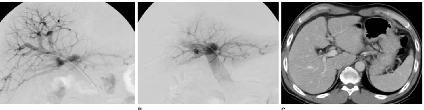
Fig. 1. A 45-year-old man with a hepatocellular carcinoma underwent preoperative portal vein embolization using only gelfoam.
A. Frontal portography before embolization shows intact right portal vein.
B. Frontal portography after embolization using gelfoam only shows occlusion of right portal vein and redirection of portal blood flow to left portal vein.
C. Abdominal CT scan obtained 4 weeks after portal vein embolization shows recanalization of right portal vein.
B
A C
1–70.9%) in GG and 36.7 ± 18.5% (range 8.4–83.2%) in GCG, respectively (p = 0.02) (Table 3).
Portal Vein Recanalization
Portal vein recanalization after PVE was observed in 15/17 after PVE in GG and from 358.4 ± 121.6 cm3 (range 174.3–
673.5 cm3) to 480.2 ± 140.3 cm3 (range 220.9–730.4 cm3) after PVE in GCG. The increased FLR volume was significantly dif- ferent between the two groups (p = 0.02). The mean % increase of FLR volume induced by PVE was 23.7 ± 23.7% (range
Table 2. Comparison of Liver Function Test between Gelfoam and Gelfoam-Coil Groups
Gelfoam Group Gelfoam-Coil Group p
AST, IU/dL
Pre-PVE 49.8 ± 22.3 44.1 ± 30.6 0.19
Post-PVE 77.2 ± 65.8 72.1 ± 59.8 0.59
ALT, IU/dL
Pre-PVE 57.3 ± 42.1 43.9 ± 27.9 0.28
Post-PVE 72.9 ± 52.1 72.9 ± 59.2 0.81
TB, mg/dL
Pre-PVE 0.8 ± 0.4 0.6 ± 0.2 0.33
Post-PVE 2.6 ± 6.7 0.9 ± 0.3 0.18
ALT = alanine aminotransferase, AST = aspartate aminotransferase, PVE = portal vein embolization, TB = total bilirubin
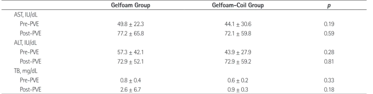
Fig. 2. A 46-year-old man with a hepatocellular carcinoma underwent preoperative portal vein embolization using gelfoam-coil combination.
A. Frontal portography before embolization shows patent right portal vein.
B. Frontal portography after embolization using gelfoam-coil combination shows complete occlusion of right portal vein.
C, D. Abdominal CT scan obtained before PVE (C) and 3 weeks after PVE (D) at the level of celiac trunk shows hypertophic change of left lobe and hepatic attenuation difference and hypotrphic change of right lobe (arrows) suggesting embolized state of right lobe of the liver. The FLRbefore PVE was 431.7 cm3 and the FLRafter PVE was 714 cm3. The % increase of FLR volume after PVE was 65.4%.
FLR = future liver remnant, PVE = portal vein embolization C
A
D B
chronic liver disease (18). These reports suggest that PVE is par- ticularly advantageous in patients with liver disease.
A few studies have compared the effectiveness of different embolic materials using animal models to determine the mate- rial that provides the highest rate of hypertrophy of the non-em- bolized portion of the liver. Huang et al. (10) reported that PVE using permanent embolic materials such as absolute ethanol or a gelfoam-coil combination can induce compensatory hypertro- phy, but gelfoam alone is an inefficient embolic material. Our study showed that total recanalization and the complete recana- lization rate of the portal vein after PVE were 88.2% and 93.3%
in GG patients, respectively. These results support the findings of previous studies that have shown the limitation of gelfoam in achieving complete PVE. De Baere et al. (9) reported that N-bu- tyl cyanoacrylate induces a significantly greater increase in the size of hepatic lobules of hypertrophy than other embolic mate- rials such as hydrophilic gel and polyvinyl alcohol. Gelfoam-coil combinations have been used clinically as embolic materials for PVE in several previous studies (11-13, 19, 20), but few studies have compared the efficacy between permanent and temporary embolic materials. Some interventional radiologists in our insti- tution performed PVE with gelfoam alone because of a few ad- vantages, including safety, low price, easy handling, and mini- mal inflammatory reaction. In addition, they thought that even if migration of gelfoam into portal branches of the contralateral lobe occurred, there would be minimal or no effect on the con- tralateral lobe because gelfoam is an absorbable biomaterial.
However, initial studies with gelfoam reported frequent recanali- zation (16, 21) and less hypertrophy compared with permanent (88.2%) patients in GG and in 8/20 (40%) patients in GCG, re-
spectively (p = 0.003). Complete recanalization of the portal vein was observed in 14/15 (93.3%) patients in GG and 3/8 (37.5%) patients in GCG. The mean % increase of FLR volume was 29.9 ± 26.12% in the recanalization group and 37 ± 15.6%
in the non-recanalization group (p = 0.219).
Changes in Tumor Size
The mean tumor size increased from 4.5 ± 2.9 cm before PVE to 5.0 ± 3.5 cm after PVE in the GG and from 4.3 ± 2.2 cm be- fore PVE to 4.7 ± 2.5 cm after PVE in the GCG. There was no significant difference in tumor size changes between the groups before and after PVE (Table 4). The mean tumor size increased from 4.2 ± 2.5 cm before PVE to 4.6 ± 2.9 cm in the recanaliza- tion group and from 4.9 ± 2.7 cm before PVE to 5.3 ± 3.2 cm in the non-recanalization group (p = 0.816).
DISCUSSION
Since its first clinical application in patients with hilar bile duct cancer to induce compensatory FLR hypertrophy (16), PVE has become a safe and effective procedure for preventing post-resec- tion liver failure due to an insufficient liver remnant (8). Al- though the normal liver is known to tolerate removal of up to 60% of its volume (5), major hepatectomy in patients with chron- ic liver disease such as chronic hepatitis or liver cirrhosis is relat- ed to the risk of hepatic failure due to impaired liver regenera- tion (17). In addition, the rate of postoperative complications is significantly reduced after preoperative PVE in patients with
Table 3. Comparison of Liver Volume Changes before and after PVE between Gelfoam and Gelfoam-Coil Groups
Gelfoam Group Gelfoam-Coil Group p
TLV (cm3) 1409 ± 260.8 1397.2 ± 307.4 0.73
FLRbefore PVE (cm3) 365.4 ± 114.8 358.4 ± 121.6 0.75
% FLR (%) 31.9 ± 10 35 ± 9.2 0.68
FLRafter PVE (cm3) 440.8 ± 114.8 480.2 ± 140.3 0.02
Mean increased volume of FLR (cm3) 75.4 ± 70.8 121.8 ± 56.1 0.02
Mean % increase of FLR volume (%) 23.7 ± 23.7 36.7 ± 18.5 0.02
FLR = future liver remnant, PVE = portal vein embolization, TLV = total liver volume
Table 4. Comparison of Tumor Size Changes before and after PVE between Gelfoam and Gelfoam-Coil Groups
Gelfoam Group Gelfoam-Coil Group p
Tumor size before PVE (cm) 4.5 ± 2.9 4.3 ± 2.2 0.78
Tumor size after PVE (cm) 5.0 ± 3.5 4.7 ± 2.5 0.80
PVE = portal vein embolization
showed that the segmental volume was overestimated by the clas- sic Couinaud’s method by up to 24% and underestimated by 13%.
In conclusion, this study demonstrates that PVE using a gel- foam-coil combination before major hepatectomy for HCC is more effective than PVE using gelfoam alone for induction of compensatory hypertrophy and there is no significant tumor growth rate after PVE between two groups.
REFERENCES
1. Parkin DM, Bray F, Ferlay J, Pisani P. Estimating the world cancer burden: Globocan 2000. Int J Cancer 2001;94:153- 156
2. Forner A, Reig ME, de Lope CR, Bruix J. Current strategy for staging and treatment: the BCLC update and future prospects. Semin Liver Dis 2010;30:61-74
3. Tanaka H, Hirohashi K, Kubo S, Shuto T, Higaki I, Kinoshita H. Preoperative portal vein embolization improves prog- nosis after right hepatectomy for hepatocellular carcino- ma in patients with impaired hepatic function. Br J Surg 2000;87:879-882
4. Denys A, Lacombe C, Schneider F, Madoff DC, Doenz F, Qanadli SD, et al. Portal vein embolization with N-butyl cyanoacrylate before partial hepatectomy in patients with hepatocellular carcinoma and underlying cirrhosis or ad- vanced fibrosis. J Vasc Interv Radiol 2005;16:1667-1674 5. Kubota K, Makuuchi M, Kusaka K, Kobayashi T, Miki K,
Hasegawa K, et al. Measurement of liver volume and he- patic functional reserve as a guide to decision-making in resectional surgery for hepatic tumors. Hepatology 1997;
26:1176-1181
6. Lee KC, Kinoshita H, Hirohashi K, Kubo S, Iwasa R. Exten- sion of surgical indications for hepatocellular carcinoma by portal vein embolization. World J Surg 1993;17:109- 115
7. Madoff DC, Hicks ME, Vauthey JN, Charnsangavej C, Mo- rello FA Jr, Ahrar K, et al. Transhepatic portal vein emboli- zation: anatomy, indications, and technical considerations.
Radiographics 2002;22:1063-1076
8. Abulkhir A, Limongelli P, Healey AJ, Damrah O, Tait P, Jackson J, et al. Preoperative portal vein embolization for major liver resection: a meta-analysis. Ann Surg 2008;247:
embolic materials (22). Therefore, the other interventional radi- ologists in our hospital added coils as embolic materials to pre- vent recanalization by occlusion of proximal portal blood flow.
In addition, an experimental study reported that proximal and complete revascularization occurred 6–8 and 12–16 days after PVE using gelfoam (23). De Baere et al. (22) demonstrated that a key factor in achieving satisfactory hypertrophy of the non- embolized liver is complete and durable occlusion of the portal vein, and showed that cyanoacrylate seemed to induce better and faster hypertrophy than gelfoam or coils alone. In the same study, coils alone showed worse results in compensatory hyper- trophy than gelfoam. The result could be explained by distal re- entry through the intraparenchymatous vascular shunt, which opened after proximal portal vein occlusion. To prevent this, the distal portal venous branch should be occluded with other em- bolic materials such as gelfoam or polyvinyl alcohol.
Our study showed that PVE using a gelfoam-coil combina- tion in patients with chronic liver disease is more likely to in- duce greater compensatory hypertrophy of the non-embolized portion of the liver than PVE using gelfoam alone. Further- more, PVE using a gelfoam-coil combination showed no signifi- cant differences in postprocedural complications. Therefore, we believe that PVE should be performed using permanent embol- ic materials in patients with liver damage.
Since the first description of the potential of intrahepatic tu- mor enlargement after PVE (24), accumulating evidence has shown that PVE stimulates tumor growth in both embolized and non-embolized liver segments (13, 25). Hayashi et al. (26) reported that the median tumor growth rate of primary liver cancer in the embolized lobe after PVE was approximately two times greater than that before PVE. Three possible mechanisms have been proposed: changes in cytokine and growth factor se- cretion, alteration in hepatic blood flow, and enhanced cellular host response that promotes local tumor growth (27). To the best of our knowledge, few studies have compared the tumor growth rate according to the embolic materials used. There was no significant difference in tumor growth rate between the two groups in this study.
The limitations of this study include the following. First, it is a retrospective study. Second, there may be a gap between the esti- mated FLR volume using CT volumetry according to Couinaud’s classification and the actual FLR volume. Fischer et al. (28)
19. Vauthey JN, Chaoui A, Do KA, Bilimoria MM, Fensterm- acher MJ, Charnsangavej C, et al. Standardized measure- ment of the future liver remnant prior to extended liver resection: methodology and clinical associations. Surgery 2000;127:512-519
20. Kim MJ, Choo SW, Do YS, Park KB, Han YH, Choo IW, et al.
Use of double-occlusion balloon catheter: preoperative por- tal vein embolization for induction of future remnant liver hypertrophy. Cardiovasc Intervent Radiol 2004;27:16-20 21. de Baere T, Roche A, Vavasseur D, Therasse E, Indushekar S,
Elias D, et al. Portal vein embolization: utility for inducing left hepatic lobe hypertrophy before surgery. Radiology 1993;188:73-77
22. de Baere T, Roche A, Elias D, Lasser P, Lagrange C, Bousson V. Preoperative portal vein embolization for extension of hepatectomy indications. Hepatology 1996;24:1386-1391 23. Lainas P, Boudechiche L, Osorio A, Coulomb A, Weber A, Pa- riente D, et al. Liver regeneration and recanalization time course following reversible portal vein embolization. J Hep- atol 2008;49:354-362
24. Elias D, De Baere T, Roche A, Mducreux, Leclere J, Lasser P.
During liver regeneration following right portal emboliza- tion the growth rate of liver metastases is more rapid than that of the liver parenchyma. Br J Surg 1999;86:784-788 25. van Gulik TM, van den Esschert JW, de Graaf W, van Lien-
den KP, Busch OR, Heger M, et al. Controversies in the use of portal vein embolization. Dig Surg 2008;25:436-444 26. Hayashi S, Baba Y, Ueno K, Nakajo M, Kubo F, Ueno S, et
al. Acceleration of primary liver tumor growth rate in em- bolized hepatic lobe after portal vein embolization. Acta Radiol 2007;48:721-727
27. de Graaf W, van den Esschert JW, van Lienden KP, van Gu- lik TM. Induction of tumor growth after preoperative por- tal vein embolization: is it a real problem? Ann Surg Oncol 2009;16:423-430
28. Fischer L, Cardenas C, Thorn M, Benner A, Grenacher L, Vetter M, et al. Limits of Couinaud’s liver segment classifi- cation: a quantitative computer-based three-dimensional analysis. J Comput Assist Tomogr 2002;26:962-967 49-57
9. de Baere T, Denys A, Paradis V. Comparison of four embolic materials for portal vein embolization: experimental study in pigs. Eur Radiol 2009;19:1435-1442
10. Huang JY, Yang WZ, Li JJ, Jiang N, Zheng QB. Portal vein embolization induces compensatory hypertrophy of rem- nant liver. World J Gastroenterol 2006;12:408-414 11. Wakabayashi H, Okada S, Maeba T, Maeta H. Effect of pre-
operative portal vein embolization on major hepatectomy for advanced-stage hepatocellular carcinomas in injured livers: a preliminary report. Surg Today 1997;27:403-410 12. Wakabayashi H, Ishimura K, Okano K, Karasawa Y, Goda F,
Maeba T, et al. Application of preoperative portal vein em- bolization before major hepatic resection in patients with normal or abnormal liver parenchyma. Surgery 2002;131:
26-33
13. Kokudo N, Tada K, Seki M, Ohta H, Azekura K, Ueno M, et al. Proliferative activity of intrahepatic colorectal metas- tases after preoperative hemihepatic portal vein emboli- zation. Hepatology 2001;34:267-272
14. Bruix J, Sherman M; American Association for the Study of Liver Diseases. Management of hepatocellular carcino- ma: an update. Hepatology 2011;53:1020-1022
15. Omary RA, Bettmann MA, Cardella JF, Bakal CW, Schwartz- berg MS, Sacks D, et al. Quality improvement guidelines for the reporting and archiving of interventional radiology procedures. J Vasc Interv Radiol 2002;13(9 Pt 1):879-881 16. Makuuchi M, Takayasu K, Takuma T, Yamazaki S, Hasegawa
H, Nishiura S, et al. Preoperative transcatheter emboliza- tion of the portal venous branch for patients receiving extended lobectomy due to the bile duct carcinoma. J Jpn Soc Clin Surg 1984;45:1558-1564
17. Lin TY, Chen CC. Metabolic function and regeneration of cirrhotic and non-cirrhotic livers after hepatic lobectomy in man. Ann Surg 1965;162:959-972
18. Farges O, Belghiti J, Kianmanesh R, Regimbeau JM, Santo- ro R, Vilgrain V, et al. Portal vein embolization before right hepatectomy: prospective clinical trial. Ann Surg 2003;
237:208-217
만성간질환 환자에서 젤폼과 젤폼-코일을 이용한 수술 전 간문맥색전술의 효과에 대한 비교 연구1
신성욱
1· 장일수
2· 주성욱
1· 도영수
1· 박홍석
1· 박광보
1· 조성기
1· 주인욱
1목적: 만성간질환 환자를 대상으로 젤폼만 사용한 경우와 젤폼-코일을 같이 사용한 수술 전 간문맥색전술의 결과를 비 교하고자 하였다.
대상과 방법: 2003년 4월부터 2007년 9월까지 간세포암으로 진단받고 수술 전 간문맥색전술을 시행 받은 37명의 환자 를 대상으로 하였고 젤폼만 사용한 경우와 젤폼과 코일을 같이 사용한 경우는 각각 17명, 20명이었다. 시술 후 2~4주 후 컴퓨터단층촬영을 통해 간부피의 변화, 간문맥의 재소통률, 종양 크기 변화를 평가하였다.
결과: 잔여 간용적의 평균 증가율은 젤폼군이 23.7 ± 23.7%, 젤폼-코일군이 36.7 ± 18.5%였다(p = 0.02). 간문맥의 재소통은 젤폼군에서 17명 중 15명, 젤폼-코일군에서는 20명 중 8명에서 발생하였다(p = 0.003). 젤폼군의 시술 전 평 균 종양 크기는 4.5 ± 2.9 cm였고 시술 후에는 5.0 ± 3.5 cm였으며 젤폼-코일군에서는 각각 4.3 ± 2.2 cm, 4.7 ± 2.5 cm로 측정되어 두 군 간에 종양 크기 증가는 차이가 없었다(p = 0.80).
결론: 만성간질환 환자에 있어서 젤폼-코일을 같이 사용하여 간문맥색전술을 시행하는 것이 젤폼만 사용하는 것에 비해 잔여 간용적을 더 크게 자라게 하는 데 효과적이다.
1성균관대학교 의과대학 삼성서울병원 영상의학과, 2건국대학교 의학전문대학원 영상의학교실